This study compared five machine learning algorithms for thyroid nodule classification using super-resolution ultrasound features. SVM performed best (accuracy 68.6%, Area Under the Curve 0.690), identifying microcalcification and post-contrast enlargement as key contributors, but the findings require external validation before clinical use.
A subscription to JoVE is required to view this content. Sign in or start your free trial.
Research Article
Quantitative Super-Resolution Ultrasound Microvascular Features for Machine Learning-Based Classification of Thyroid Nodules
210 views
⸱
September 11th, 2026
* These authors contributed equally
In This Article
Summary
Abstract
This study compared five machine learning algorithms—Random Forest (RF), Support Vector Machine (SVM), Decision Tree (DT), eXtreme Gradient Boosting (XGBoost), and Gradient Boosting (GB)—for classifying thyroid nodules using quantitative features derived from conventional ultrasound, contrast-enhanced ultrasound (CEUS), and super-resolution ultrasound (SRUS). The retrospective study included 68 thyroid nodules from 63 patients (30 benign and 38 confirmed as papillary thyroid carcinoma [PTC]) and was analyzed using 25 quantitative features via five-fold patient-grouped cross-validation, ensuring that all nodules from the same patient were assigned to the same fold. Model performance was summarized across folds and from pooled out-of-fold (OOF) predictions, and a focused OOF SHapley Additive exPlanations (SHAP) analysis using a permutation-based explainer was performed for the best-performing SVM. SVM achieved the highest mean accuracy (0.686 ± 0.120) and mean Receiver Operating Characteristic-Area Under Curve (ROC-AUC) (0.690 ± 0.164), with a pooled OOF sensitivity of 0.842 and specificity of 0.500, while microcalcification and post-contrast enlargement were identified as the most significant model-specific SHAP contributors. RF offered balanced performance (accuracy 55.9%, sensitivity 55.3%, specificity 56.7%), whereas DT performed poorly (accuracy 0.429, ROC-AUC 0.447), approaching random guessing. The study demonstrated that quantitative SRUS measurements can be incorporated into conventional classifiers; however, the findings remain exploratory and require prospective, multicenter validation with larger cohorts before clinical implementation.
Introduction
Thyroid nodules represent a highly prevalent clinical finding, with detection rates approaching 68% in asymptomatic populations when examined by ultrasound examination1. Recent meta-analyses have revealed that the global prevalence of thyroid nodules has increased from 21.53% during 2000-2011 to 29.29% during 2012-2022, affecting approximately one in every four individuals in the general population2,3. Among these nodules, 7–15% are ultimately diagnosed as thyroid cancer, of which papillary thyroid carcinoma (PTC) represents the predominant subtype4,5. PTC accounts for 80–90% of all thyroid malignancies and has become one of the fastest-growing cancer types, with incidence rates increasing from 4.8 to 14.9 per 100,000 between 1975 and 20126.
Conventional ultrasound remains the cornerstone for thyroid nodule evaluation, offering a non-invasive, cost-effective, and widely accessible diagnostic modality7. Traditional sonographic features suggestive of malignancy include marked hypo-echogenicity, irregular margins, microcalcifications, and taller-than-wide shape8,9. However, it is fundamentally limited by the acoustic diffraction barrier, which restricts spatial resolution to approximately 100 micrometers10. This limitation significantly constrains the ability to visualize microvasculature and assess fine vascular morphology, which are critical parameters for distinguishing between benign and malignant thyroid lesions11. Color Doppler flow imaging (CDFI) and contrast-enhanced ultrasound (CEUS), while improving blood flow detection, can only visualize vessels with flow velocities exceeding 1 cm/s and remain constrained by diffraction-limited resolution12,13.
Super-resolution ultrasound (SRUS), as a transformative technology, overcomes the traditional diffraction limit of conventional ultrasound systems14,15. By leveraging contrast microbubbles as point targets for localization and tracking, SRUS achieves micron-scale spatial resolution, enabling visualization and quantification of microvasculature at an unprecedented level of detail16,17. This technique can measure both microvascular density (MVD) and microvascular flow rate (MFR), providing structural and functional assessments precisely unattainable through non-invasive imaging17,18. Pilot studies have demonstrated that benign thyroid nodules exhibit significantly higher MFR than malignant ones, with mean values of 16.76 ± 6.82 mm/s versus 9.86 ± 4.54 mm/s, respectively19. The introduction of "ultrasound microvasculomics"—a high-throughput extraction of quantitative features from SRUS images—further enhances the potential for precise and personalized microvascular assessment.
Machine learning (ML) has revolutionized medical image analysis, particularly in thyroid nodule characterization20,21. Deep learning models, particularly convolutional neural networks (CNNs), have achieved Receiver Operating Characteristic-Area Under Curve (ROC-AUC) values exceeding 0.90 in previous studies22,23,24. Ensemble machine learning approaches combining multiple algorithms—including Random Forest (RF), Support Vector Machines (SVM), Decision Trees (DT), Gradient Boosting (GB), and eXtreme Gradient Boosting (XGBoost)—have shown promise in improving diagnostic accuracy and robustness across heterogeneous datasets21. As Habchi et al.25 recently reviewed comprehensively, the field of artificial intelligence (AI) for thyroid cancer has rapidly expanded to include visual converters, large language models, and hybrid architectures, reflecting the development of the field towards increasingly complex image-based diagnostic frameworks.
While conventional ultrasound features have been extensively studied in conjunction with AI algorithms, the combination of super-resolution microvascular parameters with ML models remains largely unexplored. Despite the promising potential of SRUS technology and the proven efficacy of machine learning algorithms in medical imaging, several critical questions remain unanswered. First, which machine learning architecture—whether traditional algorithms such as RF and SVM, or advanced ensemble methods like XGBoost and GB—performs optimally when applied to SRUS-derived microvascular features? Second, how do these models compare in terms of sensitivity, specificity, and overall diagnostic accuracy for differentiating benign from malignant thyroid nodules? Third, which microvascular parameters extracted from SRUS imaging contribute most significantly to classification performance? Addressing these questions is essential for establishing evidence-based frameworks for the clinical translation of SRUS-assisted thyroid nodule diagnosis.
Accordingly, this study aims to comprehensively evaluate the performance of five ML algorithms (RF, SVM, DT, XGBoost, and GB) for classifying thyroid nodules using super-resolution ultrasound microvascular imaging data. By systematically comparing these approaches and identifying the most informative microvascular biomarkers, this investigation seeks to advance the diagnostic paradigm for thyroid nodule characterization and contribute to the growing body of evidence supporting the clinical utility of SRUS technology in oncological imaging. We hypothesized that the prespecified 25 variable feature set would provide above-chance discrimination between benign nodules and nodules reported as papillary thyroid carcinoma. Patient-grouped cross-validation was used to reduce information leakage, and a focused SHAP analysis was added to describe how the selected classifier used the measured variables. Both analyses were exploratory.
Access restricted. Please log in or start a trial to view this content.
Protocol
Study design and patient population
This retrospective study analyzed thyroid ultrasound examinations obtained between 13 June 2024 and 13 January 2025. The study protocol was approved by the Ethics Committee of Beijing Friendship Hospital, Capital Medical University (approval No. BFHHZS20240300) and was conducted in accordance with the ethical principles outlined in the Declaration of Helsinki, and informed consent was waived because of the retrospective design. The dataset comprised 68 thyroid nodules from 63 patients (30 benign and 38 malignant), and the nodule was the analytic unit. Reference diagnoses were based on ultrasound-guided fine-needle aspiration cytology (FNAC). The malignant group comprised 38 nodules reported as papillary thyroid carcinoma on FNAC.
Inclusion criteria
Eligible patients were required to meet all of the following criteria: (1) underwent conventional grayscale and Doppler ultrasonography, followed by CEUS with satisfactory image quality permitting subsequent microvascular flow imaging analysis; (2) had a cytopathological diagnosis of PTC confirmed by fine-needle aspiration biopsy, with or without concomitant positive BRAFV600E mutation testing; or had a Bethesda Category III (atypia of undetermined significance) cytology result, but with concurrent positive BRAFV600E mutation testing; (3) had a cytopathological diagnosis of benign, proliferative nodules, adenomatous nodules, or benign follicular nodules in Bethesda Category II without concomitant positive BRAFV600E mutation testing.
Exclusion criteria
Patients were excluded from the study if any of the following conditions were present: (1) poor-quality CEUS images or insufficient microbubble signal precluding reliable microvascular flow assessment; (2) absence of cytological or pathological results; (3) histopathological diagnosis of a rare or special subtype of thyroid carcinoma (e.g. anaplastic variants); or (4) cytological findings suggestive of follicular neoplasm (Bethesda Category IV) or any lesion with indeterminate follicular origin, regardless of mutational status; (5) Hashimoto's thyroiditis
Ultrasound, CEUS, and SRUS acquisition
All examinations were performed with the referenced ultrasound system and a linear-array transducer. Patients were positioned supine with the neck extended. The target nodule was localized and measured on B-mode ultrasound; microcalcifications were recorded, and the same lesion-centered imaging plane was used to assess intranodular vascularity with Color Doppler.
After intravenous access was established, the system was switched to a low-mechanical-index CEUS/URM mode (URM means Ultra-Resolution Microscopy imaging). CEUS was performed with a SonoVue intravenous bolus injection of 1.2 mL immediately followed by a saline flush of 5ml; the onscreen timer and continuous cine acquisition were started with bolus administration. The probe was kept in a fixed plane with minimal pressure, and the patient was asked to avoid swallowing during wash-in and wash-out.
For quantitative CEUS, a lesion-restricted region of interest was used to derive the time-intensity parameters. For SRUS, the URM workflow localized and tracked microbubble signals after motion control and generated vessel-ratio, complexity, microvascular density, perfusion index, and flow velocity measurements. The representative image exports showed settings of VSP 4, RES 2, CTR 3, SM 2, VEN 3, and CPT 10 s; corresponding settings were unavailable for the remaining examinations.
The 25 model-input variables are listed in Table 1 and grouped by acquisition modality: age and sex; B-mode microcalcification; Color Doppler intranodular vascularity; qualitative CEUS; quantitative CEUS; and 11 SRUS microvascular measurements.
Feature selection
All 25 quantitative features were retained in the machine learning models. Only numeric features were used; patient names, registration numbers, and lesion-size fields were excluded. No data-driven feature selection was performed, and the same prespecified predictors were entered into each classifier.
Feature standardization
No transformation, imputation, or global scaling was applied before cross-validation. Standardization was applied only to the radial-basis-function SVM via a StandardScaler pipeline. The scaler was fitted on each fold's training nodules and then applied to that fold's validation nodules. Tree-based classifiers received the original numeric scales:

where x is the original feature value, µ is the training-fold mean, and σ is the training-fold standard deviation. Fold-wise preprocessing prevented validation observations from contributing to the SVM scaling parameters.
Data partitioning
Primary performance evaluation used five-fold StratifiedGroupKFold cross-validation with shuffle = True and random_state = 42. A patient identifier defined 63 groups, and all nodules from the same patient were assigned to the same fold. No patient contributed nodules to both the training and validation subsets of a fold.
Model training and validation
Training protocol
Five classifiers were evaluated: random forest (100 trees; random_state = 42), radial-basis-function SVM (C = 1.0; gamma = scale; probability = True; StandardScaler pipeline; random_state = 42), decision tree (Gini criterion; unrestricted depth; random_state = 42), XGBoost (100 estimators; learning_rate = 0.3; max_depth = 6; subsample = 1.0; colsample_bytree = 1.0; random_state = 42), and gradient boosting (100 estimators; learning_rate = 0.1; max_depth = 3; random_state = 42). No grid search, Bayesian optimization, threshold tuning, or nested model selection was performed.
Cross-validation
Within each of five patient-grouped folds, models were trained on the remaining patient groups and evaluated on the held-out groups. Accuracy, sensitivity, specificity, precision, F1-score, and ROC-AUC were calculated for each fold and summarized as mean ± SD. OOF predictions were pooled across all 68 nodules to generate one cross-validated ROC curve and one confusion matrix for each model.
Performance evaluation
Evaluation metrics
Model performance was assessed within each patient-grouped validation fold and from pooled OOF predictions using the following metrics:
Accuracy: Overall proportion of correct predictions

Sensitivity (Recall): Proportion of actual malignant nodules correctly identified


Specificity: Proportion of actual benign nodules correctly identified
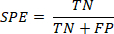
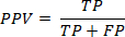
Precision (Positive Predictive Value): Proportion of predicted malignant cases that were truly malignant
F1-Score: Harmonic mean of precision and recall

Area Under the Receiver Operating Characteristic Curve (ROC-AUC): Measure of the model's ability to discriminate between benign and malignant nodules across all classification thresholds
where TP = true positives (correctly identified malignant nodules), TN = true negatives (correctly identified benign nodules), FP = false positives (benign nodules incorrectly classified as malignant), and FN = false negatives (malignant nodules incorrectly classified as benign).
Confusion matrix analysis
OOF confusion matrices were generated by pooling the predictions made for each nodule only in the fold in which that patient was held out. Thus, each nodule received a prediction from a model trained on nodules from other patients.
Feature importance analysis
For the Random Forest model, feature importance scores were calculated based on the mean decrease in Gini impurity across all decision trees. The top 15 most important features were identified and ranked to determine which microvascular parameters contributed most significantly to classification performance.
Exploratory SHAP analysis
A focused OOF SHAP analysis using a permutation-based explainer was performed for the SVM. For each held-out nodule, only the corresponding training-fold observations were used as the background distribution (128 antithetic permutations; random seed = 20260716). Mean absolute SHAP values summarized the magnitude of contributions, and signed values indicated the direction. The analysis was exploratory and not used to infer causality, identify independent biomarkers, or define clinical thresholds.
Statistical analysis
Comparative analysis
Model performance was summarized descriptively across the five patient-grouped validation folds and in pooled OOF predictions. No formal between-model hypothesis test or P-value-based ranking was performed because the folds are related and the cohort is small.
For baseline group comparisons, normality of continuous variables was assessed within each outcome group using the Shapiro-Wilk test. Welch's t test was used when both groups were compatible with normality; otherwise, a two-sided Mann-Whitney U test was used. Categorical variables were assessed with Pearson's chi-square test, with Fisher's exact test for sparse 2 x 2 tables. P values were two-sided, exploratory, and unadjusted (alpha = 0.05).
Reproducibility
Reproducibility was supported by a specified patient-group definition, fixed random seed (42), fixed classifier configurations, and fold-wise preprocessing. Identifiers were used only for grouping and were not entered as predictors or exported with model outputs.
Access restricted. Please log in or start a trial to view this content.
Results
Patient characteristics and study population
A total of 68 thyroid nodules from 63 patients were included, comprising 30 benign nodules and 38 PTCs. The nodule was the analytic unit, and patient grouping prevented nodules from the same patient from appearing in both the training and validation subsets. Baseline characteristics are summarized in Table 2.
Figure 1 shows technically successful lesion-restr...
Access restricted. Please log in or start a trial to view this content.
Discussion
Current diagnostic approaches for thyroid nodules rely primarily on conventional ultrasound, which remains inherently operator-dependent and subject to inter-observer variability5 and the inability to visualize microvasculature below the diffraction limit. To our knowledge, our study is the first comprehensive evaluation of multiple machine learning algorithms applied to super-resolution ultrasound (SRUS) microvascular imaging data, achieving an objective quantitative assessment of the risk of PTC...
Access restricted. Please log in or start a trial to view this content.
Disclosures
The authors have no conflicts of interest to declare.
Acknowledgements
The authors thank the staff of the Department of Ultrasound of Beijing Friendship Hospital, Capital Medical University, for their assistance in patient data collection.
Access restricted. Please log in or start a trial to view this content.
Materials
| Name | Company | Catalog Number | Comments |
|---|---|---|---|
| Decision Tree classifier | scikit-learn | Version 1.5.2 | Machine learning classifier |
| Fine-needle aspiration biopsy needle | Used for ultrasound-guided FNAC | ||
| Gradient Boosting classifier | scikit-learn | Version 1.5.2 | Machine learning classifier |
| Intravenous catheter | Used for contrast administration | ||
| Python | Python Software Foundation | Version 3.11.9 | Programming language used for the machine-learning analysis |
| Random Forest classifier | scikit-learn | Version 1.5.2 | Machine learning classifier |
| SHAP (permutation explainer) | SHAP Developers | Version 0.46.0 | Model explainability analysis |
| SonoVue contrast agent | Bracco | Ultrasound contrast agent for CEUS/SRUS imaging | |
| StandardScaler | scikit-learn | Version 1.5.2 | Feature standardization for the SVM pipeline |
| Sterile normal saline | Used for intravenous flush following contrast injection | ||
| StratifiedGroupKFold | scikit-learn | Version 1.5.2 | Patient-grouped cross-validation |
| Support Vector Machine (SVM) classifier | scikit-learn | Version 1.5.2 | Radial-basis-function kernel classifier |
| U5-15LE linear-array transducer | VINNO Technology | U5-15LE | Linear ultrasound probe |
| ULTIMUS 9E ultrasound system | VINNO Technology | ULTIMUS 9E | Ultrasound imaging system used for B-mode, Color Doppler, CEUS, and SRUS imaging |
| XGBoost | XGBoost Developers | Version 2.1.1 | Gradient boosting framework |
References
- Mu C, et al. Mapping global epidemiology of thyroid nodules among general population: a systematic review and meta-analysis. Front Oncol. 2022;12:1029926.
- Moon JH, et al. Prevalence of thyroid nodules and their associated clinical parameters: a large-scale, multicenter-based health checkup study. Korean J Intern Med. 2018;33(4):753-62.
- Dean DS, Gharib H. Epidemiology of thyroid nodules. Best Pract Res Clin Endocrinol Metab. 2008;22(6):901-11.
- Uppal N, Collins R, James B. Thyroid nodules: global, economic, and personal burdens. Front Endocrinol (Lausanne). 2023;14:1113977.
- Durante C, et al. The diagnosis and management of thyroid nodules: a review. JAMA. 2018;319(9):914-24.
- Lim H, Devesa SS, Sosa JA, Check D, Kitahara CM. Trends in thyroid cancer incidence and mortality in the United States, 1974-2013. JAMA. 2017;317(13):1338-48.
- Gharib H, et al. American Association of Clinical Endocrinologists, American College of Endocrinology, and Associazione Medici Endocrinologi medical guidelines for clinical practice for the diagnosis and management of thyroid nodules—2016 update. Endocr Pract. 2016;22(5):622-39.
- Tessler FN, et al. ACR Thyroid Imaging, Reporting and Data System (TI-RADS): white paper of the ACR TI-RADS Committee. J Am Coll Radiol. 2017;14(5):587-95.
- Petranović Ovčariček P, et al. The 2025 ATA guidelines for differentiated thyroid cancer—ten years of professional debate and measured progress. Eur J Nucl Med Mol Imaging. 2026;53(4):2213-6.
- Christensen-Jeffries K, et al. Super-resolution ultrasound imaging. Ultrasound Med Biol. 2020;46(4):865-91.
- Carmeliet P. Angiogenesis in health and disease. Nat Med. 2003;9(6):653-60.
- Zhang B, et al. Utility of contrast-enhanced ultrasound for evaluation of thyroid nodules. Thyroid. 2010;20(1):51-7.
- Rago T, Vitti P. Role of thyroid ultrasound in the diagnostic evaluation of thyroid nodules. Best Pract Res Clin Endocrinol Metab. 2008;22(6):913-28.
- Xia S, et al. Super-resolution ultrasound and microvasculomics: a consensus statement. Eur Radiol. 2024;34(11):7503-13.
- Song P, Rubin JM, Lowerison MR. Super-resolution ultrasound microvascular imaging: is it ready for clinical use? Z Med Phys. 2023;33(3):309-23.
- Errico C, et al. Ultrafast ultrasound localization microscopy for deep super-resolution vascular imaging. Nature. 2015;527(7579):499-502.
- Couture O, Hingot V, Heiles B, Muleki-Seya P, Tanter M. Ultrasound localization microscopy and super-resolution: a state of the art. IEEE Trans Ultrason Ferroelectr Freq Control. 2018;65(8):1304-20.
- Gao JY, Hou C. Progresses and clinical application of super-resolution ultrasound imaging: a narrative review. Ultrasound J. 2025;17(1):29.
- Zhang G, et al. Ultrasound super-resolution imaging for the differential diagnosis of thyroid nodules: a pilot study. Front Oncol. 2022;12:978164.
- Wildman-Tobriner B, et al. Using artificial intelligence to revise ACR TI-RADS risk stratification of thyroid nodules: diagnostic accuracy and utility. Radiology. 2019;292(1):112-9.
- Toro-Tobon D, et al. Artificial intelligence in thyroidology: a narrative review of the current applications, associated challenges, and future directions. Thyroid. 2023;33(8):903-17.
- Peng S, et al. Deep learning-based artificial intelligence model to assist thyroid nodule diagnosis and management: a multicentre diagnostic study. Lancet Digit Health. 2021;3(4):e250-9.
- Chen C, et al. Deep learning to assist composition classification and thyroid solid nodule diagnosis: a multicenter diagnostic study. Eur Radiol. 2024;34(4):2323-33.
- Bini F, et al. Artificial intelligence in thyroid field: a comprehensive review. Cancers (Basel). 2021;13(19):4740.
- Habchi Y, Kheddar H, Himeur Y, Ghanem MC. Machine learning and transformers for thyroid carcinoma diagnosis. J Vis Commun Image Represent. 2026;115:104668.
- Zhang M, Wang X, Deng Y, Tang K. Multimodal ultrasound integration pathways and paradigm innovations in precision diagnosis and treatment of thyroid cancer. Acad Radiol. 2026. Epub ahead of print.
- Feng JW, et al. Development and validation of the multidimensional machine learning model for preoperative risk stratification in papillary thyroid carcinoma: a multicenter, retrospective cohort study. Cancer Imaging. 2025;25(1):98.
- Gatta E, et al. Machine learning for diagnosis of malignant thyroid nodules based on thyroid ultrasound: systematic review and meta-analysis of studies with external datasets. Eur J Radiol Open. 2026;16:100716.
- Li J, Guo Q, Peng S, Tan X. Super-resolution based nodule localization in thyroid ultrasound images through deep learning. Curr Med Imaging. 2024;20(1):e15734056269264.
- He S, et al. Super-resolution ultrasound radiomics for pre-FNA prediction of nondiagnostic (Bethesda I) thyroid nodules. Front Endocrinol (Lausanne). 2026;17:1710097.
- Habchi Y, Kheddar H, Ghanem MC, Hwaidi J. Adaptive bandelet transform and transfer learning for geometry-aware thyroid cancer ultrasound classification. Diagnostics (Basel). 2026;16(4):554.
- Habchi Y, Kheddar H, Himeur Y. Adaptive bandelet transform and transfer learning for geometry-aware thyroid cancer ultrasound classification [conference paper]. In: 2024 International Conference on Telecommunications and Intelligent Systems (ICTIS). IEEE; 2024. p. 1-6.
- Xu Y, Xu M, Geng Z, Liu J, Meng B. Thyroid nodule classification in ultrasound imaging using deep transfer learning. BMC Cancer. 2025;25(1):544.
- Gou TH, et al. Application value of super-resolution ultrasound imaging in the diagnosis of American College of Radiology Thyroid Imaging Reporting and Data System category 4 and 5 thyroid nodules. Zhongguo Yi Xue Ke Xue Yuan Xue Bao. 2026. doi:10.3881/j.issn.1000-503X.16808.
Access restricted. Please log in or start a trial to view this content.