The bibliometric analysis reveals that positron emission tomography/computed tomography research in skeletal metastasis has shifted from diagnostic validation toward theranostic integration and multi-target precision imaging.
Research Article
A Bibliometric Analysis of Positron Emission Tomography/Computed Tomography in Skeletal Metastasis: Research Trends from 2016 to 2025
In This Article
Summary
Abstract
Skeletal metastasis is a common complication of advanced malignancies, and positron emission tomography/computed tomography (PET/CT) plays an increasingly important role in its diagnosis and management. Given the rapid evolution of this field over the past decade, a timely bibliometric assessment is warranted to delineate its research landscape and emerging trends. This study aimed to comprehensively analyze the global research landscape of PET/CT in skeletal metastasis from 2016 to 2025. Publications were retrieved from the Web of Science Core Collection on April 6, 2026, using a topic search strategy. After screening, 1,154 articles and reviews in English were included. Bibliometric analyses were performed using VOSviewer, CiteSpace, and Scimago Graphica. Annual publication output showed a fluctuating growth trend, peaking in 2025 with 137 papers. China ranked first in publication volume (297 papers), followed by the United States (230 papers) and Germany (150 papers). In terms of citation impact, the United States had the highest total citations (7,628), while the Netherlands achieved the highest average citations per paper (56.7). The European Journal of Nuclear Medicine and Molecular Imaging was the most productive and most cited journal. International collaboration was strongest among the United States, Germany, and the United Kingdom. The included literature covered a broad range of PET tracers, including prostate-specific membrane antigen (PSMA)-targeted agents, choline analogs, fibroblast activation protein inhibitor (FAPI), and somatostatin receptor (SSTR)-targeted tracers. Research hotspots evolved from early validation of novel tracers (68Ga-PSMA, 18F-choline) to PSMA-targeted theranostics and multi-target precision imaging, and more recently to FAPI-based imaging, neuroendocrine tumors, whole-body assessment, and treatment efficacy monitoring. Emerging topics include clinical translation of novel probes, cross-cancer applications, quantitative radiomics, and international collaboration. This evolution indicates that PET/CT research in skeletal metastasis has been moving steadily toward multi-target, cross-cancer, integrated theranostic precision medicine.
Introduction
Skeletal metastasis is a common complication of advanced malignancies, especially breast, prostate, lung, and thyroid cancers1,2,3,4,5. It often leads to skeletal-related events, including pathological fractures, spinal cord compression, hypercalcemia, and intractable pain, which impair quality of life and reduce survival6,7,8,9,10. Positron emission tomography/computed tomography (PET/CT) has emerged as a powerful molecular imaging modality that integrates functional metabolic information with anatomical localization11,12,13,14,15. Compared with conventional techniques such as bone scintigraphy, computed tomography (CT), or magnetic resonance imaging (MRI), PET/CT offers superior sensitivity and specificity for detecting bone metastases16,17,18,19,20. Commonly used tracers like 18F‑FDG, 18F‑NaF, and 68Ga‑FAPI have broadened diagnostic capabilities across different clinical scenarios21,22. In particular, prostate-specific membrane antigen (PSMA)-targeted PET and somatostatin receptor (SSTR)-targeted PET have shown significant value in detecting skeletal metastases in prostate cancer and neuroendocrine tumors, respectively, further expanding the clinical utility of PET/CT in this field23. Over the past two decades, a rapidly growing body of literature has investigated the application of PET/CT in skeletal metastasis, covering diagnostic accuracy, prognostic value, treatment response assessment, tracer development, and comparisons with other modalities24.
Despite this wealth of publications, the rapid expansion and wide dispersion of the literature make it difficult for researchers and clinicians to systematically grasp the overall structure, evolution, and emerging frontiers of the field. Bibliometric analysis provides a quantitative and visual approach to map knowledge landscapes by examining publication outputs, citation networks, co‑authorship, keyword co‑occurrence, and collaboration patterns25,26. Through quantitative analysis of publication volume, citation relationships, and author collaborations, this method can objectively and systematically identify research hotspots, highly influential works, and core collaboration networks in a given field27. To date, no comprehensive bibliometric study has specifically focused on the role of PET/CT in skeletal metastasis. Therefore, this study aims to conduct a bibliometric analysis of global research on PET/CT in skeletal metastasis, including annual trends, productive countries, institutions, journals, and authors, research hotspots, temporal evolution of frontiers, and visualized knowledge maps, thereby guiding future investigations in this field.
Protocol
Data source and retrieval strategy
The data for this study were retrieved from the Web of Science Core Collection database on April 6, 2026. The following Core Collection indexes were searched: Science Citation Index Expanded (SCI-EXPANDED) and Social Sciences Citation Index (SSCI). An advanced search was conducted, limiting the search scope to the topic field of the literature. The specific search query was constructed as follows: TS=(“bone metast” OR “skeletal metast” OR “osseous metast”) AND TS=(“Positron Emission Tomography Computed Tomography” OR “CT PET” OR “CT PET Scan” OR “PET CT Scan” OR “PET/CT” OR “PET/CT Scan” OR “Positron Emission Tomography-Computed Tomography”). The retrieval period was set from January 1, 2016, to December 31, 2025, to capture the most recent decade of research activity in this rapidly evolving field, as the past ten years have witnessed substantial advances in PET/CT technology, novel tracer development, and clinical applications in skeletal metastasis, making this timeframe most representative of the current research landscape.
Inclusion and exclusion criteria
To minimize bias in subsequent analyses, a rigorous screening process was applied. The inclusion criteria were: peer-reviewed original articles or reviews; published in English; published between 2016 and 2025. The exclusion criteria were: conference abstracts, conference papers, editorials, book chapters, and retracted publications; non-English publications; duplicate records. Preclinical studies, simulation studies, case reports, and case series were excluded from the final analysis, as the present bibliometric study focused on peer-reviewed original research and review articles that provide systematic evidence and comprehensive overviews of the field. Duplicate records were identified by comparing author names, titles, journal names, publication years, and DOIs using the “Find Duplicates” function in the bibliometric software and manual verification. Identified duplicates were removed prior to further analysis.
Data export and preprocessing
Full records and cited references of the literature meeting the inclusion criteria were exported from the database in plain text format. The exported data included core metadata fields such as author names, article titles, source publications, abstracts, keywords, reference lists, and citation counts. The exported data files were archived on local storage devices for subsequent reading and visualization by bibliometric software.
Data analysis and visualization tools
For the obtained literature dataset, multiple specialized software tools and online platforms were used collaboratively to complete the bibliometric analysis and scientific mapping. The specific analysis process was as follows: Basic bibliometric indicators were compiled and organized using Excel. Scientific knowledge maps were primarily constructed using VOSviewer and CiteSpace. For VOSviewer analysis, the full counting method was adopted. For the country collaboration network, the maximum number of displayed nodes was set to 25; for institution, journal, and author collaboration networks, it was set to 50. For iournal co-citation analysis, the minimum number of citations was set to five. The clustering resolution parameter was maintained at the default value of 1.0. VOSviewer was used for network analysis and cluster visualization of journal co-citation, country collaboration, institutional collaboration, and author co-authorship. For CiteSpace analysis, the time slicing parameter was set to 1 year per slice. The Top N per slice was set to 10. The pathfinder algorithm was adopted for network pruning, combined with the pruning sliced networks option, and all other parameters followed the software’s default recommendations. CiteSpace was employed for keyword burst detection, keyword co-occurrence time-zone evolution analysis, and reference co-citation network construction and interpretation. For the spatial visualization of country collaboration relationships, Scimago Graphica was used to generate geospatial maps to intuitively display the intensity of academic collaboration between different countries or regions. Additionally, chord diagrams illustrating country collaboration links were generated using an online bibliometric analysis platform. Journal evaluation data, including journal impact factors and Journal Citation Reports (JCR) quartile rankings, were obtained by querying and manually verifying information on the Journal Citation Reports (JCR) website. This multi-tool integrated analysis process ensured the comprehensiveness and accuracy of the analysis of research structure, hotspot evolution, and collaboration patterns in this field. The research process of this study is shown in Figure 1.
Results
Annual trends in publication and citation counts
Using the above retrieval strategy, an initial set of 1,478 records was obtained from the Web of Science Core Collection database. After refining by document type (excluding conference abstracts, conference papers, editorial materials, book chapters, and retracted articles), a total of 1,182 original articles and reviews were retained. Following further restrictions to English-language publications, 1,154 records were ultimately included for bibliometric analysis. This dataset consists of 944 original articles and 210 reviews, covering the period from January 1, 2016, to December 31, 2025, and serves as the literature foundation for all subsequent analyses and visualizations. From 2016 to 2025, the annual number of publications on PET/CT in the field of tumor skeletal metastasis showed a fluctuating growth, presenting an overall “W”-shaped trend. Three peak values occurred in 2016 (109 papers), 2022 (128 papers), and 2025 (137 papers). Citation counts evolved in two phases. Before 2021, they increased steadily from 193 to 2,867. After 2021, they continued to rise alongside fluctuations in publication output, reaching 3,710 by 2025. The fluctuating increase in publications coupled with the rapid rise in citation counts jointly reflect the continuously growing research activity and academic impact in this field (Figure 2).
Country/regional distribution and collaboration network
In terms of publication volume, China ranked first with 297 papers, followed by the United States with 230 papers. These two countries significantly outpaced other major nations such as Germany (150 papers), Italy (90 papers), and the United Kingdom (82 papers) (Figure 3A). Regarding citation counts, the United States ranked highest with 7,628 citations, followed by Germany with 7,165 citations, while China had 3,455 citations. European countries, including the Netherlands, France, Switzerland, and Italy, also performed well. Notably, Germany achieved an average of 47.8 citations per paper. In terms of international collaboration, the total link strength revealed that the United States (187), Germany (157), and the United Kingdom (127) were the central hubs in the collaboration network, whereas China had a total link strength of 46 (Table 1).
Cluster analysis divided the major countries into five collaborative groups. Cluster 1 included Australia, Canada, Denmark, the Netherlands, Sweden, Switzerland, and Turkey, forming a dispersed but closely connected Nordic-North American-Australasian collaboration circle. Cluster 2 consisted of Belgium, France, Italy, Spain, and the United Kingdom, representing a tightly knit Western European collaboration group. Cluster 3 comprised China, Japan, South Korea, and the United States, showing a cross-regional combination of East Asia and North America. Cluster 4 was a bilateral pair of Austria and Brazil, and Cluster 5 was another bilateral pair of Germany and India (Figure 3B).
Institutional impact and collaboration analysis
Southwest Medical University ranked first among all institutions with 31 publications. Memorial Sloan Kettering Cancer Center ranked second with 28 publications, followed by the National Cancer Institute with 22 publications (Figure 4A). In terms of citation count, the University of Bern ranked first with 1,466 citations, achieving an average of 146.6 citations per paper. Memorial Sloan Kettering Cancer Center ranked second with 1,424 citations, and Harvard Medical School ranked third with 1,366 citations (Figure 4B). Southwest Medical University, which had the highest publication output among Chinese institutions, received 228 citations with an average of approximately 7.4 citations per paper. Total link strength reflects the intensity of inter‑institutional collaboration. University Hospital Essen ranked first with a total link strength of 38, followed by Memorial Sloan Kettering Cancer Center (34) and The Institute of Cancer Research (31). Cluster analysis categorized 49 major institutions into six collaborative groups (Table 2).
Cluster 1 was the largest mixed cluster, encompassing Johns Hopkins University, Technical University of Munich, German Cancer Research Center, as well as Nanjing Medical University and Shandong University, representing a highly cross‑regional collaborative network. Cluster 2 mainly consisted of Chinese and American institutions, including Memorial Sloan Kettering Cancer Center, MD Anderson Cancer Center, Fudan University, and Shanghai Jiao Tong University, reflecting close Sino‑US collaboration in the field of PET/CT in tumor skeletal metastasis. Cluster 3 was dominated by European institutions, while also including Tel Aviv University (Israel) and the University of São Paulo (Brazil), indicating the radiating influence of European research on emerging countries. Cluster 4 formed a Northern Europe‑UK‑Switzerland collaborative circle comprising Aalborg University, the University of Bern, and the Institute of Cancer Research, among others. Cluster 5 exhibited trans‑Pacific collaboration between Chinese institutions—Beijing Normal University, Capital Medical University, and the Chinese Academy of Medical Sciences—and the University of Pennsylvania. Cluster 6 was a pairing of Australian and Northern European institutions, including the University of Melbourne and Uppsala University (Figure 4C).
Author collaboration network
In terms of publication output, Chen Y ranked first with 23 papers, followed by Herrmann K (19 papers), Eiber M (15 papers), and Fendler WP (14 papers). The top ten authors mainly came from Germany, the United States, China, Denmark, Japan, and Belgium (Figure 5A). Regarding citation counts, Eiber M ranked highest (1,008 citations), followed by Fanti S (958 citations) and Herrmann K (906 citations). Among the top ten most cited authors, five were German scholars (Figure 5B). Notably, there was low overlap between the top ten productive authors and the top ten highly cited authors; only Herrmann K and Eiber M appeared on both lists.
Clustering analysis of the collaboration network divided 64 major authors into six groups. Cluster 1 represented intercontinental collaboration between Europe and Africa, exemplified by cooperation between Danish and German scholars such as Petersen LJ, Zacho HD, and Haarmark C, and South African scholars including Sathekge MM, Mokoala KMG, and Lawal IO. Cluster 2 consisted mainly of researchers from the U.S. National Institutes of Health, including Choyke PL, Turkbey B, Apolo AB, Dahut WL, Lindenberg L, and Morris MJ, forming a highly intensive domestic collaborative group. Clusters 3 and 4 together constituted a core research circle centered on German scholars with outreach to Europe and America. Notably, Herrmann K, Fendler WP, Eiber M, Rowe SP, and Pomper MG frequently appeared across multiple clusters, reflecting their pivotal roles in the field. Cluster 5 was an independent cluster of Indian scholars, including Bal C, Ballal S, Tripathi M, Satapathy S, and Yadav MP, indicating that India has formed a self‑contained collaborative network in this area. Cluster 6 was primarily composed of Italian and British scholars, such as Fanti S, Koh DM, Lecouvet FE, Padhani AR, and Tunariu N, focusing on imaging evaluation of prostate cancer skeletal metastasis (Figure 5C).
Journal analysis
In terms of publication output, the European Journal of Nuclear Medicine and Molecular Imaging ranked first with 96 papers, followed by the Journal of Nuclear Medicine (59 papers), Clinical Nuclear Medicine (53 papers), Nuclear Medicine Communications (48 papers), and Frontiers in Oncology (47 papers) (Figure 6A). Among the top ten journals, six were in Q1, including authoritative journals in nuclear medicine and imaging such as Clinical Nuclear Medicine (IF 9.6), Journal of Nuclear Medicine (IF 9.1), and European Radiology (IF 4.7). In addition, Nuclear Medicine Communications (IF 1.3, Q3) and Annals of Nuclear Medicine (IF 2.5, Q2) also ranked high in publication volume (Table 3). In terms of citation count, the European Journal of Nuclear Medicine and Molecular Imaging also ranked first (3,560 citations), followed by the Journal of Nuclear Medicine (2,846 citations) and European Urology (1,886 citations). Notably, European Urology (IF 25.2, Q1) published only eight papers but achieved an average of 235.8 citations per paper; Radiology (IF 15.2) published 10 papers with 790 citations; and Theranostics (IF 13.3) published 12 papers with 620 citations (Figure 6B).
Clustering analysis of the co-citation network divided 52 journals into six groups. Cluster 1 consisted mainly of core nuclear medicine journals, including the Journal of Nuclear Medicine, European Journal of Nuclear Medicine and Molecular Imaging, Clinical Nuclear Medicine, and Seminars in Nuclear Medicine, representing the mainstream academic platform of the field. Cluster 2 encompassed comprehensive oncology journals such as Frontiers in Oncology, Cancers, and BMC Cancer, reflecting the interdisciplinary dissemination of this topic. Cluster 3 included molecular imaging and theranostics journals such as Theranostics, Molecular Imaging and Biology, and Contrast Media and Molecular Imaging, indicating an extension toward precision medicine. Clusters 4 and 5 brought together some open‑access journals and nuclear medicine physics sub‑journals such as EJNMMI Physics and EJNMMI Research, revealing an independent branch of technical and methodological research. Cluster 6 grouped traditional radiology journals, including Radiology, European Radiology, and the British Journal of Radiology, showing that this topic also attracts attention from the broader imaging community (Figure 6C).
Keyword analysis
High‑frequency keywords clearly delineate the core framework of this field. “Prostate cancer” and “breast cancer” are the predominant tumor types, while “bone metastases” and “skeletal metastasis” serve as the core outcome indicators, indicating that PET/CT research is highly concentrated on bone metastases from these two malignancies. Regarding technical approaches, “positron emission tomography”, “PET/CT”, and “F 18 FDG PET/CT” dominate, but “scintigraphy” (conventional bone scintigraphy) still appears with relatively high frequency, reflecting a research landscape where new and old technologies coexist and are often compared. From a clinical perspective, “diagnosis” and “survival” represent the two main threads of diagnostic performance and prognostic assessment, respectively (Figure 7A).
Analysis of the top 25 keywords with the strongest citation bursts reveals research hotspots and their associative cores across different years. “C 11 choline PET/CT”, “ga 68 labeled PSMA ligand”, and “biochemical recurrence” show highest centrality, indicating that choline‑based and PSMA‑based tracers are not only research hotspots but also serve as bridges connecting diagnosis, biochemical recurrence monitoring, and therapeutic decision‑making. Furthermore, the high centrality of “radium 223” and “chemotherapy” suggests that therapeutic modalities such as radionuclide therapy and chemotherapy are closely intertwined with imaging research, reflecting the theranostic integration characteristic of this field (Figure 7B, Table 4).
Timeline clustering and burst analysis illustrate the dynamic evolution of research hotspots. In the early period (2016–2018), burst keywords included “ga 68 labeled psma ligand”, “choline pet/ct”, and “f 18 fluoride pet”, with the core task being the clinical validation of novel tracers. During the middle period (2019–2023), “psma” and “psma pet” continued to exhibit bursts, marking PSMA targeting as the absolute mainstream. Notably, in recent years (2022–2025), a set of new burst keywords has emerged: “fibroblast activation protein” (FAPI) has risen as a non‑PSMA target; “neuroendocrine tumors” has expanded the research scope from prostate cancer to bone metastases of neuroendocrine tumors; and “whole body” and “efficacy” reflect the transition of imaging modalities from regional to whole‑body assessment and from qualitative evaluation to quantitative measurement of therapeutic efficacy (Figure 7C). These keywords represent the frontier directions of the field, indicating that the role of PET/CT in tumor skeletal metastasis is deepening from pure diagnosis toward therapy monitoring and precise staging.
Reference analysis
Among all cited references, the study by Hofman MS (2020) published in The Lancet ranked first with 78 citations. This study demonstrated the therapeutic value of 177Lu-PSMA-617 in metastatic castration‑resistant prostate cancer, marking a significant breakthrough in theranostic integration. The global cancer statistics by Sung H (2021) (60 citations), together with the early PSMA PET/CT diagnostic studies by Eiber M (2015) and Pyka T (2016), collectively constitute the high‑frequency knowledge base of the field (Figure 8A, Table 5). In terms of betweenness centrality, references such as Hillner BE (2015), Rauscher I (2020), and Cook GJR (2016) exhibit the highest bridging roles, connecting different modules including diagnosis, treatment decision‑making, and prognostic assessment (Table 6).
Cluster analysis divided the core references into 16 thematic groups, mainly focusing on prostate cancer, breast cancer, bone metastases, PSMA and FAPI tracers, radionuclide therapy (radium‑223, actinium‑225, lutetium‑177), biochemical recurrence, and radiomics. Among these, #15 fibroblast activation protein emerged as a novel cluster, confirming the frontier position of FAPI‑PET in skeletal metastasis imaging. Overall, the field has evolved from pure diagnostic validation into a mature landscape where theranostic integration, multi‑target probes, and individualized precision assessment coexist in parallel (Figure 8B).
Burst analysis revealed the evolution of research phases. During 2016–2019, early references such as Afshar‑Oromieh A (2014/2015) and Shen GH (2014) exhibited the highest burst intensity, establishing the technical foundation of PSMA PET/CT for skeletal metastasis detection. During 2018–2021, Eiber M (2015), Pyka T (2016), and Perera M (2016) subsequently showed bursts, validating the superiority of PSMA PET/CT over conventional bone scintigraphy. From 2021 to the present, Hofman MS (2020), Sartor O (2021), and Kratochwil C (2019) have continued to exhibit bursts, reflecting a shift of research focus toward PSMA‑targeted therapy and novel tracers (Figure 8C).
Brief summary of main findings
In summary, the results show that from 2016 to 2025, global publications on PET/CT in skeletal metastasis exhibited a fluctuating upward trend, with the highest output in 2025 (137 papers). China, the United States, and Germany were the top three contributing countries, while the United States, Germany, and the Netherlands demonstrated strong citation impact. The European Journal of Nuclear Medicine and Molecular Imaging was the most productive and most cited journal. Keyword and reference analyses revealed an evolution from early tracer validation to PSMA-targeted theranostics, and more recently to FAPI-based imaging and treatment efficacy monitoring.
DATA AVAILABILITY:
The data supporting the findings of this study have been deposited in the Science Data Bank (ScienceDB) and are publicly accessible. The reproducibility files and derived data can be accessed via the following DOI: https://doi.org/10.57760/sciencedb.46152. The literature data derived from the Web of Science Core Collection are subject to the database's licensing terms.
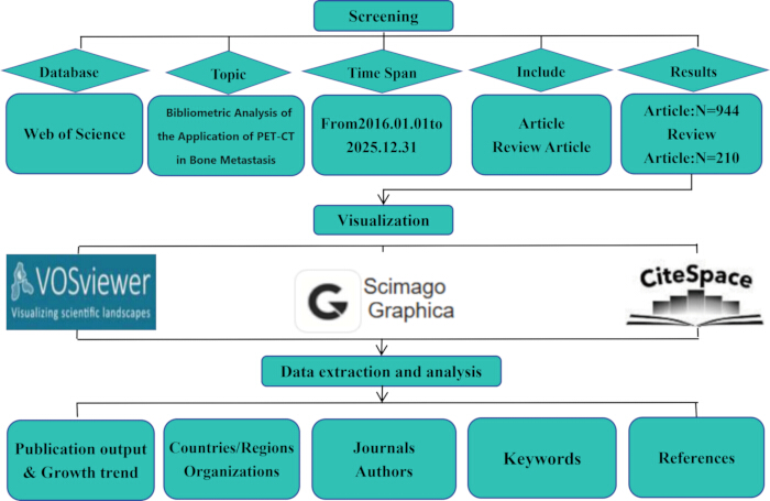
Figure 1: Flowchart of the bibliometric analysis study. This figure shows the step-by-step workflow of the study, including literature retrieval, screening, data analysis, and visualization. Please click here to view a larger version of this figure.
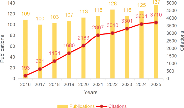
Figure 2: Annual publication and citation trends (2016–2025). This figure presents the annual number of publications (left axis) and the annual number of citations (right axis) from 2016 to 2025. Please click here to view a larger version of this figure.
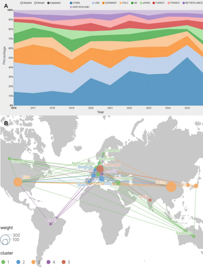
Figure 3: Country/regional distribution and collaboration network. (A) Annual publication volume proportion by country. (B) Country collaboration network. Node size denotes publication volume, edge thickness denotes collaboration strength, and colors represent different collaborative clusters. Please click here to view a larger version of this figure.
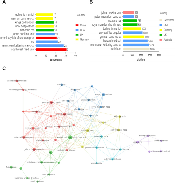
Figure 4: Institutional publication output and collaboration network. (A) Number of publications by institution, ranking institutions by publication volume. (B) Number of citations received by institution, ranking institutions by citation count. (C) Institutional collaboration network. Please click here to view a larger version of this figure.
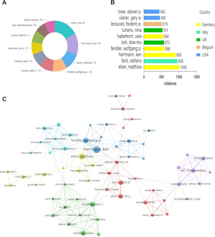
Figure 5: Author publication output and collaboration network. (A) Number of publications by author, ranking authors by publication volume. (B) Number of citations received by author, ranking authors by citation count. (C) Author collaboration network. Please click here to view a larger version of this figure.
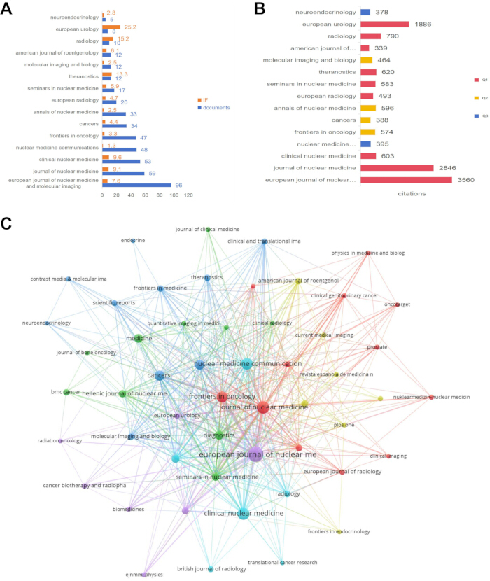
Figure 6: Journal distribution and co-citation network. (A) Number of publications and impact factors by journal, showing journal publication volumes and impact factors. (B) Number of citations received by the journal, showing journal citation counts. (C) Journal co-citation network. Please click here to view a larger version of this figure.
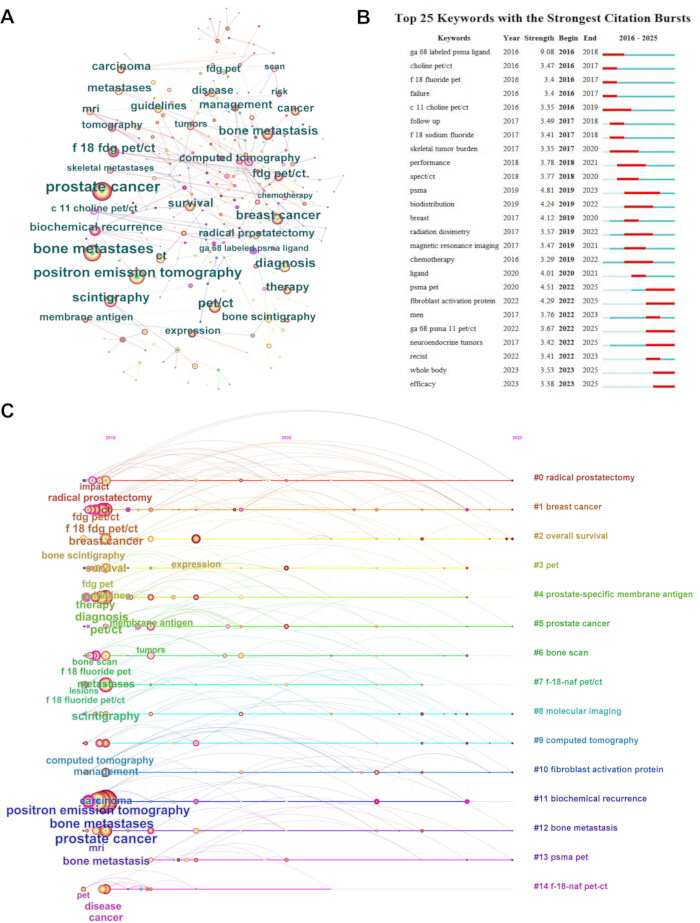
Figure 7: Evolution of research hotspots. (A) Keyword co-occurrence map, with node size representing frequency. (B) Keyword burst detection map, showing the top 25 keywords by burst intensity and their burst periods. (C) Keyword timeline map, showing the temporal evolution of keyword clusters. Please click here to view a larger version of this figure.
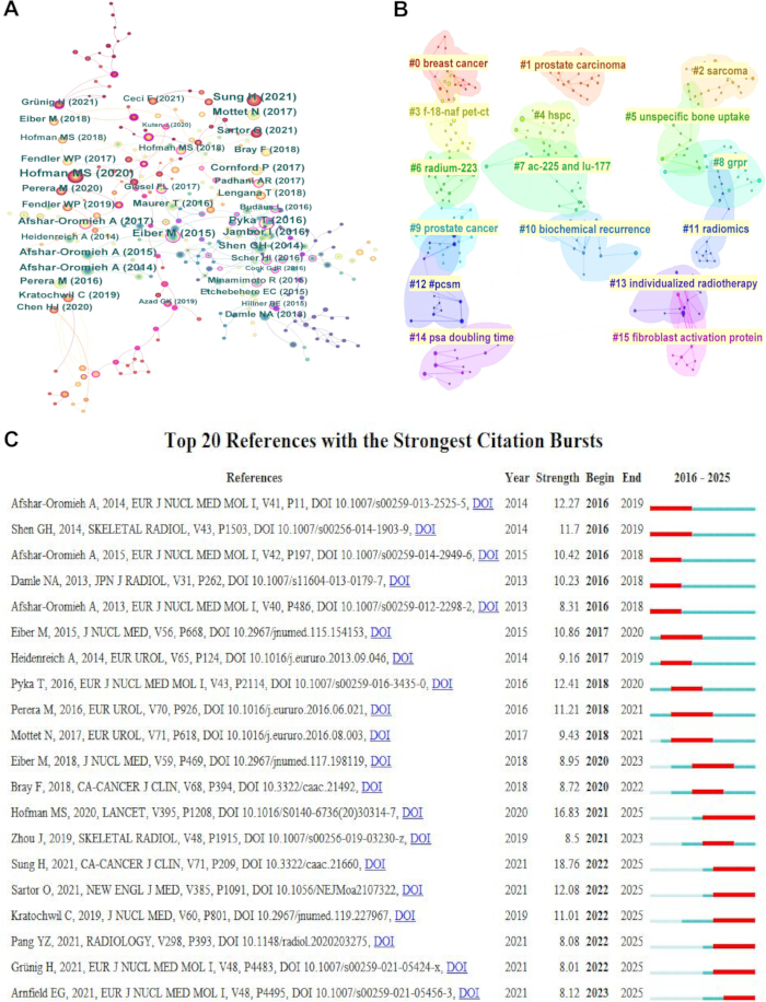
Figure 8: Reference co-citation analysis. (A) Reference co-citation map. (B) Reference keyword clustering map, showing the reference clusters labeled by high-frequency keywords. (C) Reference burst detection map, showing the top references by citation burst intensity and their burst periods. Please click here to view a larger version of this figure.
Table 1: Information on the top 10 countries ranked by number of publications. This table lists the top 10 countries by publication volume, along with their citations, average citations per paper, and total link strength (collaboration intensity). It shows that while China leads in volume, the United States and Germany have higher citation impact. Please click here to download this Table.
Table 2: Information on the top 10 institutions ranked by number of publications. This table presents the most productive institutions, their citation counts, total link strength, and country. It highlights Memorial Sloan Kettering Cancer Center as a leader in both output and impact, while Southwest Medical University, despite high output, has relatively low citations. Please click here to download this Table.
Table 3: Information on the top 10 journals ranked by number of publications. This table provides the publication count, citation count, total link strength, impact factor, and Journal Citation Reports (JCR) quartile for each journal. It shows a mix of Q1 flagship journals and lower-quartile specialized journals. Please click here to download this Table.
Table 4: Keyword count and centrality. This table lists the most frequent keywords on the left and the keywords with the highest betweenness centrality on the right. High centrality keywords such as “c 11 choline PET/CT” and “biochemical recurrence” serve as bridges connecting different research themes. Please click here to download this Table.
Table 5: Top 10 most cited references. This table ranks the most frequently cited references in the field. The top-cited study is Hofman (2020) on 177Lu-PSMA-617 therapy, followed by global cancer statistics and early PSMA PET/CT diagnostic studies. Please click here to download this Table.
Table 6: Top 10 references ranked by centrality. This table lists references with the highest betweenness centrality, indicating their role in linking different research domains such as diagnosis, treatment decision-making, and prognosis. Please click here to download this Table.
Discussion
Skeletal metastasis is one of the most common complications of advanced malignancies and severely impairs patients' quality of life and survival28,29. The most common primary tumors that metastasize to bone are breast, lung, prostate, kidney, and thyroid cancers, which account for the vast majority of skeletal metastatic lesions30. As a molecular imaging technique, PET/CT plays an increasingly important role in the diagnosis, staging, and treatment response assessment of skeletal metastasis31,32. This bibliometric study systematically reviews the development of the field from 2016 to 2025. The application of PET/CT in skeletal metastasis reveals a clear trend, shifting from early diagnostic validation toward theranostic integration. Tracers have expanded beyond the single PSMA target to include multiple options, such as FAPI and, in the context of neuroendocrine tumors, somatostatin receptor (SSTR) PET/CT. Meanwhile, assessment methods are evolving from local qualitative interpretation to whole‑body quantitative monitoring and treatment response tracking33,34. These trends confirm the general direction of “diagnostic-therapeutic integration” in nuclear medicine and provide quantitative references for future research.
At the country level, China and the United States are the two largest producers of publications on PET/CT in skeletal metastasis. Impact indicators show that the United States, Germany, and the United Kingdom occupy central positions in the global collaboration network. China's average citations per paper is 11.6, compared to 47.8 for Germany. Previous bibliometric studies have indicated that high productivity does not necessarily accompany high impact, and that the breadth and depth of international collaboration play important roles in enhancing academic influence. From the perspective of collaboration network structure, China has a relatively low total link strength, which to some extent explains the gap in average citations per paper. For Chinese researchers, while maintaining their output volume, further integration into multilateral collaboration systems, especially strengthening cooperation with core European institutions, may help improve the academic visibility and impact of their research findings. At the journal level, research on PET/CT in skeletal metastasis has formed a multi-layer publication landscape centered on nuclear medicine specialty journals, radiating to general medical journals, oncology journals, and radiology journals. Notably, high-impact journals such as European Urology published only a small number of relevant papers but achieved extremely high citations per paper, indicating that truly practice-changing research in this field tends to be published in top interdisciplinary journals. This suggests that innovative work in PET/CT skeletal metastasis research is more likely to appear in high-impact, cross-disciplinary journals.
Research hotspots in this field evolved through three phases. In the early phase (2016–2018), burst keywords included “ga 68 labeled psma ligand”, “choline pet/ct”, and “f 18 fluoride pet”. The core task was clinical validation of novel tracers, confirming their value for detecting bone metastases compared with conventional bone scintigraphy35,36. In the middle phase (2019–2023), “psma” and “psma pet” became the strongest burst keywords, and PSMA-targeted research became the mainstream direction. Research expanded from diagnosis to biochemical recurrence detection, treatment decision-making, and prognostic assessment. Treatment decision-making mainly refers to patient selection for 177Lu-PSMA-617 therapy. Meanwhile, “radium-223” and “chemotherapy” had high betweenness centrality, suggesting a substantive integration of imaging and treatment, which can be regarded as an early manifestation of the theranostic concept. In the recent phase (2022–2025), a new set of burst keywords emerged. “Fibroblast activation protein” (FAPI) rose as a non-PSMA target, filling the imaging gap for PSMA-negative or low-expressing tumors. “Neuroendocrine tumors” expanded the research scope beyond prostate and breast cancer. “Whole body” and “efficacy” reflected a shift from regional to whole-body assessment and from qualitative evaluation to quantitative monitoring of therapeutic response. This evolutionary trajectory aligns well with the changing clinical needs. The emergence of FAPI reflects the clinical demand for complementary imaging of bone metastases in PSMA-negative or low-expressing tumors, consistent with the recent exploratory application of FAPI in multiple cancers37,38. The appearance of “neuroendocrine tumors” indicates that the field is breaking through traditional cancer boundaries, while “whole body” and “efficacy” imply that the functional role of PET/CT is shifting from a qualitative diagnostic tool to a platform for quantitative whole-body tumor burden assessment and treatment response monitoring39,40. Analysis of the core keywords reveals that the application of PET/CT in skeletal metastasis is primarily reflected in three directions: the development of multi-target tracers, cross-cancer application testing, and precision therapy monitoring supported by PET/CT technology.
Skeletal metastasis differs from visceral metastasis in that its lesions are widely distributed and grow at varying rates. Relying solely on CT or MRI makes it difficult to comprehensively evaluate subtle cortical invasion or early intramedullary infiltration41. Conventional bone scintigraphy, while reasonably sensitive, lacks specificity and cannot provide metabolic activity information of the lesions. The advantage of PET/CT lies in its ability to reflect multiple pathological features of bone metastases through different tracers42,43. Prostate cancer bone metastases often present as osteoblastic changes. PSMA PET/CT detects PSMA expression on the cell membrane rather than direct changes in bone density, so it still achieves a high detection rate for osteoblastic lesions44,45. However, PSMA PET/CT may yield false-negative findings in prostatic ductal adenocarcinoma, a variant with low PSMA expression in approximately 5–10% of cases45. In cases of suspected prostate cancer bone metastases with negative PSMA PET/CT, particularly in aggressive variants such as neuroendocrine or ductal prostate cancer, complementary imaging with 18F-FDG PET/CT or FAPI PET/CT is recommended to improve diagnostic accuracy. In contrast, 18F-NaF PET/CT directly reflects osteoblast activity and is more sensitive for osteoblastic metastases, but it cannot distinguish tumor activity from reactive bone formation. For osteolytic metastases, such as those from lung or thyroid cancer, 18F-FDG PET/CT can effectively demonstrate active osteolytic lesions through glucose metabolism imaging. FAPI PET/CT targets tumor-associated fibroblasts, which are highly activated in the skeletal metastasis microenvironment of many cancer types, particularly in non-PSMA-expressing solid tumors, where FAPI shows unique complementary value. From this perspective, the shifting research hotspots in PET/CT for skeletal metastasis are essentially a technical response to the heterogeneity of bone metastases46,47. Different types of bone metastases from different primary tumors vary significantly in pathophysiology; no single tracer can fully cover all scenarios. The development of multiple types of tracers is very helpful for further PET/CT research. Researchers are gradually recognizing that skeletal metastasis is not a single disease entity but rather a manifestation of various tumors in the special microenvironment of the skeleton.
The most highly cited reference in this field, Hofman et al. (2020) in The Lancet, demonstrated the therapeutic value of 177Lu-PSMA-617 in metastatic castration-resistant prostate cancer, representing a landmark event in the widespread recognition of the theranostic concept in skeletal metastasis. For oligometastatic prostate cancer, PSMA PET/CT-guided salvage radiotherapy enables early intervention in patients with biochemical recurrence, achieving a complete clinical response in approximately 70% of cases at one year post-treatment. Additionally, PSMA PET response after stereotactic ablative radiotherapy has been shown to correlate with metastasis-free survival in oligometastatic castration-sensitive prostate cancer48. Meanwhile, the high betweenness centrality of “radium-223” and “chemotherapy”, as well as the theranostic potential of FAPI as a non-PSMA target, all indicate that this integrative trend is expanding from prostate cancer to more cancer types and more targets. Overall, the role of PET/CT in skeletal metastasis has gradually evolved from an adjunct diagnostic tool to a comprehensive management platform that supports treatment decision-making, response monitoring, and prognostic assessment. Beyond its diagnostic role, PET/CT, particularly with FAPI tracers, has emerged as a critical tool for assessing patient eligibility for targeted radionuclide therapy. A recent systematic review by Ruzzeh et al. demonstrated that FAPI radioligand therapy achieved disease control rates ranging from 18.2% to 83.3% across various metastatic cancers, with a favorable safety profile49,50. This transformation not only reflects technological advances in nuclear medicine but also meets the demand for individualized, dynamic information in precision oncology.
This study provides a systematic bibliometric analysis of PET/CT in skeletal metastasis research from 2016 to 2025. It reveals a clear evolutionary trajectory from diagnostic validation to theranostic integration, from single-target to multi-target imaging, and from qualitative assessment to whole-body quantitative monitoring. China and the United States lead in publication volume, but the United States, Germany, and the United Kingdom are at the core of academic impact and international collaboration networks; China needs to strengthen its international cooperation. Research hotspots have shifted from early tracer validation, through PSMA-targeted theranostics, to current frontiers including FAPI, neuroendocrine tumors, whole-body assessment, and treatment efficacy monitoring. The concept of theranostic integration runs throughout this evolution and is expanding toward multiple targets and across cancer types. Future efforts should focus on promoting the clinical translation of novel probes, expanding cross-cancer applications, developing quantitative radiomics and artificial intelligence-based assessment methods, and enhancing global academic influence through deeper international collaboration. This study has several limitations. First, the literature data were obtained solely from the Web of Science Core Collection; although this database is comprehensive, some high-quality studies published in non-indexed journals or in languages other than English may have been missed. Second, the study included only original articles and reviews, excluding conference abstracts, case reports, and clinical trial protocols. This may have omitted some preliminary or novel findings that have not yet been formally published as full papers. Third, bibliometric analyses inherently depend on the chosen software and analytical parameters; different thresholds, counting methods, and clustering algorithms may yield slightly different network structures and cluster assignments. Fourth, data normalization across different citation windows and subject categories was not performed, which may affect cross-country and cross-journal citation comparisons.
Disclosures
The authors declare no conflicts of interest.
No artificial intelligence tools were used in the preparation of this manuscript, including the writing, data analysis, or figure generation. All work was performed independently by the authors.
Materials
| Name | Company | Catalog Number | Comments |
|---|---|---|---|
| Bibliometric Online Analysis Platform | bibliometric.com | https://bibliometric.com | Used for generating chord diagrams illustrating country/regional collaboration networks. |
| CiteSpace | Chaomei Chen, Drexel University | version 6.4.R1; https://citespace.podia.com/ | Used for keyword burst detection, co-citation analysis, and time-zone evolution analysis. |
| Journal Citation Reports (JCR) | Clarivate Analytics | https://jcr.clarivate.com | Used for obtaining journal impact factors and JCR quartile rankings. |
| Microsoft Excel | Microsoft Corporation | Microsoft 365; https://www.microsoft.com/microsoft-365/excel | Used for descriptive statistical analysis and data management. |
| Scimago Graphica | SCImago Lab | version 1.0.25; https://graphica.scimago.es/ | Used for geospatial visualization of country/regional collaboration networks. |
| VOSviewer | Centre for Science and Technology Studies (CWTS), Leiden University | version 1.6.19; https://www.vosviewer.com/ | Used for constructing and visualizing co-authorship, co-occurrence, and collaboration networks. |
| Web of Science Core Collection | Clarivate Analytics | https://www.webofscience.com | Literature database used for data acquisition and search strategy. |
References
- Clézardin P, et al. Bone metastasis: mechanisms, therapies, and biomarkers. Physiol Rev. 2021;101(3):797-855.
- Sui L, Wang J, Jiang WG, Song X, Ye L. Molecular mechanism of bone metastasis in breast cancer. Front Oncol. 2024;14:1401113.
- Munavvir M, Asadur Rahman M, M M. Pharmacological management of prostate cancer-bone metastasis and recent advancements: A comprehensive review. J Oncol Pharm Pract. 2025;31(8):1307-1315.
- Xu Y, et al. Prediction of Lung Cancer Metastasis Risk Based on Single-Cell Metabolic Profiling of Circulating Tumor Cells. Adv Sci (Weinh). 2025;12(39):e08878.
- Tan LC, et al. Different clinicopathologic features predispose to different patterns of distant metastasis with heterogeneous short-term prognosis in patients with differentiated thyroid cancer. Clin Endocrinol (Oxf). 2022;96(3):402-412.
- Takei D, Tagami K. Management of cancer pain due to bone metastasis. J Bone Miner Metab. 2023;41(3):327-336.
- Marchal C, et al. Epidemiology of bone metastasis in France between 2009 and 2018. JBMR Plus. 2025;9(8):ziaf081.
- Chebli N, et al. Symptomatic spinal cord compression in spinal epidural metastasis of a testicular germ cell tumor: a case report with review of the literature. Ann Transl Med. 2022;10(23):1286.
- Yetiskul E, et al. Hypercalcemia and bone metastasis in a Case of Large Cell Neuroendocrine Carcinoma With Unknown Primary. Case Rep Oncol Med. 2024;2024:8792291.
- Gravel G, Nobileau A, Guth A, Mellot F, Roussel A. Interventional Radiology Management of bone metastasis Pain: Strategies and Techniques. Cardiovasc Intervent Radiol. 2025;48(7):907-918.
- Murtazaliev S, et al. Positron Emission Tomography/Computed Tomography Transformation of Oncology: Multiple Myeloma. PET Clin. 2024;19(2):249-260.
- Alwan M, et al. Cardiac positron emission tomography/Computed tomography (PET/CT) in current cardiology guidelines. J Nucl Cardiol. 2025;48:102231.
- Pei Y, et al. Improving MRI-Negative Epilepsy Localization: Synergy of Dual-Probe PET/MR (18F-FDG/11C-FMZ). J Vis Exp. 2025;(226):e69386.
- Lajtos M, Raedt R, Neyt S, De Herdt V, Vanhove C. Studying Metabolic Brain Connectivity Using 2-Deoxy-2-[18F]Fluoro-D-Glucose Dynamic Positron Emission Tomography at the Single-subject Level. J Vis Exp. 2025;(215):e67458.
- Yang Z, et al. UniPET: A universal network for high-quality PET image denoising across varied dose reduction factors. Med Image Anal. 2026;111:104059.
- Gerke O, et al. Diagnosing Bone Metastases in Breast Cancer: A Systematic Review and Network Meta-Analysis on Diagnostic Test Accuracy Studies of 2-[18F]FDG-PET/CT, 18F-NaF-PET/CT, MRI, Contrast-Enhanced CT, and Bone Scintigraphy. Semin Nucl Med. 2025;55(1):137-151.
- Chai L, et al. Synthesizing [18F]PSMA-1007 PET bone images from CT images with GAN for early detection of prostate cancer bone metastases: a pilot validation study. BMC Cancer. 2025;25(1):907.
- Wang X, Xu Y, Jing J. [18F]FDG PET/CT versus Bone Scintigraphy for the Diagnosis of bone metastasis in Breast Cancer: A Systematic Review and Meta-Analysis. Curr Med Chem. 2025.
- Zamani-Siahkali N, et al. SPECT/CT, PET/CT, and PET/MRI for Response Assessment of Bone Metastases. Semin Nucl Med. 2024;54(3):356-370.
- Luan X, et al. Bifunctional Nanoassembly Enables Metabolism-Driven Microfluidic Blood Screening Guided by MRI Localization for Cancer Monitoring. Anal Chem. 2025;97(6):3395-3403.
- Li K, et al. 68Ga-FAPI PET imaging monitors response to combined TGF-βR inhibition and immunotherapy in metastatic colorectal cancer. J Clin Invest. 2024;134(4):e170490.
- Ruan D, et al. 68Ga-FAPI-46 PET/CT in the evaluation of gliomas: comparison with 18F-FDG PET/CT and contrast-enhanced MRI. Theranostics. 2024;14(18):6935-6946.
- Sheikh GT, et al. PSMA PET/CT response after metastasis-directed radiotherapy of bone oligometastases in prostate cancer. EJNMMI Rep. 2024;8(1):25.
- Sheikhbahaei S, et al. 18F-NaF-PET/CT for the detection of bone metastasis in prostate cancer: a meta-analysis of diagnostic accuracy studies. Ann Nucl Med. 2019;33(5):351-361.
- Chen G, Yan Z, Wang Y, Tao Q. Rheumatoid Arthritis and Fibromyalgia Syndrome: A Bibliometric and Bioinformatics Perspective on Comorbidity Research. J Multidiscip Healthc. 2025;18:6811-6827.
- Wang Y, et al. Research Trends and Hotspots in JAK Inhibitors for Ulcerative Colitis: A Bibliometric Analysis From 2015 to 2024. J Multidiscip Healthc. 2025;18:6755-6771.
- Wang YF, Ma LJ, Han YX, Chen GY, Ma X. Global trends and hotspots of artificial intelligence in pain management: A bibliometric analysis. Digit Health. 2026;12:20552076261460708.
- Yang W, Pan Q, Huang F, Hu H, Shao Z. Research progress of bone metastases: From disease recognition to clinical practice. Front Oncol. 2023;12:1105745.
- Hamash K, Walker SL. Comprehensive Clinical Literature Review of Managing Bone Metastases in Breast Cancer: Focus on Pain and Skeletal-Related Events. Clin J Oncol Nurs. 2023;27(6):615-628.
- Zacharia B, Subramaniam D, Joy J. Skeletal Metastasis-an Epidemiological Study. Indian J Surg Oncol. 2018;9(1):46-51.
- Orcajo-Rincon J, et al. Review of imaging techniques for evaluating morphological and functional responses to the treatment of bone metastases in prostate and breast cancer. Clin Transl Oncol. 2022;24(7):1290-1310.
- Shen CT, Qiu ZL, Han TT, Luo QY. Performance of 18F-fluoride PET or PET/CT for the detection of bone metastases: a meta-analysis. Clin Nucl Med. 2015;40(2):103-110.
- Yang Y, et al. Research hotspots, context, and future directions of ulcerative colitis comorbid with depression from 2006 to 2025: a bibliometric and visualization analysis. Front Med. 2026;13:1868648.
- Ji W, et al. Macrophages in Systemic Lupus Erythematosus: A Bibliometric Analysis of Research Trends and Emerging Frontiers. J Vis Exp. 2026;(234):e72238.
- Andring LM, Teh BS, Butler EB, Farach AM. Focal versus whole gland salvage brachytherapy for recurrent prostate cancer in the prostate specific membrane antigen PET era: a narrative review. Chin Clin Oncol. 2023;12(3):26.
- Kesch C, et al. High fibroblast-activation-protein expression in castration-resistant prostate cancer supports the use of FAPI-molecular theranostics. Eur J Nucl Med Mol Imaging. 2021 Dec;49(1):385-389.
- Rodrigues M, et al. Diagnostic performance of [18F] FDG PET/CT compared to bone scintigraphy for the detection of bone metastases in lung cancer patients. Q J Nucl Med Mol Imaging. 2016;60(1):62-68.
- Feng S, Zhang Y, Wang Y, Gao Y, Song Y. Harnessing Gene Editing Technology for Tumor Microenvironment Modulation: An Emerging Anticancer Strategy. Chem Eur J. 2024;30:e202402485.
- Bai L, et al. The Application of 68Ga-Somatostatin Analog and 18F-FDG PET/CT for bone metastasis from Neuroendocrine Tumors. Neuroendocrinology. 2024;114(8):775-785.
- Borges N, et al. Effect of bone marrow disease on hematologic toxicity and response to [177Lu]Lu-PSMA-617 therapy: insights from PSMA PET/CT imaging. Eur J Nucl Med Mol Imaging. 2026;53(3):1559-1569.
- Zoeller G, Sandrock D, Munz DL, Ringert RH. Bone marrow immunoscintigraphy versus conventional bone scintigraphy in the diagnosis of skeletal metastases in urogenital malignancies. Eur Urol. 1994;26(2):141-144.
- Guo S, et al. Metabolic-Related Gene Prognostic Index for Predicting Prognosis, Immunotherapy Response, and Candidate Drugs in Ovarian Cancer. J Chem Inf Model. 2024;64(3):1066-1080.
- Kunikowska J, Lewington V, Krolicki L. Optimizing Somatostatin Receptor Imaging in Patients With Neuroendocrine Tumors: The Impact of 99mTc-HYNICTOC SPECT/SPECT/CT Versus 68Ga-DOTATATE PET/CT Upon Clinical Management. Clin Nucl Med. 2017;42(12):905-911.
- Janssen JC, et al. [68Ga]PSMA-HBED-CC Uptake in Osteolytic, Osteoblastic, and Bone Marrow Metastases of Prostate Cancer Patients. Mol Imaging Biol. 2017 Dec;19(6):933-943.
- Pepe P, Pepe L, Curduman M, Pennisi M, Fraggetta F. Ductal prostate cancer staging: Role of PSMA PET/CT. Arch Ital Urol Androl. 2024;96(1):12132.
- Zeng Y, et al. Fisetin Micelles Precisely Exhibit a Radiosensitization Effect by Inhibiting PDGFRβ/STAT1/STAT3/Bcl-2 Signaling Pathway in Tumor. Chin Chem Lett. 2025;36:109734.
- Zhu L, et al. Molecular imaging reveals the heterogeneous progression of tumor cells and tumor stroma: a practice of FDG PET and FAPI PET in diagnosing PSMA-negative bone metastases of progressive prostate cancer. Am J Nucl Med Mol Imaging. 2024;14(1):13-21.
- Tamburo M, et al. Salvage Radiotherapy PSMA PET/CT-guided in Men With PSA Recurrence. Anticancer Res. 2024;44(5):2205-2210.
- Ruan D, et al. Evaluation of FAPI PET imaging in gastric cancer: a systematic review and meta-analysis. Theranostics. 2023;13(13):4694-4710.
- Ruzzeh S, et al. Therapeutic potential of FAPI RLT in oncology: A systematic review. Theranostics. 2025;15(9):4084-4100.