This retrospective comparative cohort study evaluated whether concomitant arthroscopic capsular release was associated with earlier functional recovery and a lower burden of stiffness-related treatment after rotator cuff repair in patients with preoperative shoulder stiffness. No statistically significant increase in retear was observed at 12 months.
Research Article
Recovery After Arthroscopic Rotator Cuff Repair With or Without Capsular Release in Patients With Preoperative Stiffness: A Cohort Study
In This Article
Summary
Abstract
Preoperative shoulder stiffness may delay functional recovery after arthroscopic rotator cuff repair, but the incremental value of concomitant capsular release remains uncertain. This retrospective comparative cohort study evaluated 124 patients with reparable rotator cuff tears and preoperative stiffness who underwent arthroscopic repair with concomitant capsular release (CR group, n = 62) or without capsular release (No CR group, n = 62). Shoulder function, pain, active forward elevation, and active external rotation were assessed preoperatively and at 3, 6, and 12 months postoperatively. Additional outcomes included estimated time to functional range of motion, persistent stiffness, secondary stiffness-related interventions, patient satisfaction, complications, and repair integrity on 12-month magnetic resonance imaging. Multivariable analyses were used to account for measured baseline differences associated with nonrandom treatment allocation. Compared with the CR group, the No CR group had a 3.71-point lower adjusted American Shoulder and Elbow Surgeons score at 6 months and had a 3.80-week longer estimated time to functional range of motion. Any secondary stiffness-related intervention was required in 1.6% of the CR group and 16.1% of the No CR group. Persistent stiffness and retear were numerically less frequent in the CR group, but the adjusted associations were not statistically significant. By 12 months, functional outcomes had largely converged. Concomitant capsular release was associated primarily with earlier recovery and a lower stiffness-related treatment burden rather than superior final functional status. No statistically significant increase in retear was observed at 12 months, but the small number of events limits conclusions regarding structural safety.
Introduction
Rotator cuff tears are a common cause of shoulder pain, weakness, and functional limitation in adults, particularly in older populations, and their clinical burden generally increases with the extent and progression of structural tendon disease1. Arthroscopic rotator cuff repair is widely used for symptomatic reparable tears that remain unresponsive to nonoperative treatment. However, successful recovery depends not only on restoration of tendon continuity but also on the progressive recovery of shoulder motion, pain control, and functional use of the affected limb. These considerations are especially important in patients with clinically meaningful shoulder stiffness before surgery. Preoperative motion restriction may complicate rehabilitation, delay active use of the shoulder, and increase the burden of postoperative treatment even when the repaired tendon remains structurally intact2.
Postoperative stiffness remains a clinically important component of recovery after rotator cuff repair. Prospective evidence has shown that stiffness may affect approximately 31% of patients at 3 months and 20% at 6 months after surgery3. The development and persistence of stiffness may also be influenced by patient-specific biological and metabolic factors. Elevated preoperative glycated hemoglobin, for example, has been associated with an increased risk of postoperative stiffness after posterior-superior rotator cuff repair4. These observations indicate that postoperative motion loss is not simply a transient mechanical consequence of surgery. Rather, it may reflect an interaction among pre-existing capsular contracture, patient susceptibility, tendon pathology, operative treatment, postoperative protection, and rehabilitation exposure.
Concomitant arthroscopic capsular release is one strategy used to address pre-existing capsular restriction during rotator cuff repair. The procedure is intended to improve capsular compliance, restore clinically useful joint excursion, and reduce the persistence of stiffness during rehabilitation. Nevertheless, capsular release is generally recommended selectively rather than routinely. A Delphi consensus suggested that capsular release should be reserved for clinically stiff shoulders instead of being applied broadly during arthroscopic cuff repair5. Previous studies have shown that one-stage rotator cuff repair in patients with concurrent stiffness can produce final range of motion, patient-reported outcomes, and retear rates comparable to those obtained after conventional repair in patients without stiffness6. Sustained improvements in pain, function, and motion have also been reported after combined repair, manipulation, and capsular release, although recurrent stiffness and residual loss of external rotation may still occur7.
The decision to release the capsule must also account for the biological requirements of tendon healing. Early postoperative stiffness has been associated in some studies with greater tendon integrity despite worse early pain and function, raising the possibility that rapid restoration of motion may not always be fully aligned with protection of the healing repair8. In addition, the available evidence remains limited in scale and consistency. A systematic review of treatment for stiffness after rotator cuff repair identified only five eligible studies involving 177 patients9. Much of the existing literature has compared stiff and non-stiff shoulders or evaluated combined one-stage treatment without isolating the incremental contribution of capsular release among patients who were already stiff before surgery.
The unresolved clinical question is therefore not simply whether rotator cuff tears and shoulder stiffness can be treated during the same operation. The more specific question is whether adding capsular release to arthroscopic rotator cuff repair provides measurable benefit over repair alone within a consistently defined population with preoperative stiffness. Such benefit may be reflected less in the final 12-month score than in the trajectory of recovery, including earlier restoration of functional motion, fewer persistent stiffness events, and reduced need for subsequent manipulation or injection. These potential advantages must be considered together with operative burden, complications, and structural tendon healing. Recent comparative evidence has approached this question, but recovery trajectory, subsequent treatment burden, and repair integrity have not always been evaluated within the same longitudinal framework10.
The present retrospective comparative cohort study, therefore, applied a standardized clinical, operative, rehabilitation, and longitudinal outcome-assessment protocol to patients with reparable rotator cuff tears and preoperative shoulder stiffness. Arthroscopic rotator cuff repair with concomitant capsular release was compared with repair without capsular release. The primary hypothesis was that capsular release would be associated with a more favorable early-to-intermediate recovery trajectory and earlier achievement of functional range of motion, rather than a substantially superior final functional outcome. It was further hypothesized that capsular release would reduce the need for secondary stiffness-related intervention without an observed increase in complications or retear at 12 months.
Protocol
The study protocol was approved by the Institutional Review Board of The Affiliated Hospital of Xuzhou Medical University before data extraction and analysis (Approval No. XYFY2026-KL188-01). The study was conducted in accordance with the Declaration of Helsinki and its subsequent amendments. Given the retrospective nature of the study and the use of routinely collected clinical data, the requirement for participant-informed consent was waived by the Institutional Review Board of The Affiliated Hospital of Xuzhou Medical University. All direct patient identifiers were removed before analysis.
Study design and cohort establishment
This single-center retrospective comparative cohort study included consecutive patients treated between January 2018 and December 2024 for reparable rotator cuff tears accompanied by preoperative shoulder stiffness. Patients who underwent arthroscopic rotator cuff repair with concomitant capsular release were assigned to the capsular release group (CR group), whereas those who underwent repair without capsular release were assigned to the no capsular release group (No CR group). No formal a priori sample-size calculation was performed because this was a retrospective cohort study. All consecutive patients who met the eligibility criteria during the predefined study period were included, and the available cohort therefore determined the final sample size of 124 patients.
Treatment allocation was not randomized and reflected clinical and intraoperative judgment. Active range-of-motion thresholds were used to identify the eligible clinically stiff cohort, but active restriction alone was not considered proof of capsular contracture because pain and rotator cuff-related weakness could also limit active motion. The decision to perform capsular release was based on the combined assessment of preoperative active motion restriction, persistent passive limitation during examination under anesthesia, and arthroscopic evidence of clinically relevant capsular contracture. Examination under anesthesia was used to reduce the influence of voluntary guarding and pain during assessment of passive motion. These findings informed treatment selection but did not constitute a randomized allocation rule. The same operative indications were applied throughout 2018–2024. Measured baseline differences associated with clinical treatment selection were addressed in the statistical analysis, although residual confounding could not be excluded. The screening process, eligibility assessment, exclusions, and final group allocation are presented in Figure 1.
Patient population and eligibility criteria
The source population consisted of adults who underwent arthroscopic treatment for symptomatic rotator cuff tears after unsuccessful nonoperative management, including medication, activity modification, supervised rehabilitation, or local injection. Preoperative physiotherapy was considered an important component of conservative treatment and, when clinically appropriate, included supervised pain-limited range-of-motion exercises and shoulder rehabilitation. Surgery was considered only when clinically meaningful pain, functional limitation, or shoulder stiffness persisted despite these measures. Because treatment was individualized in routine clinical practice, the duration and intensity of preoperative physiotherapy were not completely standardized.
Patients were eligible if they were at least 18 years old; had a symptomatic rotator cuff tear confirmed by magnetic resonance imaging and arthroscopy; had persistent pain or functional limitation despite nonoperative treatment; met the definition of preoperative shoulder stiffness; had a reparable tear treated during the index procedure; completed clinical assessments at baseline and 3, 6, and 12 months after surgery; and underwent structural assessment of the repair by magnetic resonance imaging at approximately 12 months. Preoperative shoulder stiffness was defined by at least one of the following findings in the affected shoulder: active forward elevation below 120°, active external rotation below 30° with the arm at the side, or internal rotation behind the back below the L3 vertebral level. Eligibility was based on preoperative active motion to identify patients with clinically meaningful motion restriction; this operational definition did not by itself establish the presence of capsular contracture. Because active motion could also be limited by pain or cuff-related weakness, persistent passive limitation during examination under anesthesia and arthroscopic evidence of capsular contracture were used to support the operative decision regarding capsular release. Active forward elevation and external rotation were analyzed longitudinally. Internal rotation was retained for stiffness classification but was not analyzed as a repeated continuous variable.
Patients were excluded for an irreparable massive rotator cuff tear, previous surgery on the affected shoulder, advanced glenohumeral osteoarthritis, acute shoulder fracture or dislocation, inflammatory arthropathy, active infection, neurologic disease affecting shoulder motion, or isolated adhesive capsulitis without a reparable rotator cuff tear. Patients with incomplete perioperative records, incomplete follow-up, or unavailable 12-month structural assessment were also excluded.
Preoperative clinical assessment
Baseline variables included age, sex, body mass index, diabetes mellitus, current smoking status, dominant-arm involvement, and symptom duration. Symptom duration was calculated from the documented onset of persistent symptoms to the date of surgery or obtained from the preoperative clinical record when an exact onset date was unavailable. For patients with diabetes mellitus, preoperative glycated hemoglobin (HbA1c) values obtained within 3 months before surgery were extracted when available. However, HbA1c testing was not performed within a uniform preoperative time window for all patients because of the retrospective study design. Diabetes status was therefore included as a prespecified covariate, whereas HbA1c was not entered into the primary multivariable models because of incomplete availability. Patients with diabetes received routine perioperative glucose management according to institutional clinical practice.
Shoulder function was assessed using the American shoulder and elbow surgeons (ASES) score and the Constant score, each ranging from 0 to 100, with higher scores indicating better function. Assessments were completed before surgery and at 3, 6, and 12 months postoperatively. Pain was measured using a 10-point visual analog scale (VAS), with 0 indicating no pain and 10 indicating the worst pain imaginable. VAS scores were recorded at baseline and at each postoperative assessment.
Range-of-motion assessment
Active forward elevation and external rotation were measured at baseline and at 3, 6, and 12 months after surgery using the same institutional examination protocol. Measurements were recorded in degrees. For forward elevation, the patient was positioned standing or seated with the trunk upright and asked to elevate the affected arm actively without assistance. Natural scapular movement was permitted, while compensatory trunk extension and lateral bending were minimized. The maximum active elevation was recorded. For external rotation, the arm was maintained against the trunk with the elbow flexed to 90°. The patient rotated the forearm externally while keeping the upper arm at the side. The angle from the neutral forearm position to maximum active external rotation was recorded.
Internal rotation was assessed by asking the patient to place the hand behind the back and move the thumb superiorly along the spine. The highest vertebral level reached was documented. Measurements were repeated when compensatory movement occurred or the required position was not maintained. Range-of-motion assessors were not consistently blinded to treatment allocation, creating a potential risk of detection bias.
Preoperative imaging and tear characterization
Preoperative magnetic resonance imaging was used to confirm the rotator cuff tear and evaluate tear location, size, morphology, and apparent reparability. Imaging findings were verified arthroscopically. When imaging and operative findings differed, the arthroscopic assessment was used for final classification. Tear size was classified according to the DeOrio and Cofield system as small (<1 cm), medium (1–3 cm), large (>3–5 cm), or massive (>5 cm). Irreparable massive tears were excluded. Tear morphology was classified as crescent-shaped, U-shaped, or L-shaped. Tendon involvement was recorded as isolated supraspinatus involvement, combined supraspinatus and infraspinatus involvement, or partial subscapularis involvement.
Surgical procedures
All operations were performed arthroscopically under general anesthesia, with or without an interscalene regional block, by two fellowship-trained shoulder and elbow surgeons, each with more than 10 years of independent experience in arthroscopic shoulder surgery. After anesthesia induction, passive forward elevation, external rotation, and internal rotation were reassessed to determine whether restriction persisted after minimizing voluntary guarding and pain. Passive restriction under anesthesia was interpreted together with the preoperative examination and arthroscopic capsular findings rather than as an isolated criterion.
Standard posterior viewing and anterior working portals were established. Diagnostic glenohumeral arthroscopy was used to inspect the articular surfaces, labrum, long head of the biceps tendon, rotator interval, capsule, and articular surface of the cuff. The subacromial space was then examined to assess tear configuration, tendon mobility, tissue quality, and footprint characteristics. Rotator cuff repair was performed after tendon mobilization and preparation of the greater tuberosity footprint. A single-row or double-row construct was selected according to tear size, configuration, mobility, and tissue quality. Suture anchors were inserted into the prepared footprint, and sutures were passed through viable tendon tissue to restore tendon-to-bone contact without excessive tension. Repair configuration, anchor number, tear characteristics, and tendon-mobilization procedures were recorded.
In the CR group, capsular release was performed after diagnostic arthroscopy and before final cuff fixation. Release initially involved the rotator interval and anterior capsule. When the restriction remained, the release was extended cautiously toward the inferior capsule. During inferior release, instruments were maintained close to the glenoid rim to reduce the risk of axillary nerve injury. Posterior release was performed only when clinically relevant posterior restriction persisted. Complete circumferential release was not routinely performed, and no separate forceful manipulation under anesthesia was used.
In the No CR group, rotator cuff repair was completed without deliberate rotator interval release or capsulotomy. Standard debridement, subacromial bursectomy, tendon mobilization, and footprint preparation were permitted but were not classified as stiffness-directed capsular release. Biceps tenotomy or tenodesis and acromioplasty were performed when indicated by intraoperative findings. Repair configuration, associated procedures, operative time, and estimated blood loss were recorded.
Postoperative management
The same staged rehabilitation protocol was applied to both groups. The operated shoulder was protected in a sling immediately after surgery, and supervised rehabilitation generally began during the first postoperative week. Early management included pain and edema control, movement of the elbow, wrist, and hand, scapular exercises, and protected passive shoulder motion within surgeon-approved limits.
The sling was maintained for approximately 4–6 weeks according to tear size, repair configuration, tissue quality, and clinical progress. Passive forward elevation and external rotation were advanced gradually without forceful stretching. After sling removal, patients progressed from passive to active-assisted and then active motion, generally between 4 and 8 weeks postoperatively. Resisted strengthening usually began after 12 weeks when active motion and repair stability were considered adequate. Return to unrestricted overhead activity or heavy loading required satisfactory motion, strength, pain control, and surgeon approval. The CR and No CR groups followed the same rehabilitation stages and motion goals. Rehabilitation progression was modified according to pain, tear characteristics, fixation stability, and clinical assessment rather than treatment allocation. Clinical evaluations were performed at approximately 3, 6, and 12 months after surgery. Pain, active range of motion, functional scores, complications, and additional stiffness-related treatments were recorded at each visit. Repair integrity was assessed by magnetic resonance imaging at approximately 12 months.
Outcome assessment
The principal outcomes were postoperative shoulder function and active range-of-motion recovery. The American Shoulder and Elbow Surgeons (ASES) score, Constant score, active forward elevation, and active external rotation were assessed before surgery and at 3, 6, and 12 months postoperatively. Pain intensity was measured using the 10-point visual analog scale at the same time points.
Estimated time to functional range of motion was defined as the postoperative interval required to achieve both active forward elevation of at least 140° and active external rotation of at least 30°. The earliest documented date on which both criteria were met was recorded. When no interval assessment was available, the corresponding scheduled follow-up visit was used. Consequently, this outcome represented an estimated, visit-based recovery interval rather than an exact, continuously observed event time, and actual attainment could have occurred between documented assessments.
Persistent stiffness at 12 months was defined by at least one of the following findings: active forward elevation below 120°, active external rotation below 30°, or internal rotation behind the back below the L3 vertebral level. Secondary stiffness-related interventions were recorded separately as manipulation under anesthesia, therapeutic injection, or another unplanned procedure performed to manage persistent stiffness or delayed recovery after the index operation. A composite outcome of any secondary stiffness-related intervention was also reported. Routine rehabilitation, scheduled follow-up, and analgesic adjustment were not classified as secondary interventions.
Repair integrity was assessed on 12-month magnetic resonance imaging using the Sugaya classification. Types I–III were classified as intact repairs, whereas types IV and V were classified as retears. Magnetic resonance images were reviewed by two musculoskeletal radiologists with 8 and 12 years of post-training experience, respectively, who were blinded to treatment allocation and clinical outcomes. Imaging findings were reassessed when the descriptive report and recorded Sugaya category were inconsistent, and disagreements were resolved by consensus after joint image review. Postoperative complications included infection, neurologic injury, symptomatic hematoma, repair-related reoperation, and other procedure-related adverse events documented during follow-up. Patient satisfaction was assessed at 12 months using a 0–10 scale, with higher scores indicating greater satisfaction.
Data collection and quality control
Study variables were entered into a structured patient-level dataset using predefined field names and coding rules. Recorded variables included demographic characteristics, comorbidities, symptom duration, preoperative assessments, functional and pain scores, range-of-motion measurements, tear characteristics, repair configuration, concomitant procedures, operative time, estimated blood loss, postoperative interventions, complications, satisfaction, and 12-month structural outcomes. The ASES score, Constant score, visual analog scale pain score, active forward elevation, and active external rotation were stored separately for the preoperative, 3-month, 6-month, and 12-month assessments. Treatment group, diabetes mellitus, smoking status, dominant-arm involvement, persistent stiffness, retear, and complications were coded as categorical variables. Manipulation under anesthesia, therapeutic injection, other secondary procedures, and the composite outcome of any secondary stiffness-related intervention were coded separately. Continuous variables were retained in their original units.
Before analysis, the dataset was checked for duplicate records, missing data, implausible values, visit misclassification, and inconsistencies with the source records. Functional scores and pain values outside their allowable ranges were verified against the original assessments. Range-of-motion measurements, tear classifications, operative procedures, secondary interventions, complications, and 12-month magnetic resonance imaging findings were cross-checked against the clinical, operative, and imaging records. Missing values were not replaced with generated observations.
Statistical analysis
Continuous variables were summarized as mean ± standard deviation or median with interquartile range according to their distributions. Categorical variables were summarized as numbers and percentages. The CR and No CR groups were compared using the independent-samples t-test for approximately normally distributed continuous variables and the Mann–Whitney U test for skewed continuous variables. Categorical variables were compared using the chi-square test or Fisher’s exact test, as appropriate. Baseline demographic and clinical characteristics, tear characteristics, repair configuration, concomitant procedures, operative time, and estimated blood loss were compared between groups. The ASES score, Constant score, visual analog scale pain score, active forward elevation, and active external rotation were summarized at baseline and at 3, 6, and 12 months. Unadjusted between-group comparisons were performed at each assessment, and group-specific mean values with standard deviations were used to display recovery trajectories.
Prespecified multivariable models were used to account for measured baseline differences associated with nonrandom treatment allocation. The core adjustment variables were age, sex, symptom duration, diabetes mellitus, tear size, preoperative active forward elevation, and preoperative active external rotation. Preoperative ASES score was additionally included in the models for postoperative ASES outcomes. Covariates were selected a priori on the basis of clinical relevance and their potential association with treatment selection or postoperative recovery rather than by univariable p-value screening. Diabetes mellitus and tear size were included in the prespecified core models. Tear morphology and repair configuration were not added to the primary models because their distributions were similar between groups, repair configuration was identical across groups, and expanded models for outcomes with limited event counts would have increased the risk of overfitting and unstable estimates.
Multiple linear regression was used to analyze the ASES score at 6 and 12 months and the estimated time to functional range of motion. The recorded or visit-assigned recovery interval was analyzed as a continuous outcome; no interval-censoring model was applied. Accordingly, the estimated effect on recovery time should be interpreted as a difference in visit-based documented attainment rather than an exact time-to-event effect. Results were reported as adjusted mean differences with 95% confidence intervals. Binary logistic regression was used for persistent stiffness and retear at 12 months, with results reported as adjusted odds ratios and 95% confidence intervals. Manipulation under anesthesia, therapeutic injection, other secondary procedures, the composite secondary-intervention outcome, complications, and other binary outcomes with limited event counts were compared using unadjusted categorical analyses. The CR group was coded as 0 and the No CR group as 1. Adjusted mean differences, therefore, represented No CR minus CR, and adjusted odds ratios represented the odds in the No CR group relative to the CR group. A negative adjusted difference in ASES score favored the CR group, whereas a positive difference in estimated time to functional range of motion indicated slower recovery in the No CR group.
Exploratory subgroup analyses of the adjusted 6-month ASES difference were performed according to age (<55 versus ≥55 years), sex, diabetes mellitus, tear size (small or medium versus large), and dominant-arm involvement. The subgroup-defining variable was omitted from the corresponding adjusted model. These analyses were interpreted as exploratory. Linear-model assumptions were assessed using residual diagnostics, and collinearity among adjustment variables was examined before model interpretation. A two-sided p-value below 0.05 was considered statistically significant. Effect estimates were reported with 95% confidence intervals. Statistical analyses were performed using SPSS Statistics, version 26.0.
Results
Cohort formation and baseline characteristics
A total of 168 patients were screened, of whom 44 were excluded for not meeting the eligibility criteria or for lacking the required follow-up or structural assessment. The final cohort included 124 patients: 62 underwent arthroscopic rotator cuff repair with concomitant capsular release, and 62 underwent repair without capsular release. The screening and cohort-allocation process is shown in Figure 1. Baseline demographic and preoperative clinical characteristics are presented in Table 1. Age, sex, body mass index, diabetes mellitus, smoking status, dominant-arm involvement, symptom duration, preoperative pain, and preoperative ASES score did not differ significantly between groups. Mean preoperative forward elevation was 92.0 ± 14.7° in the CR group and 96.1 ± 14.0° in the No CR group (p = 0.112). Preoperative external rotation was lower in the CR group than in the No CR group (16.7 ± 7.9° versus 20.4 ± 7.2°, p = 0.007). The CR group also had a lower preoperative Constant score (41.8 ± 7.1 versus 44.5 ± 7.1, p = 0.034), whereas preoperative ASES scores were comparable (30.1 ± 6.1 versus 31.9 ± 5.4, p = 0.078). Preoperative HbA1c values were not consistently available within a uniform 3-month preoperative window among patients with diabetes; therefore, no formal group-level HbA1c summary or between-group comparison was performed.
Tear and operative characteristics
Tear characteristics, repair configurations, and operative variables are summarized in Table 2. Tear-size distribution, tear morphology, and tendon involvement were similar between groups. Small, medium, and large tears accounted for 29.0%, 38.7%, and 32.3% of cases in the CR group and 29.0%, 41.9%, and 29.0% in the No CR group, respectively. Crescent-shaped tears were present in 58.1% of patients in each group. Single-row repair was performed in 41 patients (66.1%) in each group, and double-row repair was performed in 21 patients (33.9%) in each group. The frequencies of biceps procedures and acromioplasty did not differ significantly. Operative time was longer in the CR group than in the No CR group (88.9 ± 13.5 min versus 75.2 ± 12.3 min, p < 0.001), and estimated blood loss was higher (39.1 ± 9.0 mL versus 35.4 ± 9.9 mL, p = 0.033).
Recovery of shoulder motion
Changes in active forward elevation and external rotation are summarized in Table 3. Active forward elevation progressively improved in both groups (Figure 2A). In the CR group, forward elevation increased from 92.0 ± 14.7° preoperatively to 140.7 ± 16.5° at 3 months, 159.9 ± 14.6° at 6 months, and 165.1 ± 13.5° at 12 months. Corresponding values in the No CR group were 96.1 ± 14.0°, 135.7 ± 15.5°, 159.5 ± 15.5°, and 165.7 ± 14.6°. Active external rotation also progressively improved in both groups (Figure 2B). In the CR group, external rotation increased from 16.7 ± 7.9° preoperatively to 34.8 ± 8.7° at 3 months, 41.0 ± 8.6° at 6 months, and 48.6 ± 8.6° at 12 months. In the No CR group, external rotation changed from 20.4 ± 7.2° to 32.2 ± 8.9°, 41.6 ± 8.2°, and 48.0 ± 8.1°, respectively. No significant between-group difference in forward elevation or external rotation was observed at any postoperative assessment.
Functional recovery and pain
Longitudinal changes in ASES score, Constant score, and VAS pain score are shown in Figure 3A, Figure 3B, and Figure 3C, respectively. Mean ASES score in the CR group increased from 30.1 ± 6.1 preoperatively to 55.0 ± 8.9 at 3 months, 66.7 ± 8.1 at 6 months, and 72.0 ± 7.4 at 12 months. Corresponding values in the No CR group were 31.9 ± 5.4, 52.4 ± 7.4, 64.5 ± 9.0, and 73.6 ± 7.0. Unadjusted between-group differences in ASES score were not significant at 3, 6, or 12 months. The Constant score increased from 41.8 ± 7.1 preoperatively to 59.8 ± 8.7, 70.5 ± 7.8, and 76.3 ± 8.5 at 3, 6, and 12 months in the CR group. Corresponding values in the No CR group were 44.5 ± 7.1, 61.2 ± 8.1, 71.9 ± 10.0, and 79.0 ± 8.6. No significant postoperative between-group differences were observed. Mean VAS pain score in the CR group decreased from 5.9 ± 0.7 preoperatively to 3.8 ± 0.8 at 3 months, 2.4 ± 1.0 at 6 months, and 1.8 ± 0.9 at 12 months. Corresponding values in the No CR group were 5.9 ± 0.6, 3.8 ± 1.0, 2.6 ± 0.9, and 1.9 ± 0.9. Pain scores did not differ significantly between groups at any postoperative time point. The complete unadjusted numerical results for shoulder motion, functional scores, and pain at baseline and follow-up are provided in Table 3.
Adjusted analyses
Multivariable-adjusted results are presented in Table 4. After adjustment for age, sex, symptom duration, diabetes mellitus, tear size, preoperative forward elevation, and preoperative external rotation, the No CR group had a lower ASES score at 6 months than the CR group (adjusted mean difference, −3.71; 95% CI, −6.03 to −1.39; p = 0.002). Preoperative ASES score was additionally included in the postoperative ASES models. Although the unadjusted 6-month ASES comparison was not statistically significant, the adjusted estimate became more pronounced after accounting for measured baseline differences, including the greater preoperative external-rotation restriction in the CR group. The adjusted 3.71-point difference was statistically significant but modest; because no study-specific threshold for clinical importance was prespecified, its clinical relevance should be interpreted cautiously. At 12 months, the adjusted difference in ASES score was not significant (0.13; 95% CI, −1.64 to 1.90; p = 0.887).
The unadjusted mean estimated time to functional range of motion was 17.7 ± 3.4 weeks in the CR group and 20.3 ± 2.9 weeks in the No CR group, corresponding to an unadjusted between-group difference of 2.6 weeks. After multivariable adjustment for age, sex, symptom duration, diabetes mellitus, tear size, preoperative active forward elevation, and preoperative active external rotation, the No CR group had a 3.80-week longer estimated time to functional range of motion than the CR group (adjusted mean difference, 3.80 weeks; 95% CI, 2.87–4.74; p < 0.001). The adjusted odds ratio for persistent stiffness at 12 months was 3.05 for No CR versus CR (95% CI, 0.96–9.68; p = 0.058), and the adjusted odds ratio for retear was 1.86 (95% CI, 0.56–6.14; p = 0.307). Neither association reached statistical significance, and the wide confidence intervals indicate limited precision. Exploratory subgroup estimates for the adjusted 6-month ASES difference are shown in Figure 4. The estimates generally favored the CR group across strata defined by age, sex, diabetes mellitus, tear size, and dominant-arm involvement, although several confidence intervals included zero.
Secondary outcomes and 12-month repair integrity
Secondary outcomes are summarized in Table 5. Retear occurred in 5 of 62 patients (8.1%) in the CR group and 9 of 62 patients (14.5%) in the No CR group (p = 0.395). Persistent stiffness at 12 months occurred in 5 patients (8.1%) and 13 patients (21.0%), respectively (p = 0.072). Any secondary stiffness-related intervention was required in 1 patient (1.6%) in the CR group and 10 patients (16.1%) in the No CR group (p = 0.008). When examined separately, manipulation under anesthesia occurred in the CR group in 0 patients and the No CR group in 2 patients, therapeutic injection occurred in the CR group in 1 patient, and the No CR group in 8 patients, and other unplanned stiffness-related procedures occurred in neither group (0 patients in each group). Mean estimated time to functional range of motion was 17.7 ± 3.4 weeks in the CR group and 20.3 ± 2.9 weeks in the No CR group (p < 0.001). These values reflect the earliest documented or visit-assigned attainment of the functional range-of-motion criteria rather than continuously monitored event times.
Mean patient-satisfaction scores at 12 months were 8.3 ± 0.9 in the CR group and 8.5 ± 0.8 in the No CR group (p = 0.134). One superficial infection occurred in each group, and no cases of complex regional pain syndrome were recorded. Retear rate, persistent stiffness, estimated time to functional range of motion, and operative time are displayed in Figure 5A, Figure 5B, Figure 5C, and Figure 5D, respectively.
DATA AVAILABILITY:
The raw data underlying the findings of this study, including the patient-level data used for statistical analyses and the numerical values used to generate the figures and tables, are publicly available in Figshare at https://doi.org/10.6084/m9.figshare.33170429.v1.
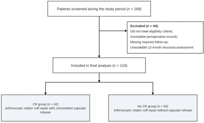
Figure 1: Study flowchart and cohort allocation. Flow diagram showing patient screening, eligibility assessment, exclusion, and final group allocation. A total of 168 patients were screened, 44 were excluded, and 124 were included in the final analysis. Sixty-two patients underwent arthroscopic rotator cuff repair with concomitant capsular release (CR group), and 62 underwent repair without capsular release (No CR group). Please click here to view a larger version of this figure.
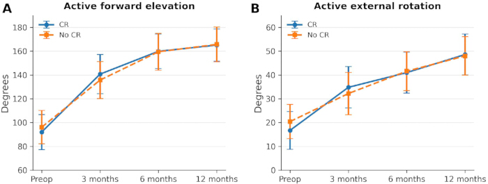
Figure 2: Recovery trajectories of active shoulder range of motion. (A) Active forward elevation before surgery and at 3, 6, and 12 months after surgery in the CR and No CR groups. (B) Active external rotation at the same assessment points. Points represent means, and error bars indicate standard deviations. CR, capsular release; No CR, no capsular release. Please click here to view a larger version of this figure.
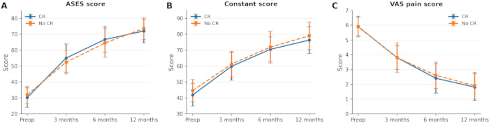
Figure 3: Longitudinal changes in shoulder function and pain. (A) American Shoulder and Elbow Surgeons score, (B) Constant score, and (C) visual analog scale pain score before surgery and at 3, 6, and 12 months after surgery in the CR and No CR groups. Points represent means, and error bars indicate standard deviations. Higher functional scores indicate better shoulder function, whereas lower visual analog scale scores indicate less pain. ASES, American Shoulder and Elbow Surgeons; CR, capsular release; No CR, no capsular release; VAS, visual analog scale. Please click here to view a larger version of this figure.
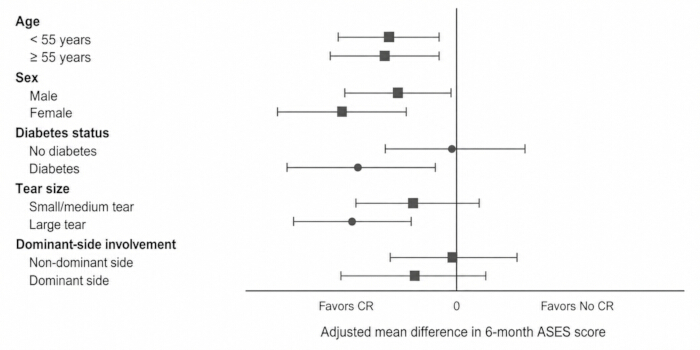
Figure 4: Exploratory subgroup analysis of the adjusted 6-month ASES score difference. Forest plot showing adjusted mean differences in the 6-month American Shoulder and Elbow Surgeons score for the No CR group relative to the CR group across strata defined by age, sex, diabetes mellitus, tear size, and dominant-arm involvement. Points indicate adjusted mean differences, and horizontal lines indicate 95% confidence intervals. Adjusted mean differences were calculated as No CR minus CR; therefore, negative values favor the CR group, whereas positive values favor the No CR group. Abbreviations: ASES = American Shoulder and Elbow Surgeons; CR = capsular release; No CR = no capsular release. Please click here to view a larger version of this figure.
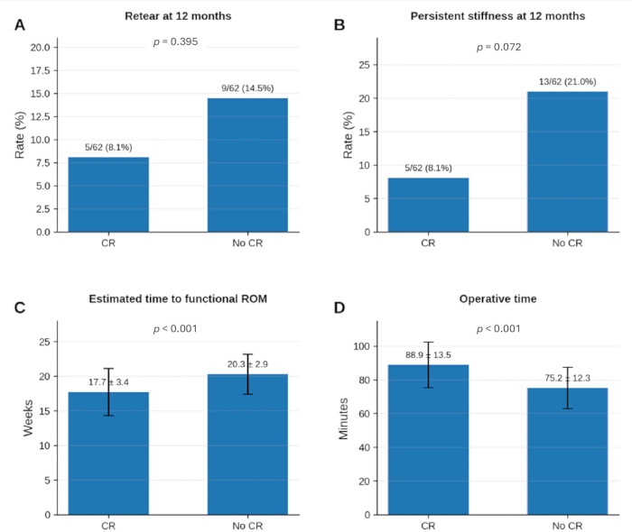
Figure 5: Selected secondary outcomes and operative burden according to treatment group. (A) Retear rate at 12 months. (B) Persistent stiffness rate at 12 months. (C) The estimated time to functional range of motion, defined as the documented achievement of both active forward elevation of at least 140° and active external rotation of at least 30°. (D) Operative time. Panels A and B are presented as numbers/totals and percentages. In panels C and D, bars represent means, and error bars indicate standard deviations. Abbreviations: CR = capsular release; No CR = no capsular release; ROM = range of motion. Please click here to view a larger version of this figure.
| Characteristic | CR | No CR | p value |
| Age, years | 55.3 ± 7.4 | 56.5 ± 7.7 | 0.361 |
| Female sex | 33 (53.2%) | 35 (56.5%) | 0.857 |
| BMI, kg/m² | 25.9 ± 3.3 | 26.5 ± 2.5 | 0.258 |
| Diabetes mellitus | 17 (27.4%) | 24 (38.7%) | 0.252 |
| Current smoker | 8 (12.9%) | 7 (11.3%) | 1 |
| Dominant side involved | 39 (62.9%) | 30 (48.4%) | 0.148 |
| Symptom duration, months | 5.8 (3.8–8.9) | 7.4 (4.5–9.9) | 0.201 |
| Preoperative forward elevation, ° | 92.0 ± 14.7 | 96.1 ± 14.0 | 0.112 |
| Preoperative external rotation, ° | 16.7 ± 7.9 | 20.4 ± 7.2 | 0.007 |
| Preoperative VAS pain | 5.9 ± 0.7 | 5.9 ± 0.6 | 1 |
| Preoperative ASES score | 30.1 ± 6.1 | 31.9 ± 5.4 | 0.078 |
| Preoperative Constant score | 41.8 ± 7.1 | 44.5 ± 7.1 | 0.034 |
Table 1: Baseline demographic and preoperative clinical characteristics. Comparison of demographic characteristics, comorbidities, symptom duration, preoperative active range of motion, pain, and functional scores between the CR and No CR groups. Continuous variables are presented as mean ± standard deviation or median with interquartile range, and categorical variables are presented as number and percentage. Preoperative HbA1c was not consistently available within a standardized 3-month time window among patients with diabetes and was therefore not formally compared between groups. Abbreviations: ASES = American Shoulder and Elbow Surgeons; BMI = body mass index; CR = capsular release; No CR = no capsular release; VAS = visual analog scale.
| Variable | CR | No CR | p value |
| Tear size: Small | 18 (29.0%) | 18 (29.0%) | 1 |
| Tear size: Medium | 24 (38.7%) | 26 (41.9%) | 0.855 |
| Tear size: Large | 20 (32.3%) | 18 (29.0%) | 0.846 |
| Crescent pattern | 36 (58.1%) | 36 (58.1%) | 1 |
| U-shaped pattern | 15 (24.2%) | 19 (30.6%) | 0.546 |
| L-shaped pattern | 11 (17.7%) | 7 (11.3%) | 0.445 |
| Supraspinatus only | 40 (64.5%) | 38 (61.3%) | 0.853 |
| Supra+Infra | 17 (27.4%) | 19 (30.6%) | 0.843 |
| Partial subscapularis | 5 (8.1%) | 5 (8.1%) | 1 |
| Single-row repair | 41 (66.1%) | 41 (66.1%) | 1 |
| Double-row repair | 21 (33.9%) | 21 (33.9%) | 1 |
| Biceps procedure | 14 (22.6%) | 18 (29.0%) | 0.539 |
| Acromioplasty | 30 (48.4%) | 33 (53.2%) | 0.72 |
| Operative time, min | 88.9 ± 13.5 | 75.2 ± 12.3 | <0.001 |
| Estimated blood loss, mL | 39.1 ± 9.0 | 35.4 ± 9.9 | 0.033 |
Table 2: Tear characteristics and operative findings. Comparison of tear size, tear morphology, tendon involvement, repair configuration, concomitant procedures, operative time, and estimated blood loss between the CR and No CR groups. Continuous variables are presented as mean ± standard deviation, and categorical variables are presented as number and percentage. Abbreviations: CR = capsular release; No CR = no capsular release; Supra+Infra = combined supraspinatus and infraspinatus involvement.
| Outcome | Time point | CR | No CR | Between-group p |
| Forward elevation, ° | Preop | 92.0 ± 14.7 | 96.1 ± 14.0 | 0.112 |
| Forward elevation, ° | 3 months | 140.7 ± 16.5 | 135.7 ± 15.5 | 0.084 |
| Forward elevation, ° | 6 months | 159.9 ± 14.6 | 159.5 ± 15.5 | 0.872 |
| Forward elevation, ° | 12 months | 165.1 ± 13.5 | 165.7 ± 14.6 | 0.839 |
| External rotation, ° | Preop | 16.7 ± 7.9 | 20.4 ± 7.2 | 0.007 |
| External rotation, ° | 3 months | 34.8 ± 8.7 | 32.2 ± 8.9 | 0.117 |
| External rotation, ° | 6 months | 41.0 ± 8.6 | 41.6 ± 8.2 | 0.709 |
| External rotation, ° | 12 months | 48.6 ± 8.6 | 48.0 ± 8.1 | 0.668 |
| VAS pain | Preop | 5.9 ± 0.7 | 5.9 ± 0.6 | 1 |
| VAS pain | 3 months | 3.8 ± 0.8 | 3.8 ± 1.0 | 0.923 |
| VAS pain | 6 months | 2.4 ± 1.0 | 2.6 ± 0.9 | 0.175 |
| VAS pain | 12 months | 1.8 ± 0.9 | 1.9 ± 0.9 | 0.566 |
| ASES score | Preop | 30.1 ± 6.1 | 31.9 ± 5.4 | 0.078 |
| ASES score | 3 months | 55.0 ± 8.9 | 52.4 ± 7.4 | 0.075 |
| ASES score | 6 months | 66.7 ± 8.1 | 64.5 ± 9.0 | 0.152 |
| ASES score | 12 months | 72.0 ± 7.4 | 73.6 ± 7.0 | 0.199 |
| Constant score | Preop | 41.8 ± 7.1 | 44.5 ± 7.1 | 0.034 |
| Constant score | 3 months | 59.8 ± 8.7 | 61.2 ± 8.1 | 0.359 |
| Constant score | 6 months | 70.5 ± 7.8 | 71.9 ± 10.0 | 0.413 |
| Constant score | 12 months | 76.3 ± 8.5 | 79.0 ± 8.6 | 0.079 |
Table 3: Longitudinal clinical outcomes at baseline and follow-up. Active forward elevation, active external rotation, visual analog scale pain score, American Shoulder and Elbow Surgeons score, and Constant score before surgery and at 3, 6, and 12 months after surgery in the CR and No CR groups. Values are presented as mean ± standard deviation. Between-group p values are derived from unadjusted comparisons at each assessment. Abbreviations: ASES = American Shoulder and Elbow Surgeons; CR = capsular release; No CR = no capsular release; Preop = preoperative; VAS = visual analog scale.
| Dependent outcome | Estimate type | Point estimate | 95% CI | p value |
| ASES at 6 months | Adjusted mean difference for No CR vs CR | -3.71 | -6.03 to -1.39 | 0.002 |
| ASES at 12 months | Adjusted mean difference for No CR vs CR | 0.13 | -1.64 to 1.90 | 0.887 |
| Estimated time to functional ROM | Adjusted mean difference for No CR vs CR (weeks) | 3.8 | 2.87 to 4.74 | <0.001 |
| Persistent stiffness at 12 months | Adjusted OR for No CR vs CR | 3.05 | 0.96 to 9.68 | 0.058 |
| Retear at 12 months | Adjusted OR for No CR vs CR | 1.86 | 0.56 to 6.14 | 0.307 |
Table 4: Multivariable-adjusted analyses of key postoperative outcomes. Adjusted associations of treatment group with the American Shoulder and Elbow Surgeons score at 6 and 12 months, estimated time to functional range of motion, persistent stiffness at 12 months, and retear at 12 months. Adjusted mean differences and odds ratios are reported for the No CR group relative to the CR group with 95% confidence intervals. Models were adjusted for age, sex, symptom duration, diabetes mellitus, tear size, preoperative active forward elevation, and preoperative active external rotation. Preoperative ASES score was additionally included in models of postoperative ASES outcomes. Negative adjusted differences in ASES score favor the CR group, whereas a positive difference in estimated time to functional range of motion indicates slower recovery in the No CR group. Abbreviations: ASES = American Shoulder and Elbow Surgeons; CI = confidence interval; CR = capsular release; No CR = no capsular release; OR = odds ratio; ROM = range of motion.
| Outcome | CR | No CR | p value |
| Retear at 12 months | 5 (8.1%) | 9 (14.5%) | 0.395 |
| Persistent stiffness at 12 months | 5 (8.1%) | 13 (21.0%) | 0.072 |
| Manipulation under anesthesia | 0 (0.0%) | 2 (3.2%) | 0.496 |
| Therapeutic injection | 1 (1.6%) | 8 (12.9%) | 0.032 |
| Other unplanned stiffness-related procedure | 0 (0.0%) | 0 (0.0%) | 1.000 |
| Any secondary stiffness-related intervention | 1 (1.6%) | 10 (16.1%) | 0.008 |
| Superficial infection | 1 (1.6%) | 1 (1.6%) | 1.000 |
| Complex regional pain syndrome | 0 (0.0%) | 0 (0.0%) | 1.000 |
| Estimated time to functional ROM, weeks | 17.7 ± 3.4 | 20.3 ± 2.9 | <0.001 |
| Satisfaction at 12 months, 0-10 | 8.3 ± 0.9 | 8.5 ± 0.8 | 0.134 |
Table 5: Secondary outcomes and 12-month repair integrity. Comparison of retear, persistent stiffness, manipulation under anesthesia, therapeutic injection, other unplanned stiffness-related procedures, any secondary stiffness-related intervention, postoperative complications, estimated time to functional range of motion, and patient satisfaction between the CR and No CR groups. Continuous variables are presented as mean ± standard deviation, and categorical variables are presented as number and percentage. Abbreviations: CR = capsular release; No CR = no capsular release; ROM = range of motion.
Discussion
This retrospective comparative cohort study evaluated the incremental value of concomitant capsular release during arthroscopic rotator cuff repair in patients with preoperative shoulder stiffness. The adjusted 6-month ASES result favored the CR group even though the unadjusted postoperative comparison was not statistically significant. Adjustment accounted for measured baseline differences associated with nonrandom treatment selection, including the greater preoperative external-rotation restriction in the CR group, and therefore changed the estimated early group effect. Nevertheless, the adjusted 3.71-point ASES difference was modest, and its clinical importance remains uncertain because no study-specific threshold for a clinically important difference was prespecified. Capsular release was also associated with earlier estimated attainment of functional range of motion and fewer composite secondary stiffness-related interventions. By 12 months, adjusted functional outcomes were similar between groups. Persistent stiffness and retear were numerically less frequent in the CR group, but neither adjusted association reached statistical significance. The small number of events and wide confidence intervals preclude a definitive conclusion regarding structural safety.
Previous studies have shown that one-stage treatment of rotator cuff tears accompanied by shoulder stiffness can produce satisfactory improvements in pain, motion, and shoulder function11. Longer-term evaluations of combined rotator cuff repair, manipulation, and capsular release have also reported sustained clinical improvement, although residual or recurrent motion restriction may persist in some patients12. The present study extends these observations by directly comparing repair with and without capsular release within a population already defined by preoperative stiffness. The results suggest that final outcome scores alone may not fully reflect the clinical course because patients who achieve similar 12-month function may differ in the duration of restricted motion and the need for additional stiffness-related treatment.
The lower frequency of any secondary stiffness-related intervention in the CR group supports the clinical relevance of the earlier recovery pattern, although manipulation under anesthesia and therapeutic injection should be considered separately because they differ in invasiveness and clinical implication. The findings do not justify routine capsular release during all rotator cuff repairs. Patients most likely to benefit may include those with substantial preoperative active restriction that persists during passive examination under anesthesia and is accompanied by arthroscopic evidence of capsular contracture, particularly when earlier functional restoration and avoidance of a subsequent stiffness-related procedure are important priorities. Repair without capsular release may be sufficient for patients with milder restriction, no persistent passive limitation under anesthesia, concern regarding additional operative burden, or a willingness to accept a slower early recovery because functional outcomes largely converged by 12 months. These clinical-selection implications are hypothesis-generating and require prospective confirmation. Persistent stiffness was numerically more frequent in the No CR group, but the adjusted confidence interval included the null; therefore, the present data do not establish a statistically significant reduction in persistent stiffness. This selective approach is consistent with recommendations that stiffness-directed release should be reserved for appropriately defined stiff shoulders rather than applied routinely13.
Structural safety must be considered when earlier mobilization is expected after capsular release. Postoperative stiffness has previously been associated with favorable tendon integrity in some cohorts, raising concern that rapid restoration of motion could conflict with protection of the repaired tendon14. In the present study, no statistically significant increase in retear was observed in the CR group at 12 months. However, only 14 retears occurred overall, and the adjusted estimate had a wide confidence interval. The findings therefore indicate an absence of an observed 12-month increase in retear rather than definitive evidence that capsular release is structurally safe or improves tendon healing. Capsular release was also associated with longer operative time and modestly greater estimated blood loss. The additional procedural burden may be less acceptable in patients with mild stiffness but may be justified when earlier functional restoration and avoidance of secondary intervention are important treatment priorities15.
Several limitations should be acknowledged. First, treatment allocation was nonrandom and reflected clinical and intraoperative judgment, leaving the possibility of residual confounding despite multivariable adjustment. The CR group had greater preoperative external-rotation restriction and a lower Constant score, and unmeasured factors such as capsular severity, tendon quality, rehabilitation adherence, surgeon preference, and calendar period may have influenced treatment selection and recovery. Both groups were treated by the same two surgeons across the study period, and no protocol change or calendar-period allocation was identified; however, individual surgeon judgment may still have contributed to treatment selection. Second, cohort eligibility was based on active range of motion, which may have been limited by pain or cuff-related weakness as well as capsular restriction. Examination under anesthesia and arthroscopic findings strengthened the operative assessment of capsular contracture but did not eliminate the possibility of clinical misclassification. Third, range-of-motion assessors were not consistently blinded, creating potential detection bias, whereas magnetic resonance imaging reviewers were blinded to treatment allocation and clinical outcomes. Fourth, the estimated time to functional range of motion was derived from scheduled assessments and interval clinical records rather than continuous monitoring. The true event could have occurred at any point between observations, and the linear-regression approach did not account for interval censoring; the 3.80-week adjusted difference should therefore be interpreted as a visit-based estimate. Fifth, the single-center sample was modest, particularly for persistent stiffness, retear, individual secondary interventions, and complications, resulting in imprecise estimates and limited ability to establish structural safety. Tendon integrity was assessed only at approximately 12 months, and the subgroup analyses were exploratory and not sufficiently powered to establish treatment-effect heterogeneity. Future multicenter prospective studies should apply standardized indications for capsular release, blinded outcome assessment, separate reporting of secondary intervention types, interval-censored or more frequently measured recovery outcomes, and longer structural follow-up. Randomized trials or carefully designed propensity-adjusted cohorts may further clarify which patients derive the greatest benefit16. Sixth, no formal a priori sample-size calculation was performed; the sample size was determined by the available consecutive cohort, which may have limited the precision of estimates for outcomes with few events. Seventh, preoperative HbA1c measurements were not consistently available within a standardized time window, so the independent association between the degree of glycemic control and postoperative recovery could not be evaluated reliably. Finally, the study was not designed or powered to determine the independent prognostic effects of diabetes mellitus, tear morphology, or repair configuration, particularly for binary outcomes with limited event counts.
In conclusion, concomitant capsular release during arthroscopic rotator cuff repair was associated with earlier estimated functional recovery and fewer composite secondary stiffness-related interventions in patients with preoperative shoulder stiffness, but it was not associated with superior adjusted function at 12 months. The differences in persistent stiffness and retear were not statistically significant. No statistically significant increase in retear was observed at 12 months, but the small number of events and wide confidence interval do not establish definitive structural safety. These findings support selective consideration of capsular release for patients with clinically meaningful active restriction, persistent passive limitation under anesthesia, and arthroscopic evidence of capsular contracture, rather than routine use to produce a superior final short-term outcome.
Disclosures
The author declares no competing financial interests.
Acknowledgements
The author thanks the clinical staff, nursing team, rehabilitation personnel, and radiology team of the Department of Orthopedics, The Affiliated Hospital of Xuzhou Medical University, for their support in patient care, follow-up assessment, rehabilitation guidance, and imaging evaluation. The author also thanks all patients who participated in this cohort study.
Materials
| Name | Company | Catalog Number | Comments |
|---|---|---|---|
| Baseline plastic goniometer, 360° head, 12-inch arms | Fabrication Enterprises Inc. | 12-1000 | Transparent plastic goniometer used to measure active forward elevation and active external rotation in degrees. |
| 4.75 mm BioComposite SpeedBridge implant system | Arthrex, Inc. | AR-2600SBS-4 | Four-anchor double-row repair system containing two 4.75 mm BioComposite SwiveLock C anchors with preloaded FiberTape loops for the medial row, two 4.75 mm BioComposite SwiveLock C anchors for the lateral row, and a disposable punch. |
| Corkscrew FT II suture anchor, 5.5 mm × 16.3 mm, titanium | Arthrex, Inc. | AR-1928SNF-2 | Titanium suture anchor supplied with two No. 2 FiberWire sutures and four 26 mm half-circle needles; used for single-row rotator cuff repair. |
| DualWave arthroscopy pump | Arthrex, Inc. | AR-6480 | Arthroscopy pump used to maintain controlled fluid inflow, joint distension, and visualization during glenohumeral and subacromial procedures. |
| Excalibur, 4.0 mm × 13 cm | Arthrex, Inc. | AR-8400EX | Disposable arthroscopic shaver blade used for soft-tissue debridement and subacromial bursectomy. |
| IBM SPSS Statistics | IBM Corp. | Version 26.0 | Statistical software used for descriptive analyses, between-group comparisons, multiple linear regression, binary logistic regression, confidence intervals, and subgroup analyses. Additional Info- RRID:SCR_002865 |
| MAGNETOM Skyra 3 T MRI system | Siemens Healthineers | MAGNETOM Skyra | 3 T magnetic resonance imaging system used for preoperative rotator cuff assessment and approximately 12-month evaluation of repair integrity according to the Sugaya classification. |
| Shoulder Large 16 / Shoulder Small 16 coil kit | Siemens Healthineers | Shoulder Large 16 / Shoulder Small 16 | Two dedicated 16-channel shoulder receive coils for high-resolution imaging of small and large shoulder anatomy; compatible with MAGNETOM Skyra. |
| Shoulder Arthroscopy Instrument System | Arthrex, Inc. | AR-8405S | Reusable shoulder arthroscopy instrument set used for portal access, tissue manipulation, suture passage and retrieval, cutting, knot management, and anchor-site preparation. |
| Shaver HP, APS II, Handcontrol | Arthrex, Inc. | AR-8330H | Powered arthroscopic shaver handpiece used with compatible blades and the Synergy Resection shaver console. |
| Synergy Resection Shaver Console | Arthrex, Inc. | AR-8305 | Powered arthroscopic resection console used to operate compatible shaver handpieces, blades, and burrs. |
| SynergyRF Console | Arthrex, Inc. | AR-9800 | Bipolar radiofrequency generator used with a compatible ApolloRF probe for capsular release, tissue ablation, and coagulation in the capsular release group. |
| SynergyUHD4 4K Console with Matrix | Arthrex, Inc. | AR-3200-0020 | 4K arthroscopic imaging console used with a compatible camera head and rigid arthroscope for visualization and image recording. |
| Twist-In Cannula, 8.25 mm × 7 cm | Arthrex, Inc. | AR-6530 | Disposable working cannula used for arthroscopic instrument insertion and suture management through the working portal. |
| UltraSling PRO shoulder immobilizer | Enovis (DonJoy) | 11-0447-9 | Universal postoperative shoulder immobilizer with a removable contoured abduction cushion, used during the initial protection period after rotator cuff repair. |
References
- Hinsley H, Ganderton C, Arden NK, Carr AJ. Prevalence of rotator cuff tendon tears and symptoms in a Chingford general population cohort, and the resultant impact on UK health services: a cross-sectional observational study. BMJ Open. 2022;12(9):e059175.
- Wang S, Zhang P, Xie J, Jiang C. Risk factors of postoperative shoulder stiffness after rotator cuff repair: a systematic review and meta-analysis. JBJS Rev. 2025;13(10):e25.00134.
- Salas M, et al. Incidence and risk factors for shoulder stiffness after open and arthroscopic rotator cuff repair. Arch Orthop Trauma Surg. 2024;144(5):2047-55.
- Dannaway J, et al. Is preoperatively elevated glycated hemoglobin (HbA1c) a risk factor for postoperative shoulder stiffness after posterior-superior rotator cuff repair? JSES Int. 2024;8(1):47-52.
- Saccomanno MF, et al. High degree of consensus on diagnosis and management of rotator cuff tears: a Delphi approach. Knee Surg Sports Traumatol Arthrosc. 2023;31(10):4594-600.
- Yao L, et al. One-stage rotator cuff repair in stiff shoulders shows comparable range of motion, clinical outcome and retear rates to non-stiff shoulders: a systematic review. J Orthop Surg Res. 2023;18(1):613.
- Cognetti DJ, Hartzler RU, Burkhart SS. Arthroscopic rotator cuff repair, manipulation under anesthesia, and capsular release for concurrent rotator cuff tear and adhesive capsulitis maintain improvements in outcomes and range of motion at a minimum 5-year follow-up. Arthrosc Sports Med Rehabil. 2023;5(3):e597-605.
- Takahashi R, et al. Early postoperative stiffness after arthroscopic rotator cuff repair correlates with improved tendon healing. Arthroscopy. 2024;40(8):2186-94.
- Saade F, et al. Management of shoulder stiffness following rotator cuff repair: a systematic review and meta-analysis. JSES Rev Rep Tech. 2023;3(3):324-30.
- Brush PL, et al. Arthroscopic treatment of shoulder stiffness with rotator cuff repair yields similar outcomes to isolated rotator cuff repair. Arthrosc Sports Med Rehabil. 2023;5(4):100751.
- Cho CH, et al. Clinical outcomes of rotator cuff repair with arthroscopic capsular release and manipulation for rotator cuff tear with stiffness: a matched-pair comparative study between patients with and without stiffness. Arthroscopy. 2015;31(3):482-7.
- McGrath JP, Lam PH, Tan MT, Murrell GA. The effect of concomitant glenohumeral joint capsule release during rotator cuff repair: a comparative study. J Shoulder Elbow Surg. 2016;25(5):714-22.
- Park HS, Choi KH, Lee HJ, Kim YS. Rotator cuff tear with joint stiffness: a review of current treatment and rehabilitation. Clin Shoulder Elb. 2020;23(2):109-17.
- McNamara WJ, Lam PH, Murrell GA. The relationship between shoulder stiffness and rotator cuff healing: a study of 1,533 consecutive arthroscopic rotator cuff repairs. J Bone Joint Surg Am. 2016;98(22):1879-89.
- Chuang TY, et al. Arthroscopic treatment of rotator cuff tears with shoulder stiffness: a comparison of functional outcomes with and without capsular release. Am J Sports Med. 2012;40(9):2121-27.
- Lee HJ, et al. Postoperative corticosteroid injection after arthroscopic rotator cuff repair in patients with stiffness has a similar clinical outcome compared with repair combined with capsular release: a prospective randomized clinical trial. Arthroscopy. 2025;41(8):2809-18.