This work presents a protocol for obtaining cloned embryos of an endangered deer species using enucleated oocytes from cattle. Here, we describe the detailed methodology of interspecific somatic cell nuclear transfer and the use of zona pellucida-free oocytes as cytoplasts. Additionally, the results obtained are discussed in detail.
Method Article
Interspecific Somatic Cell Nuclear Transfer Using Bovine (Bos taurus) Zona Pellucida-Free Oocytes and Deer (Mazama americana) Fibroblasts
In This Article
Summary
Abstract
Reproductive biotechnologies used in animals make it possible to increase species populations with high genetic value. Furthermore, these technologies aid in the conservation, preservation, and dissemination of genetic material from individuals with superior characteristics. The genetic rescue of selected individuals has led to numerous studies in domestic species aiming to extrapolate the findings to other animal populations or specific individuals. One such approach is through the use of somatic cell nuclear transfer (SCNT), or cloning, which is used to produce embryos with genetic material identical to that of an existing specimen. Among the limitations of this technique is the restricted availability of oocytes from wild animals. Thus, as an alternative, interspecific somatic cell nuclear transfer (iSCNT) emerged using a somatic cell (karyoplast) from the species to be cloned as a nuclear donor and its insertion into an enucleated oocyte (cytoplast) obtained from a species belonging to a different order or taxon. This approach makes it possible to obtain viable cloned embryos. In Brazil, there are nine species of deer, and many of them are endangered due to indiscriminate hunting, habitat loss, and other causes. The red brocket deer (Mazama americana) is one of the species suffering from these conditions. Therefore, this work presents a methodology for obtaining cloned embryos from red brocket deer using the iSCNT technique, in order to establish an embryo bank for subsequent transfer to recipient females and thereby help ensure the survival of the species.
Introduction
The Cervidae family currently comprises 55 species of ungulate mammals with true antlers – bony structures exposed on the upper part of the skulls of males with the exception of the reindeer (Rangifer tarandus), in which both sexes possess them1, and the Chinese water deer (Hydropotes inermis), in which neither sex possesses them2. Cervids belong to the order Cetartiodactyla, which also includes the families of pronghorn, bovids, pigs, taiassuids, camels, tangs, giraffes and hippos. In general, cervids are herbivorous ruminants that inhabit a wide variety of habitats throughout the globe, except for the Antarctic continent3.
Nine species belonging to the Cervidae family are currently recognized in Brazil: Odocoileus virginianus (white-tailed deer), Blastocerus dichotomus (marsh deer), Ozotoceros bezoarticus (pampas deer), Mazama americana, Mazama rufa, Mazama jucunda, Mazama nana, Subulo gouazoubira, and Passalites nemorivagus4.
This taxonomy reflects recent molecular studies showing that the genus Mazama is polyphyletic5, which led to the recognition of Subulo6 and Passalites7 as separate valid genera. Many of these Neotropical cervids are increasingly threatened by anthropogenic pressures, including habitat loss, landscape fragmentation, and the consequent reduction in genetic diversity associated with population isolation8.
Over the past 100 years, assisted reproductive techniques have been successfully implemented initially in laboratory mammals, and are gradually being transferred to wild species with high conservation priority9. Consequently, the vast majority of reproductive biotechnologies have evolved using the mammalian model, and therefore, this group has received the majority of research attention. Several reproductive technologies have been successfully adapted for mammals, including artificial insemination, embryo transfer, in vitro fertilization, gamete and embryo sexing, and the cryopreservation of genetic resources10. However, one technique in particular, the somatic cell nuclear transfer (SCNT), has stood out from the others due to its results and potential impact when used in endangered species11.
Among the available reproductive biotechnologies, cloning enables the generation of individuals that are genetically identical to the donor animal. Somatic cell nuclear transfer (SCNT) is the main strategy used for cloning, in which a somatic cell nucleus is introduced into an enucleated oocyte. In the context of wildlife species, however, its application is frequently limited by the scarcity of oocyte donors and the technical difficulties associated with oocyte recovery12. These constraints have led to the development of interspecies somatic cell nuclear transfer (iSCNT) as a complementary approach in conservation programs, in which oocytes from domestic animals are used as recipient cytoplasts for nuclei derived from endangered species (Figure 1). The feasibility of this approach has been demonstrated by the successful production of live offspring from several wild species using iSCNT13,14,15.
Different strategies have been proposed for the production of cloned embryos, depending on how oocytes and donor cells are manipulated. In the conventional approach, oocytes with an intact zona pellucida are enucleated, after which the donor cell is introduced into the perivitelline space and fused by electrofusion16. Another possibility is the direct injection of the somatic nucleus into a zona-enclosed oocyte using micromanipulation techniques such as microinjection17. In contrast, zona pellucida-free (ZP-free) systems simplify the cloning procedure by avoiding several micromanipulation steps, as enucleation can be performed using a blunt pipette without the need for complex equipment18. In this assay, we propose to use the ZP-free oocytes as cytoplasts during the iSCNT to obtain cloned embryos using bovine cytoplast and deer karyoplast.
Protocol
A red brocket deer adult female was recovered by the environmental authorities in Guaramiranga, Ceará, Brazil (4°13'53.0'' S, 38°56'42.6'' W). The animal was received dead at the Laboratory for the Conservation of Neotropical Cervids (LACCERNE), State University of Ceará (UECE), Fortaleza, Brazil. Necropsy findings indicated that death resulted from multiple traumatic injuries caused by a firearm projectile that entered through the left dorsolateral abdominal region. The use of this specimen was authorized by Ethics Committee in Animal Experimentation of State University of Ceará (CEUA-UECE, protocol number 450620/2022) and Brazilian Institute of the Environment and Renewable Natural Resources (IBAMA, protocol number 02027.014734/99-34). Additionally, the use of bovine ovaries was approved by CEUA/UECE (protocol number 31032.004671/2024-81). The details of all the reagents and the equipment used are listed in the Table of Materials. data supporting this study are available from Zenodo at DOI number 10.5281/zenodo.22129414.
1. Solutions and culture media
NOTE: Perform all filtering operations through a 0.22 µm sterile filter and store the filtrate at 4 °C for up to one week. Store stock solution at -20 °C.
- Transportation medium: Add 8.1 g of NaCl and 0.045 g of pentabiotic and make up to 900 mL with distilled water.
- Phosphate-buffered saline (PBS): Add 0.4 g of NaCl, 0.01 g of KCl, 0.077 g of NaH2PO4, and 0.01 g of KH2PO4, and make up to 50 mL with ultrapure water.
- Cell culture medium: Dulbecco's Modified Eagle Medium (DMEM) supplemented with 10% (v/v) fetal bovine serum (FBS) and 1.0% (v/v) penicillin, streptomycin, and amphotericin B solution (ATB).
- Fibroblast cryopreservation solution: DMEM supplemented with 10% (v/v) FBS and 10% (v/v) dimethyl sulfoxide (DMSO), and 2.0% (v/v) ATB solution.
- Trypsin-EDTA (0.25%).
- Manipulation medium: HEPES-buffered Tyrode’s medium containing albumin, lactate, and pyruvate (H-TALP). Use this medium for all procedures performed outside the incubator. For 1 L, add 6.763 g NaCl, 0.239 g KCl, 0.294 g CaCl2·2H2O, 0.048 g MgCl2, 0.168 g NaHCO3 and 3.575 g HEPES, to 981.86 mL sterile embryo tested water. Swirl and add 10 mL antibiotic–antimycotic, 1 mL phenol red, 1.44 mL sodium lactate, 1 mL of 100 mM sodium pyruvate stock, and 3 g bovine serum albumin (BSA). Adjust the osmolality to 280 mOsm and the pH to a range of 7.2 to 7.4. Divide into 50-mL aliquots and store at 4 °C for one month.
- In vitro maturation medium: Medium 199 (TCM-199) supplemented with 10% (v/v) FBS, 10 ng/mL epidermal growth factor (EGF), 1 mM sodium pyruvate, 100 µM cysteamine, 10 µg/mL follicle-stimulating hormone (FSH), 1 µg/mL estradiol, and 1% (v/v) ATB. Adjust pH to 7.2–7.4.
- Hyaluronidase solution (final concentration: 1 mg/mL): Dissolve 10 mg of hyaluronidase in 10 mL of H-TALP. Prepare 200 µL aliquots in 1.5-mL tubes, ready for use.
- Pronase solution (final concentration: 1.5 mg/mL): Dissolve 15 mg of pronase in 10 mL of H-TALP. Prepare 200 µL aliquots in 1.5-mL tubes, ready for use.
- Demecolcine (final concentration: 0.4 µg/mL): Dissolve 5 mg of demecolcine (DMC) in 125 µL of DMSO and prepare 5 µL aliquots (stock solution). Freeze the aliquots until use. Dilute them by mixing 2 µL of the stock solution with 1998 µL of TCM to obtain the working stock solution. Before use, add 5 µL of the working stock solution to 495 µL of synthetic oviduct fluid (SOF).
- Hoechst (final concentration: 10 µg/mL): Dissolve 1 mg/mL of Hoechst 33342 in H-TALP. Prepare 5 µL aliquots and 1:100 dilutions in SOF culture medium.
CAUTION: Hoechst 33342, used to visualize nuclear DNA under UV light, is an intercalating agent and a strong mutagen. Handle it with appropriate personal protective equipment. - Phytohemagglutinin (final concentration: 1 mg/mL): Dissolve 5 mg of phytohemagglutinin (PHA) in 500 µL of H-TALP. Prepare 10 µL aliquots in 200 µL tubes. To use, add 90 µL of H-TALP.
- Cell fusion media: 0.3 M mannitol, 0.1 mM MgSO4, 0.05 mM CaCl2, HEPES 0.5 mM and 1 mg/mL polyvinyl alcohol (PVA) in ultrapure water. Use the same day of preparation.
- Ionomycin (final concentration: 5 µM): Add 1 mL of DMSO to the ionomycin vial (1 mg/vial). Add 1.67 mL of TCM-199. Freeze 10 µL aliquots in 1.5 mL tubes at -20 °C. To use, add 990 µL of H-TALP to the contents of each 1.5 mL tube. Protect from light.
- 6-Dimethylaminopurine (final concentration: 2 mM): Dissolve 10 mg of 6-dimethylaminopurine (6-DMAP). Freeze 50 µL aliquots in 1.5-mL tubes at -20 °C. To use, add 950 µL of SOF to the contents of each 1.5 mL tube.
- Embryo culture medium: 0.2 mM L-glutamine, 0.2 mM sodium pyruvate, 2% essential amino acids Basal Medium Eagle (BME), 1% non-essential amino acids MEM 100×, 1.0% (v/v) ATB solution, 2.77 mM myo-inositol, 2.5% (v/v) FBS, 10 µL/mL ITS (mixture of insulin, human transferrin, and sodium selenite) and 3 g/L BSA.
2. Collection and manipulation of donor tissue sample
- Wash and shave the surface of the donor female's ear with soap and water; then, disinfect the area with alcohol (70%).
- Remove a small ear sample (1 × 2 cm) with a scalpel blade, place it in a PBS solution containing 4% ATB, and transport it to the laboratory while keeping it at 4 °C.
- Cut the biopsy specimen into small pieces using a scalpel blade and place them in a 35 mm Petri dish covered with culture medium. Culture at 38.5 °C under saturated humidity with 5% CO₂.
- Replace the culture medium every 48 h until the cells reach confluence, in order to prevent contamination.
- Remove the culture medium from the confluent plate and add 1 mL of 0.25% trypsin-EDTA to a 35 mm Petri dish for 2 min.
- To inactivate the trypsin, add 2 mL of cell culture medium. Transfer the cell suspension to a 15 mL tube and centrifuge it at 200 x g for 10 min at room temperature (≈ 25 °C).
- Remove the supernatant and resuspend the cell pellet in 10 mL of culture medium. Passage the fibroblast culture every 5 days.
- Cryopreserve the fibroblasts by resuspending the cell pellet in 1 mL of cryopreservation solution. Transfer the cell suspension to cryovials, place them at -80 °C in a controlled-rate freezing container for 18 h, and then transfer them to a liquid nitrogen tank (-196 °C) until use. Following fibroblast cryopreservation, cell manipulation can be paused and restarted only at the time of cell cycle synchronization.
- Thaw and culture the fibroblasts 1-2 weeks before their use for iSCNT.
3. Cell cycle synchronization
- Synchronize the cell cycle by confluence at the G0/G1 phase.
- Remove the culture medium from the confluent plate and add 1 mL of 0.25% trypsin-EDTA to a 35 mm Petri dish for 2 min in the CO₂ incubator at 38.5 °C. Add 2 mL of culture medium to inactivate the trypsin.
- Transfer the cell suspension to a 15 mL tube and centrifuge at 200 x g for 10 min.
- Remove the supernatant and resuspend the cell sediment in 1-3 mL of cell culture medium for iSCNT.
4. Oocyte collection
- Collect bovine ovaries from heifers or cows immediately after slaughter and transport them to the laboratory in a thermos containing transport medium, maintaining a temperature of 25-30 °C.
- Wash the ovaries twice with culture medium and place them in a beaker in a water bath at 38.5 °C.
- Aspirate follicular fluid from follicles measuring 2-8 mm in diameter using an 18-gauge needle attached to a 10 mL syringe containing H-TALP medium.
- Transfer the aspirated follicular fluid to a 50-mL conical tube and let it stand for at least 15 min in a water bath at 38.5 °C.
5. Oocyte maturation and denuding
- After 15 min, carefully transfer the entire pellet using a plastic Pasteur pipette to a Petri dish (100 × 15 mm).
- Use a stereomicroscope to select cumulus-oocyte complexes (COCs) that have at least three compact layers of cumulus cells and correspond to grades I through III (Table 1)19.
- Wash the COCs three times with half H-TALP and then twice with in vitro maturation medium.
- Transfer 20-25 COCs to each 100 µL drop of in vitro maturation medium covered with mineral oil. Incubate them at 38.5 °C in an atmosphere containing 5% CO₂ and at saturated humidity for 20-22 h.
- Remove the cells from the COC cluster by gently pipetting them in 200 µL of hyaluronidase solution for 2 min.
- Wash the oocytes free of cumulus cells in 2 mL of H-TALP medium.
- Under a stereomicroscope, select the oocytes showing extrusion of the first polar body (metaphase II - MII) for subsequent steps.
6. Zona pellucida removal, enucleation and reconstruction of zona pellucida-free oocytes
- Incubate the MII-stage oocytes in a 100-µL drop of pronase solution at 38.5 °C for 1-2 min. Observe under a stereomicroscope until the zona pellucida begins to thin.
- Immediately retrieve the oocytes and place them in culture medium supplemented with 20% FBS to inactivate pronase.
- Incubate the oocytes without the zona pellucida in SOF medium containing 0.4 µg/mL of demecolcine for 20 min to induce a cytoplasmic protrusion containing DNA, followed by a second 20-min incubation in the same medium supplemented with 10 µg/mL of Hoechst.
- Transfer the zona pellucida-free oocytes to drops of H-TALP and perform enucleation using micromanipulators attached to an inverted microscope.
- Place the oocyte next to the holding pipette.
- Rotate the oocyte until the protrusion is positioned at the three o'clock position and aspirate the protrusion along with 5-10% of the cytoplasm using a 25 µm blunt-tip injection pipette.
- Confirm successful enucleation by viewing the aspirated cytoplasm under ultraviolet light (Figure 2).
CAUTION: Exposure of oocytes to DNA stains and ultraviolet light damages genetic material and induces oxidative stress. Minimize the exposure time.
7. Cell fusion
- Assemble the fusion chamber under a stereomicroscope and fill it with 2 mL of fusion medium at pH 7.
- In a 60 mm Petri dish, prepare five 100-µL drops and one 30-µL drop of H-TALP, and add 50 µL of PHA.
- Add 5 µL of fibroblasts to the 30 µL droplet of H-TALP, select the uniform, refractive cells, and transfer them to a 100 µL droplet of H-TALP.
- Transfer the enucleated oocytes to a 100 µL droplet of H-TALP.
- Expose the oocyte to the PHA droplet for 4 s, place it on a cell, and confirm that the two have adhered. Then transfer the complex to a TALP-H droplet that is free of any contents.
- After the oocyte and cell have adhered, transfer them to a plate containing 2 mL of fusion medium to allow them to settle. Then, place the complex in the fusion chamber between the electrodes, positioning the cell at the six or 12 o'clock position.
CAUTION: The activation buffers used during electrofusion contain ionomycin, which disrupts cellular calcium homeostasis. High-voltage electrical fields pose a risk of electrical shock or arcing if the equipment is wet or mishandled. - Perform the fusion using a microfusion chamber with a 0.2 mm electrode gap. Using an electroporator, apply a single pulse train consisting of two unidirectional direct-current pulses at 0.72 kV/cm with each pulse lasting 30 µs and an interval of 0.1 s between pulses.
- After applying the pulse, transfer each oocyte-cell pair individually to 5-µL microdroplets of SOF supplemented with 2.5% FBS and culture them at 38.5 °C in a 5% CO₂ atmosphere.
- Assess fusion individually at 30 min under a stereomicroscope.
- Subject any unfused structures to a second round of fusion.
- Consider the fused complexes as reconstituted embryos without a zona pellucida and incubate them for 2 h until activation (reprogramming time).
8. Oocyte activation and embryo culture
- Incubate the reconstructed structures in 100 µL drops of H-TALP medium containing 5 µM ionomycin for 4 min.
- Wash the reconstructed structures in four drops of 100 µL H-TALP medium.
- Incubate the reconstructed structures in 100 µL drops SOF medium containing 2 mM 6-DMAP for 3 h.
- Wash the reconstructed structures in four drops of 100 µL of SOF medium.
- Produce microwell plates using a heated glass capillary lightly pressed to the bottom of a 35 × 10 mm Petri dish. Produce between 20 and 25 microwells, each 500 µm in diameter and 250 µm in depth20.
- Place the reconstituted embryos individually in modified microplate wells, cover them with 100 µL of SOF medium, and finally cover with 3 mL of mineral oil. Place the microplates in a tri-gas incubator with 5% CO₂, 5% O₂, and 90% N₂ for 7 days.
Results
The results were analyzed using R software (v.4.5.2). Cleavage and blastocyst rates were compared between pairs (deer/cattle vs cattle/cattle) using Fisher's exact test. Ten replicates were performed: four of deer/cattle pair and six of cattle/cattle pair. Additionally, for all analyses, the significance level was set at 5%.
During the assay, 472 bovine oocytes obtained from a local slaughterhouse were used as cytoplast recipients. However, after in vitro maturation and subsequent electrofusion, 73 structures failed to fuse and were excluded from further steps, resulting in 399 successfully fused karyoplast/cytoplast structures. From this total, the following pairs (karyoplast/cytoplast) were used: four replicates of deer/cattle (n = 175) and six replicates of cattle/cattle (n = 224) (Table 2).
To assess the impact of iSCNT, zona pellucida-free reconstructed embryos were cultured in microwells until day 7. Representative microphotographs of morula and blastocyst without zona pellucida are showed in the Figure 3.
Table 2 presents the cleavage and blastocyst rates obtained in this experiment. The cleavage rate showed no significant differences (P = 0.74) between the groups (deer/cattle and cattle/cattle pairs). On the other hand, regarding the blastocyst formation rate, a statistical difference (P=0.02) was observed, with a higher percentage in the intraspecific cloning group. However, it is important to highlight the power limitation for this type of experiment, in which the number of interspecific clones is commonly small.
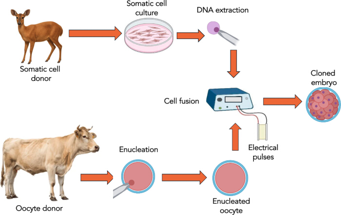
Figure 1: Schematic stages of interspecific Somatic Cell Nuclear Transfer. Cows are used as cytoplast donors, while deer are used as karyoplast donors. After obtaining the cloned embryo, it can be transferred to recipient females or cryopreserved for future transfer. Please click here to view a larger version of this figure.
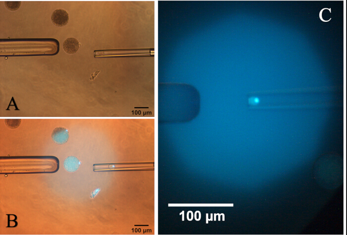
Figure 2: Photomicrography of activated oocyte with a cytoplasm protrusion. (A) Rotation of the oocyte to place the protrusion in the three o’clock position. (B) Aspiration of the protrusion with 5-10% of the cytoplasm under exposure to ultraviolet light. (C) Cytoplasm and its protrusion in the aspiration pipette under ultraviolet light. Please click here to view a larger version of this figure.
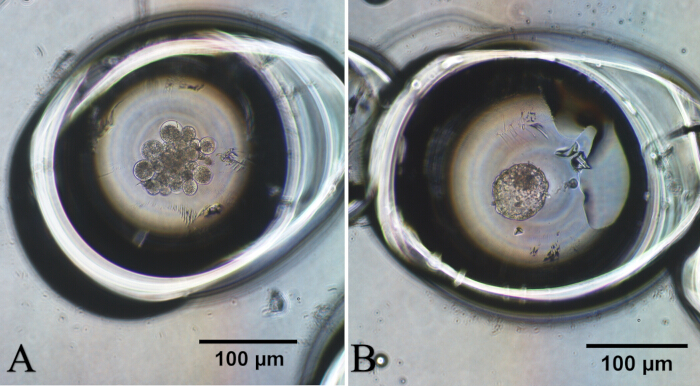
Figure 3: Photomicrography of deer embryos produced by interspecific Somatic Cell Nuclear Transfer. (A) Cloned embryo at morula stage. (B) Cloned embryo at blastocyst stage. Please click here to view a larger version of this figure.
| Grade | No. of cumulus layers | Expansion of cumulus |
| I | > 3 | Complete cumulus cover with several compacts and slightly loose cell layers |
| II | 2 - 3 | Slight expansion in outer cell layers |
| III | 1 | Partial cumulus cover and loose cell layers |
| IV | 0 | No cumulus |
Table 1: Classification of bovine oocytes to be subsequently enucleated and used as cytoplasts during interspecific somatic cell nuclear transfer (Adapted from Aguila et al.)19.
| Groups (carioplast/cytoplast) | No. of replicates | No. of reconstructed embryos | No. of cleaved (%) | No. of blastocyst (%) |
| Deer/cattle | 4 | 175 | 122 (69.7)a | 8 (4.6)a |
| Cattle/cattle | 6 | 224 | 160 (71.4)a | 24 (10.7)b |
| Different letters in the same column indicate a statistical difference. | ||||
| Cleavage rate: P = 0.74 | ||||
| Blastocyst rate: P = 0.02 | ||||
Table 2: In vitro development of red brocket deer cloned embryos using bovine cytoplast. Ten replicates were performed: four of deer/cattle pair and six of cattle/cattle pair. The cleavage and blastocyst rates were calculated using the number of reconstructed embryos as the denominator.
Discussion
It is already well documented that the genome of endangered species can be reprogrammed using oocytes from farm animals, especially cattle and pigs, which are the largest source of cytoplasts21. One of the greatest advances achieved in iSCNT was the cloning of the Pyrenean ibex (Capra pyrenaica pyrenaica), a subspecies that had disappeared following the death of its final known individual. The reconstructed embryos were obtained by fusing epithelial cells of ibex with goat (Capra hircus) oocytes. The authors obtained the birth of a female, without external morphological abnormalities. However, offspring presented severe respiratory failure and died a few minutes after birth22.
One of the stronger candidates for “resurrection” is the woolly mammoth (Mammuthus primigenius). For the cloning to be possible, the availability of viable tissue with identifiable nuclei is fundamental, which is difficult for a species that disappeared approximately 10,000 years ago. Even then, mammoth bodies found in the frozen earth layer meet the minimum requirements for iSCNT23. However, perhaps endangered species are the more suitable candidates to benefit from the use of this technique.
The segmentation rate observed in our study demonstrated that activation and the first embryonic division were not affected by the interspecific combination of nucleus and cytoplasm. During the reconstruction phase, the manipulation and fusion of the structures (karyoplast/cytoplast) presented the same technical ease in both groups, suggesting structural compatibility between species. Concerning the blastocyst rate, the results suggests that, although the reconstruction and early activation of the iSCNT embryo are not a limiting factor, there is a nuclear-cytoplasmic incompatibility that manifests itself in later stages of development likely related to incomplete epigenetic reprogramming or the maternal-zygotic transition affecting the hybrid embryo’s ability to reach the blastocyst stage24. The efficiency of iSCNT tends to be greater when karyoplasts and cytoplasts are derived from phylogenetically related species, as the natural production of viable hybrid offspring indicates a degree of nucleocytoplasmic compatibility between the two species23. Thus, inter-subspecies SCNT has already been shown to produce healthy offspring. The authors used bitch cytoplast and wolf fibroblast and transferred the embryos to 12 females, resulting in the birth of an offspring25.
The use of iSCNT faces a major "power limitation" in its biological and developmental efficiency, which drops drastically as the evolutionary distance between the donor nucleus and the recipient oocyte increases. While it succeeds between closely related species (e.g., domestic cattle and gaur)26, it routinely fails to produce viable offspring or stable embryonic cell lines when attempting to cross more distant taxonomic boundaries.
Cloning represents an important tool for animal breeding and conservation programs, as it enables the multiplication of genetically superior individuals, supports the generation of transgenic animals, and contributes to the long-term preservation of valuable genetic resources through gene banks. Nevertheless, the adoption of cloning in livestock breeding programs is still constrained by the low number of live animals produced relative to the number of embryos transferred18. Current approaches for cloned embryo production are generally divided into two categories: conventional cloning and ZP-free cloning. In the conventional approach, a micromanipulator is required to remove the oocyte nucleus and introduce the donor somatic cell into the recipient cytoplast26. Unlike conventional cloning, the ZP-free method is carried out without micromanipulation27. The results obtained in our study were very similar to those verified with the use of iSCNT, whether for cleavage rate or blastocyst production20,21.
Finally, taking into account the different steps of this procedure, it is important to highlight the critical points and practical troubleshooting. Concerning the ZP removal, ZP-free cloning offers several advantages, including ease of operation, cost-effectiveness, and a comparatively improved live birth rate per embryo transferred28. Confirming complete nuclear removal (enucleation) without inducing fatal cytoplast damage determines whether the reconstructed structure can successfully support a cloned embryo. Although UV exposure is known to have harmful effects29, it has been routinely used in the majority of cloning laboratories. Finally, ZP-free embryo culture lacks the protective outer glycoprotein shell, leading to major problems like high physical fragility during handling and blastomere dissociation30.
In conclusion, the results obtained in our study should be interpreted as preliminary evidence of the technical feasibility of the iSCNT protocol in the deer/cattle combination, rather than as definitive proof of its productive efficacy. Thus, additional studies with a larger number of replicates are needed to confirm these findings.
Disclosures
The authors have no conflicts of interest to disclose.
Acknowledgements
This work was supported by Cearense Foundation for Scientific and Technological Development Support (FUNCAP, Fortaleza, Brazil, grant number 11212233/2022) and National Institutes of Science and Technology (INCT/CNPq, grant number 406866/2022-8).
Materials
| Name | Company | Catalog Number | Comments |
|---|---|---|---|
| Aspiration Needle (18G) | LABIMPORT | 353601 | Disposable needle, short cut |
| 6-Dimethylaminopurine (6-DMAP) | Sigma-Aldrich | D2629 | |
| Antibiotic–antimycotic | Gibco, Thermo Fisher Scientific | 15240062 | |
| Bovine serum albumin | Sigma-Aldrich | A9647 | |
| Calcium Chloride Dihydrate (CaCl2·2H2O) | Sigma-Aldrich | C7902 | |
| Cell Culture Flask T-25 | Corning | 430639 | |
| CellTram Microinjector | Eppendorf | 5196 000.021 | |
| Centrifuge | Eppendorf | 5702 | Used for centrifugation of cells at 200 × g for 10 minutes to pellet and wash samples prior to nuclear transfer procedures. |
| Centrifuge Tubes (50mL) | Corning | CLS430291 | |
| Cryovials (1.8 mL) | Nunc / Thermo Fisher | 368632 | |
| Cysteamine | Sigma-Aldrich | M9768 | |
| Demecolcine | Sigma-Aldrich | D1925 | |
| Distilled water. | _ | _ | |
| DMSO (cell freezing) | Sigma-Aldrich | D2650 | 10% cryoprotectant for donor cell cryopreservation |
| Dulbecco's Modified Eagle Medium (DMEM) | Gibco / Thermo Fisher | 11995065 | |
| Enucleation Pipette (20–25 µm ID) | Narishige | GD-1 | |
| Epidermal Growth Factor (EGF) | Sigma-Aldrich | E4127 | |
| Essential amino acids BME | Sigma-Aldrich | M5550 | |
| Estradiol | Sigma-Aldrich | E8875 | |
| EVE Incubator (5% O2, 5% CO2, 90% N2) | WTA medical | REF. 23578 | |
| Fetal Bovine Serum (FBS) | Bio Nutrientes, Induslab | FEN-500 | |
| FSH (Folltropin-V) | Vetoquinol | Q-7090-124 | |
| Glass Capillary with Filament | Narishige | GD-1 | This glass capillary is used to make microwells in the petri dish |
| Heated Plate | WTA medical | 17656 | |
| Hepes | Sigma-Aldrich | H6147 | |
| Hoechst 33342 (DNA stain) | Sigma-Aldrich | B2261 | |
| Holding Pipette (120 μm OD) | Narishige | GD-1 | |
| Hyaluronidase | Sigma-Aldrich | H4272 | |
| Inverted Microscope ECLIPSE | Nikon | TE2000-U | |
| Ionomycin (Calcium Ionophore A23187) | Sigma-Aldrich | I0634 | |
| ITS | Sigma-Aldrich | I2771 | |
| KCl | Sigma-Aldrich | P5405 | |
| KH2PO4 | Sigma-Aldrich | P5655 | |
| Laminar Flow Cabinet Isocide | Esco | 2120765 | Laminar flow cabinet providing a sterile working environment for embryo and cell culture procedures. |
| L-Glutamine | Sigma-Aldrich | G8540 | |
| Liquid Nitrogen Storage Dewar | Nitroagro | Varies | |
| Magnesium Sulfate (MgSO4) | Sigma-Aldrich | M2643 | |
| Mannitol Fusion Medium (0.3M) | Sigma-Aldrich | M9546 | |
| MgCl2 | Sigma-Aldrich | M8266 | |
| Microforge MF-900 | Narishige | MF-900 | |
| Micromanipulator System | Narishige | MM-89 / TransferMan 4r | |
| Micropipette Puller | Narishige | Narishige PC-100 | |
| Micropipettes (10 µL, 20 µL, 100 µL, 200 µL, 1000 µL) | Eppendorf | Research Plus Series | |
| Microfusion Chamber | Eppendorf | 12816532 | Is a specialized 0.2 mm gap accessory designed for electroporation applications |
| Mineral Oil (embryo-tested) | Sigma-Aldrich | M8410 | |
| Multiporator | Eppendorf | 4308 000.015 | |
| Myo-inositol | Sigma-Aldrich | I7508 | |
| NaCl | Sigma-Aldrich | S5886 | |
| NaH2PO4 | Sigma-Aldrich | S5011 | |
| NaHCO3 | Sigma-Aldrich | S4219 | |
| Non-essential amino acids MEM | Sigma-Aldrich | M7145 | |
| Pentabiotic | Zoetis | 10005048 | 2,400,000 IU vial. |
| Petri Dish 35mm (embryo-tested) | Corning | 430165 | |
| Petri Dish 60mm (embryo-tested) | Corning | 430196 | |
| Phenol red | Sigma-Aldrich | P0290 | |
| Pipette Tips 1000 µL | Eppendorf | 9401030 | |
| Pipette Tips 200 µL | Eppendorf | 94052300 | |
| Pipette Tips Without Filter (0.5–10 µL) | Axygen | T-300 | |
| Poly(vinyl alcohol) (PVA) | Sigma-Aldrich | P8136 | |
| Pronase (Protease from S. griseus) | Sigma-Aldrich | P8811 | |
| Sodium lactate | Sigma-Aldrich | L7900 | |
| Sodium pyruvate | Sigma- Aldrich | P4562 | |
| Stereomicroscope | Nikon | SMZ800 | |
| Sterile embryo tested water | Sigma-Aldrich | W3500 | |
| Sterile filter (0.22 μm) | Corning | 431154 | |
| TCM-199 | Sigma-Aldrich | M2154 | |
| Tri-gas Incubator (5% O2, 5% CO2, 90% N2) | WTA medical | REF. 23578 | |
| Trypsin-EDTA (0.25%) | Gibco / Thermo Fisher | 25200056 | |
| Ultrapure wáter | Sigma-Aldrich | W3500 | |
| UV Lamp / Fluorescence Module | Leica / Olympus | Varies | Brief UV exposure to verify enucleation; minimize photo-toxicity |
References
- Flagstad O, Roed KH. Refugial origins of reindeer (Rangifer tarandus.) inferred from mitochondrial DNA sequences. Evolution. 2003;57(3):658-70.
- Schilling AM, Rössner GE. The (sleeping) beauty in the beast—a review on the water deer, Hydropotes inermis. Hystrix It J Mamm. 2017;28(2):121-33.
- Newman BA, D’Angelo GJ. A review of cervidae visual ecology. Animals (Basel). 2024;14(3):420.
- Rola LD. Integrating cryobiology and conservation: the role of biobanks for neotropical deer in Latin America. Biopreserv Biobank. 2025;23(6):575-88.
- Duarte JMB, González S, Maldonado JE. The surprising evolutionary history of South American deer. Mol Phylogenet Evol. 2008;49:17-22.
- Bernegossi AM, et al. Resurrection of the genus Subulo. for the gray brocket deer, with designation of a neotype. J Mammal. 2023;104(3):619-33.
- Morales-Donoso JA, et al. Revalidation of Passalites Gloger, 1841 for the Amazon brown brocket deer P. nemorivagus. (Cuvier, 1817) (Mammalia, Artiodactyla, Cervidae). ZooKeys. 2023;1167:241-64.
- El Bizri HR. Exposing illegal hunting and wildlife depletion in the world's largest tropical country through social media data. Conserv Biol. 2024;38:e14334.
- Pasquariello R, et al. Use of assisted reproductive technologies (ARTs) to shorten the generational interval in ruminants: current status and perspectives. Theriogenology. 2024;225(1):16-32.
- Herrick JR. Assisted reproductive technologies for endangered species conservation: developing sophisticated protocols with limited access to animals with unique reproductive mechanisms. Biol Reprod. 2019;100:1158-70.
- Iqbal A, et al. Conservation of endangered species through somatic cell nuclear transfer (SCNT). Conserv Genet Resour. 2021;13:349-57.
- Hwang I. Successful cloning of coyotes through interspecies somatic cell nuclear transfer (iSCNT) using domestic dog oocytes. Biol Reprod. 2012;87(1):56.
- Loi P, et al. Genetic rescue of an endangered mammal by cross-species nuclear transfer using post-mortem somatic cells. Nat Biotechnol. 2001;19:962-4.
- Gómez MC, et al. Birth of African wildcat cloned kittens born from domestic cats. Cloning Stem Cells. 2004;6:247-58.
- Wani NA, Vettical BS, Hong SB. First cloned Bactrian camel (Camelus bactrianus.) calf produced by interspecies somatic cell nuclear transfer: a step towards preserving the critically endangered wild Bactrian camels. PLoS One. 2017;12:e0177800.
- Ross PJ, Cibelli JB. Bovine somatic cell nuclear transfer. Methods Mol Biol. 2010;636:155-77.
- Choi YH, Velez IC, Macias-Garcia B, Hinrichs K. Timing factors affecting blastocyst development in equine somatic cell nuclear transfer. Cell Reprogram. 2015;17:124-30.
- Herrid H, Van Eenennaam H. Application of zona-free cloning technologies to livestock breeding programmes. Anim Reprod Sci. 2026;287:108107.
- Aguila L, et al. Oocyte selection for in vitro embryo production in bovine species: noninvasive approaches for new challenges of oocyte competence. Animals. 2020;10:2196.
- Vajta G, et al. New method for culture of zona-included or zona-free embryos: the well of the well (WOW) system. Mol Reprod Dev. 2000;55(3):256-64.
- Lagutina I, Fulka H, Lazzari G, Galli C. Interspecies somatic cell nuclear transfer: advancements and problems. Cell Reprogram. 2013;15:374-84.
- Folch J, et al. First birth of an animal from an extinct subspecies (Capra pyrenaica pyrenaica.) by cloning. Theriogenology. 2009;71:1026-34.
- Loi P, Saragusty J, Ptak G. Cloning the mammoth: a complicated task or just a dream. Adv Exp Med Biol. 2014;753:489-502.
- Mastromonaco GF, et al. The influence of nuclear content on developmental competence of gaur cattle hybrid in vitro fertilized and somatic cell nuclear transfer embryos. Biol Reprod. 2007;76:514-23.
- Kim MK, et al. Endangered wolves cloned from adult somatic cells. Cloning Stem Cells. 2007;9:130-7.
- Lanza RP, et al. Cloning of an endangered species (Bos gaurus.) using interspecies nuclear transfer. Cloning. 2000;2(2):79-90.
- Hill J, et al. The effect of donor cell serum starvation and oocyte activation compounds on the development of somatic cell cloned embryos. Cloning. 1999;1:201-8.
- Verma G, et al. Handmade cloning: recent advances, potential and pitfalls. J Anim Sci Biotechnol. 2015;6:43.
- Takenaka M, Horiuchi T, Yanagimachi R. Effect of light on development of mammalian zygote. Proc Natl Acad Sci U S A. 2007;104:14289-93.
- Madani S, Machaty Z, Vajta G. An alternative way to improve mammalian embryo development in vitro: culture of zona pellucida-free embryos. Cell Reprogram. 2022;24(3):111-7.
Reprints and Permissions
This article has been published
Video Coming Soon