This retrospective cohort study of 136 postoperative endometrial cancer patients evaluated Astragalus Injection with paclitaxel-carboplatin chemotherapy. Adjunctive Astragalus Injection was associated with greater improvements in quality of life, CD4⁺/CD8⁺ ratio, and performance status, fewer selected chemotherapy-related adverse events, and greater reductions in exploratory serum biomarkers.
Research Article
Quality of Life, Immune Function, and Treatment Outcomes with Astragalus Injection in Endometrial Cancer: A Retrospective Cohort Study
* These authors contributed equally
In This Article
Summary
Abstract
Adjuvant chemotherapy with paclitaxel and carboplatin is commonly used for high-risk postoperative endometrial cancer but is associated with significant toxicity. Astragalus Injection has immunomodulatory properties, yet clinical evidence regarding its adjunctive role remains limited. This retrospective cohort study compared quality of life, immune parameters, exploratory serum biomarkers, performance status, and adverse events between patients receiving paclitaxel-carboplatin with or without adjunctive Astragalus Injection. A total of 136 patients treated between January 2022 and March 2023 were included in an observation group (chemotherapy plus Astragalus Injection, n = 68) or control group (chemotherapy alone, n = 68). Treatment consisted of six 21-day cycles. Outcomes included European Organization for Research and Treatment of Cancer Quality of Life Questionnaire-Core 30 (EORTC QLQ-C30) scores, CD4⁺ and CD8⁺ T-cell counts, CD4⁺/CD8⁺ ratio, squamous cell carcinoma antigen (SCC-Ag), cytokeratin 19 fragment (CYFRA21-1), adverse events, and Karnofsky Performance Status (KPS) scores. Changes in all five EORTC QLQ-C30 functional domain scores were significantly greater in the observation group than in the control group (P < 0.001). The increase in CD4⁺/CD8⁺ ratio was also significantly greater in the observation group (P < 0.001). SCC-Ag and CYFRA21-1 decreased more substantially in the observation group (P = 0.003 and P < 0.001, respectively). The incidences of grade ≥ 2 gastrointestinal toxicity (14.71% vs. 32.35%, P = 0.015) and bone marrow suppression (11.76% vs. 27.94%, P = 0.019) were lower in the observation group. KPS improvement was greater in the observation group (P < 0.001). Adjunctive Astragalus Injection was associated with improvements in quality of life, immune parameters, performance status, selected chemotherapy-related adverse events, and exploratory serum biomarkers. These findings are hypothesis-generating and require confirmation in prospective studies.
Introduction
Endometrial cancer is one of the most common gynecological malignancies worldwide, with rising incidence rates in both developed and developing countries. For patients with surgically resected endometrial cancer at intermediate or high risk of recurrence, adjuvant chemotherapy is recommended to reduce recurrence risk and improve survival outcomes1,2,3. Paclitaxel combined with carboplatin is a commonly used chemotherapy regimen for patients with high-risk or advanced endometrial cancer, including selected patients receiving postoperative adjuvant treatment4. However, this regimen is associated with substantial toxicity, including myelosuppression, gastrointestinal reactions, peripheral neuropathy, and hepatic impairment, which may adversely affect patients’ quality of life and compromise treatment completion and long-term outcomes5,6. Therefore, strategies to improve the tolerability of paclitaxel-carboplatin chemotherapy while maintaining its therapeutic benefit represent an important clinical need.
Astragalus Injection (Huangqi Injection) is a preparation derived from the root of Astragalus membranaceus (Fisch.) Bunge, a medicinal herb that has been used in traditional Chinese medicine for thousands of years. The primary active components of Astragalus include astragalosides, Astragalus polysaccharides, and various flavonoids7. Preclinical studies have suggested that Astragalus-derived preparations exhibit multiple pharmacological activities relevant to cancer therapy, including immunomodulatory effects, such as enhancement of T-cell proliferation, natural killer cell activity, and macrophage function; hematopoietic protection against chemotherapy-induced myelosuppression; anti-inflammatory and antioxidant properties; and potential antitumor effects through induction of apoptosis and inhibition of tumor cell proliferation8,9. Astragalus Injection and Astragalus-derived preparations have been reported to exert immunomodulatory effects and influence immune parameters in patients receiving cancer therapy8,9. A recent retrospective cohort study demonstrated that astragalus polysaccharides (PG2) alleviated chemotherapy-induced fatigue in patients with gynecologic malignancies, including endometrial cancer10. However, high-quality prospective evidence specifically evaluating Astragalus Injection as an adjunct to paclitaxel-carboplatin chemotherapy in endometrial cancer remains limited.
Despite these findings, clinical evidence regarding the adjunctive use of Astragalus Injection in patients with endometrial cancer receiving paclitaxel-carboplatin chemotherapy remains limited. The present study was therefore designed to evaluate outcomes associated with adding Astragalus Injection to standard paclitaxel-carboplatin adjuvant chemotherapy in postoperative patients with endometrial cancer. We hypothesized that adjunctive Astragalus Injection would be associated with improved immune function, reduced chemotherapy-related toxicity, and better quality of life compared with chemotherapy alone. This retrospective cohort analysis compares outcomes between patients receiving paclitaxel-carboplatin plus Astragalus Injection and those receiving chemotherapy alone over six cycles of treatment.
Protocol
Ethics Statement
This study was approved by the Ethics Committee of Liyuan Hospital, Tongji Medical College, Huazhong University of Science and Technology (Approval No. LY15971, approved in January 2022). The requirement for informed consent was waived by the Ethics Committee due to the retrospective nature of the study and the use of anonymized clinical data from routine medical records. All procedures were conducted in accordance with the Declaration of Helsinki and the International Conference on Harmonisation of Good Clinical Practice (ICH-GCP) guidelines. The study involved no more than minimal risk to subjects, and the waiver did not adversely affect the rights or welfare of the subjects. Astragalus Injection was prescribed by attending physicians based on clinical judgment independently of this retrospective study. All patients provided standard clinical informed consent for chemotherapy and adjunctive therapies before treatment initiation, in accordance with institutional practice.
Study Design and Participants
This retrospective cohort study included 136 postoperative patients with primary endometrial cancer treated at Liyuan Hospital between January 2022 and March 2023. All diagnoses were confirmed by histopathological review of surgical specimens. Patients with cervical primary malignancies or other concurrent active malignancies were excluded. Based on the treatment received in routine clinical practice, patients were classified into an observation group (Astragalus Injection combined with paclitaxel-carboplatin, n = 68) and a control group (paclitaxel-carboplatin alone, n = 68). The decision to add Astragalus Injection to the chemotherapy regimen was made by the attending physicians based on comprehensive clinical evaluation, including the patient’s baseline performance status, anticipated chemotherapy tolerance, and individual preferences, rather than by randomization. The retrospective grouping was determined solely by the actual treatment received. The overall study design and patient flow are summarized in Figure 1.
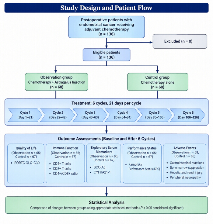
Figure 1. Study Design and Patient Flow. Schematic overview of the retrospective cohort study of 136 postoperative patients with endometrial cancer treated between January 2022 and March 2023. Patients were retrospectively classified according to treatment received in routine clinical practice into an observation group receiving paclitaxel-carboplatin chemotherapy plus Astragalus Injection (n = 68) and a control group receiving paclitaxel-carboplatin chemotherapy alone (n = 68). Treatment consisted of six 21-day chemotherapy cycles. Four patients discontinued treatment early; therefore, post-treatment efficacy analyses included 65 patients in the observation group and 67 patients in the control group, whereas all 136 patients were included in the safety analysis. Quality of life, immune function, exploratory serum biomarkers, and performance status were assessed at baseline and after six treatment cycles, and treatment-related adverse events were monitored throughout treatment. EORTC QLQ-C30, European Organization for Research and Treatment of Cancer Quality of Life Questionnaire-Core 30; SCC-Ag, squamous cell carcinoma antigen; CYFRA21-1, cytokeratin 19 fragment; KPS, Karnofsky Performance Status. Please click here to view a larger version of this figure.
Inclusion criteria were: (1) histopathologically confirmed primary endometrial cancer; (2) completion of surgical treatment for endometrial cancer; (3) age between 18 and 75 years; (4) first receipt of paclitaxel-carboplatin adjuvant chemotherapy; (5) expected survival >6 months; and (6) availability of complete clinical records for the study period. Expected survival >6 months was determined by the attending physicians based on comprehensive clinical assessment, including disease stage, histopathological grade, surgical resection status, baseline performance status, and overall clinical condition at the time of treatment planning.
Exclusion criteria were: (1) concurrent active malignant tumors of other types; (2) pregnancy or lactation; (3) psychiatric disorders or cognitive impairment; (4) significant organ dysfunction involving the heart, liver, kidneys, or brain; (5) myocardial infarction or unstable angina within the preceding 6 months; (6) known hypersensitivity to paclitaxel, carboplatin, or Astragalus Injection; and (7) incomplete clinical records for the study period. Significant organ dysfunction was defined as follows: (a) cardiac dysfunction, New York Heart Association (NYHA) class ≥III or left ventricular ejection fraction < 50%; (b) hepatic dysfunction, alanine aminotransferase (ALT) or aspartate aminotransferase (AST) > 2.5 × the upper limit of normal (ULN), or total bilirubin > 1.5 × ULN; (c) renal dysfunction, serum creatinine > 1.5 × ULN or estimated glomerular filtration rate (eGFR) < 60 mL/min/1.73 m2; and (d) cerebral dysfunction, a history of cerebrovascular accident with significant neurological deficits.
Surgical Procedures
Surgical procedures were selected according to disease stage, tumor characteristics, and institutional practice. All patients underwent total hysterectomy with bilateral salpingo-oophorectomy. Pelvic lymph node assessment, including lymphadenectomy or sentinel lymph node biopsy, was performed when any of the following criteria were met: (1) preoperative imaging (pelvic MRI or CT) suggested lymph node enlargement (short-axis diameter ≥ 10 mm); (2) intraoperative palpation revealed suspicious pelvic lymph nodes; or (3) high-risk histological features, including non-endometrioid subtype, high grade, or deep myometrial invasion ≥ 1/2, were identified on frozen section. Para-aortic lymph node dissection was performed in patients with high-risk features or suspected nodal involvement. High-risk features were defined as: (1) non-endometrioid histological subtypes (serous, clear cell, or carcinosarcoma); (2) histological grade 3; (3) myometrial invasion > 1/2; (4) cervical stromal invasion; or (5) positive peritoneal cytology. Suspected nodal involvement was defined as preoperative imaging (CT or MRI) showing para-aortic lymph nodes with a short-axis diameter ≥10 mm or intraoperative findings of enlarged, firm, or fixed para-aortic lymph nodes.
Chemotherapy Regimen
Adjuvant chemotherapy was initiated within 4 weeks after surgery based on pathological staging and clinical features. Pathological staging was performed according to the International Federation of Gynecology and Obstetrics (FIGO) 2009 staging system. Adjuvant chemotherapy was recommended for patients with: (1) FIGO stage IB with high-risk features (grade 3, lymphovascular space invasion, or non-endometrioid histology); (2) FIGO stage II–IV; or (3) any stage with positive pelvic or para-aortic lymph nodes. The chemotherapy regimen consisted of paclitaxel combined with carboplatin administered in 21-day cycles for a total of six cycles. Paclitaxel was administered at 175 mg/m2 by intravenous infusion over 3 h on day 1 of each cycle. Carboplatin was administered at a target area under the concentration-time curve (AUC) of 5–6 by intravenous infusion over 2 h on day 1 of each cycle. The carboplatin dose was calculated using the Calvert formula: dose (mg) = target AUC × (GFR + 25). Renal function for this calculation was estimated using the Cockcroft-Gault formula for creatinine clearance (CrCl): CrCl (mL/min) = [(140 − age) × weight (kg)]/[72 × serum creatinine (mg/dL)] × 0.85 for female patients. Serum creatinine was measured within 72 h before each chemotherapy cycle.
Astragalus Injection Administration
Patients in the observation group received Astragalus Injection in addition to the above chemotherapy regimen. Astragalus Injection was administered at a fixed dose of 20 mL (two 10-mL ampoules, each equivalent to 20 g of crude herb), diluted in 250 mL of 5% glucose solution, and administered by intravenous infusion over 60–90 min once daily for 5 consecutive days during each chemotherapy cycle. The product administered was Astragalus Injection (National Drug Approval No. Z19993281; Shanghai No. 1 Biochemical & Pharmaceutical Co., Ltd., China), formulated as a sterile aqueous solution containing 2 g of crude herb per mL in accordance with National Drug Standard WS3-B-3335-98. Batch numbers were verified from pharmacy records.
On day 1, the Astragalus Injection infusion was initiated 30 min before paclitaxel administration and was administered daily through day 5 of each 21-day cycle. All patients in the observation group received the same fixed dose, with no dose adjustments. Astragalus Injection was administered as a standalone infusion and was not mixed with other medications in the same infusion bag. Heart rate, respiratory rate, blood pressure, and oxygen saturation were recorded at baseline before infusion, every 15 min during the first hour, every 30 min thereafter, and until 30 min after completion of the infusion. Any infusion-related reactions were documented.
Supportive Care
All patients received standardized symptomatic supportive care during chemotherapy according to the institutional standardized chemotherapy supportive care protocol (Liyuan Hospital, version 2022). Prophylactic antiemetic treatment consisted of ondansetron 8 mg intravenously 30 min before chemotherapy on day 1, followed by ondansetron 4 mg orally twice daily on days 2–3, with dexamethasone 10 mg intravenously before paclitaxel administration as premedication. Reduced glutathione 1.2 g or a glycyrrhizic acid preparation was administered intravenously once daily on days 1–3 of each cycle to patients with baseline hepatic dysfunction or elevated transaminase levels during treatment. Intravenous fluids and electrolyte correction were administered based on daily clinical assessment and electrolyte panel results according to institutional electrolyte replacement protocols. Adverse events were graded according to the Common Terminology Criteria for Adverse Events (CTCAE), version 5.0. For grade 1–2 nausea or vomiting, metoclopramide 10 mg orally or ondansetron 4 mg orally was administered as needed. For grade ≥3 nausea or vomiting, granisetron 3 mg or ondansetron 8 mg was administered intravenously with 500–1,000 mL of normal saline, with dose adjustments based on clinical response. Antiemetic selection and dosing followed institutional supportive care guidelines. Febrile neutropenia was defined as a single oral temperature of ≥38.3°C or a temperature of ≥38.0°C sustained for ≥1 h, with an absolute neutrophil count (ANC) < 0.5 × 10⁹/L. Empiric broad-spectrum antibiotic treatment with piperacillin-tazobactam 4.5 g intravenously every 6 h or cefepime 2 g intravenously every 8 h was initiated within 1 h of fever detection according to institutional antimicrobial stewardship guidelines. Granulocyte colony-stimulating factor (G-CSF; filgrastim 5 µg/kg/day subcutaneously) was administered for severe neutropenia (ANC < 0.1 × 10⁹/L) or prolonged neutropenia (>7 days) according to institutional guidelines.
Outcome Measurements
Quality of life was assessed using the European Organization for Research and Treatment of Cancer Quality of Life Questionnaire-Core 30 (EORTC QLQ-C30, version 3.0) at baseline (within 1 week before the first chemotherapy cycle, after surgical recovery) and after six cycles of treatment (within 1 week after completion of the sixth cycle). The questionnaire was administered by trained research nurses in a quiet setting, and patients completed the questionnaire independently. The five functional domain scores (Physical, Role, Cognitive, Emotional, and Social Function) were calculated according to the EORTC scoring manual (version 3.0) using a linear transformation to a 0–100 scale, with higher scores indicating better functioning. All questionnaire data were collected as part of routine clinical assessment and transcribed to electronic case report forms.
Immune function was evaluated by measuring peripheral blood CD4⁺ and CD8⁺ T-cell counts using flow cytometry. Peripheral blood samples (5 mL) were collected in EDTA anticoagulation tubes from all patients at baseline (within 3 days before the first chemotherapy cycle) and after six cycles (within 3 days after completion of the sixth cycle). All post-treatment samples were collected between 8:00 and 10:00 AM under standardized conditions after overnight fasting. Flow cytometry was performed using a BD FACSCanto II flow cytometer (BD Biosciences, San Jose, CA, USA; RRID: SCR_018056) within 6 h of sample collection. The following fluorochrome-conjugated monoclonal antibodies were used: FITC anti-human CD4 (BD Biosciences, Cat. No. 555346; RRID: AB_395751) and PE anti-human CD8 (BD Biosciences, Cat. No. 555367; RRID: AB_395770). Briefly, 100 µL of peripheral blood was incubated with the appropriate antibodies for 30 min at room temperature in the dark, followed by red blood cell lysis using BD FACS Lysing Solution (BD Biosciences, Cat. No. 349202) according to the manufacturer’s instructions. Data were acquired using BD FACSDiva software (version 8.0.1) and analyzed using FlowJo software (version 10.8, BD Biosciences). Lymphocytes were gated based on forward- and side-scatter characteristics. CD4⁺ and CD8⁺ T-cell counts were expressed as absolute counts (cells/µL) and percentages, and the CD4⁺/CD8⁺ ratio was calculated.
Exploratory serum biomarkers SCC-Ag and CYFRA21-1 were measured using enzyme-linked immunosorbent assay (ELISA) at baseline and after six cycles. Serum was separated by centrifugation at 800 × g for 10 min at 4°C within 2 h of collection, aliquoted, and stored at −80°C until batch analysis. SCC-Ag levels were measured using a human SCC-Ag ELISA kit (Cloud-Clone Corp., Houston, TX, USA; Cat. No. SEC893Hu; intra-assay CV < 8%, inter-assay CV < 10%). CYFRA21-1 levels were measured using a human CYFRA21-1 ELISA kit (Cloud-Clone Corp.; Cat. No. SEB246Hu; intra-assay CV < 8%, inter-assay CV < 10%). All assays were performed in duplicate according to the manufacturer's instructions. Absorbance was measured at 450 nm using a BioTek ELx808 microplate reader (Agilent Technologies, Santa Clara, CA, USA). Concentrations were calculated from standard curves using a four-parameter logistic regression model in Gen5 software (version 3.04, Agilent Technologies).
All treatment-related adverse events were graded according to the National Cancer Institute Common Terminology Criteria for Adverse Events (CTCAE), version 5.0. Adverse events, including gastrointestinal reactions, bone marrow suppression, hepatic and renal injury, and peripheral neuropathy, were monitored throughout treatment. Adverse events were identified and recorded retrospectively through systematic review of electronic medical records, nursing notes, laboratory results, and physician progress notes by two independent data abstractors. Gastrointestinal reactions were defined as nausea, vomiting, diarrhea, or constipation. Bone marrow suppression was assessed using weekly complete blood count results and included leukopenia, neutropenia, anemia, and thrombocytopenia. Hepatic injury was assessed using serum ALT, AST, and total bilirubin levels, and renal injury was assessed using serum creatinine, BUN, and eGFR measured using an AU5800 automated biochemical analyzer (Beckman Coulter, Brea, CA, USA). Hepatic injury was defined as ALT or AST > ULN or total bilirubin > ULN. Renal injury was defined as serum creatinine > ULN or eGFR < 90 mL/min/1.73 m2. The severity of these adverse events was graded according to CTCAE v5.0.
Performance status was evaluated using the KPS scale at baseline and after six cycles. KPS assessments were performed by the attending physicians as part of routine clinical evaluation at baseline (within 3 days before the first chemotherapy cycle) and after completion of the sixth cycle (within 7 days after the last chemotherapy dose). All assessors were trained in KPS administration according to institutional standards.
Statistical Analysis
Statistical analyses were performed using SPSS version 26.0 (IBM Corp., Armonk, NY, USA; RRID: SCR_019096). Continuous variables are presented as mean ± standard deviation (SD). Between-group comparisons of continuous variables at a single time point were performed using independent-samples t-tests, whereas within-group comparisons before and after treatment were performed using paired t-tests. Changes from baseline were calculated as post-treatment minus baseline values and compared between groups using independent-samples t-tests. Baseline characteristics were compared using independent-samples t-tests for continuous variables and chi-square tests for categorical variables. Categorical variables are presented as frequencies (percentages) and were analyzed using chi-square tests or Fisher’s exact test, as appropriate. Fisher’s exact test was used when any expected cell count was <5 in a 2 × 2 contingency table; otherwise, the chi-square test was applied. For quality of life, immune function, KPS, and the exploratory serum biomarkers SCC-Ag and CYFRA21-1, analyses were based on changes from baseline to post-treatment. Parametric test assumptions were assessed before analysis. Normality was evaluated using the Shapiro–Wilk test, and homogeneity of variance was evaluated using Levene's test. For variables with mild departures from normality (skewness or kurtosis within ±2), parametric tests were retained because of their robustness with sample sizes > 30. For variables with substantial departures from normality (Shapiro–Wilk P <0.05 with skewed distributions), the Mann–Whitney U test for between-group comparisons and Wilcoxon signed-rank test for within-group comparisons were performed as sensitivity analyses. These analyses yielded results consistent with the parametric analyses and are therefore not reported separately. Four patients who discontinued treatment early (three in the observation group and one in the control group) were excluded from all post-treatment efficacy analyses, including the QLQ-C30, immune-function, serum-biomarker, and KPS analyses, but were retained in the safety analysis of adverse events. Thus, post-treatment efficacy analyses included 65 patients in the observation group and 67 patients in the control group. No imputation was performed for missing data. All tests were two-sided, and P < 0.05 was considered statistically significant.
Results
Baseline Characteristics
A total of 136 patients were included in this analysis, with 68 patients classified into each group. Baseline characteristics were well balanced between the two groups (all P > 0.05) (Table 1). In the observation group, the mean age was 41.28 ± 5.67 years (range, 30–52 years), and the mean weight was 58.15 ± 6.31 kg (range, 51–70 kg). Histopathological subtypes confirmed by pathology review included 60 endometrioid adenocarcinomas, 5 adenosquamous carcinomas, and 3 other subtypes (including 2 serous carcinomas and 1 clear cell carcinoma). Clinical staging according to the FIGO 2009 system included 20 stage I, 24 stage II, and 24 stage III cases. In the control group, the mean age was 40.47 ± 6.02 years (range, 29–53 years), and the mean weight was 59.03 ± 6.78 kg (range, 50–72 kg). Histopathological subtypes included 61 endometrioid adenocarcinomas, 4 adenosquamous carcinomas, and 3 other subtypes (including 2 serous carcinomas and 1 carcinosarcoma). Clinical staging included 22 stage I, 23 stage II, and 23 stage III cases. There were no cases of pure primary squamous cell carcinoma of the endometrium in either group.
| Characteristic | Observation Group (n = 68) | Control Group (n = 68) | P |
| Age (years) | 41.28 ± 5.67 | 40.47 ± 6.02 | 0.421 |
| Weight (kg) | 58.15 ± 6.31 | 59.03 ± 6.78 | 0.435 |
| FIGO stage | 0.933 | ||
| Stage I | 20 (29.4%) | 22 (32.4%) | |
| Stage II | 24 (35.3%) | 23 (33.8%) | |
| Stage III | 24 (35.3%) | 23 (33.8%) | |
| Histological subtype | 0.942 | ||
| Endometrioid adenocarcinoma | 60 (88.2%) | 61 (89.7%) | |
| Adenosquamous carcinoma | 5 (7.4%) | 4 (5.9%) | |
| Other | 3 (4.4%) | 3 (4.4%) | |
| Baseline KPS | 70.42 ± 8.56 | 69.98 ± 8.62 | 0.766 |
| Baseline CD4⁺/CD8⁺ ratio | 1.20 ± 0.32 | 1.19 ± 0.30 | 0.851 |
| Baseline SCC-Ag (ng/mL) | 3.25 ± 1.15 | 3.35 ± 1.35 | 0.643 |
| Baseline CYFRA21-1 (ng/mL) | 3.58 ± 1.42 | 3.56 ± 1.33 | 0.933 |
Table 1: Baseline Characteristics of Study Participants. Data are presented as mean ± SD or n (%). P values were calculated using independent-samples t-tests for continuous variables and chi-square tests for categorical variables. FIGO, International Federation of Gynecology and Obstetrics; KPS, Karnofsky Performance Status; SCC-Ag, squamous cell carcinoma antigen; CYFRA21-1, cytokeratin 19 fragment; SD, standard deviation.
Patient Disposition
All enrolled patients received at least one cycle of treatment and were included in the safety analysis (n = 136). Three patients in the observation group (4.4%) discontinued treatment early due to grade 3–4 adverse events (two with febrile neutropenia and one with grade 3 peripheral neuropathy). One patient in the control group (1.5%) discontinued treatment due to grade 3 gastrointestinal toxicity. These four patients were excluded from the post-treatment analyses of QLQ-C30, immune function, exploratory serum biomarkers, and KPS but were included in the safety analysis.
Quality of Life Scores
Changes from baseline to post-treatment in EORTC QLQ-C30 functional domain scores were significantly greater in the observation group than in the control group (Table 2; Figure 2). The mean changes (post-treatment minus baseline) were as follows: physical function, +15.2 ± 6.8 in the observation group versus +8.5 ± 7.2 in the control group (P < 0.001); role function, +14.1 ± 6.5 versus +7.8 ± 7.0 (P < 0.001); cognitive function, +13.5 ± 6.8 versus +7.1 ± 7.5 (P < 0.001); emotional function, +12.8 ± 7.2 versus +6.9 ± 8.1 (P < 0.001); and social function, +16.2 ± 5.9 versus +8.5 ± 7.8 (P < 0.001).
| Domain | Observation Group (n = 65) | Control Group (n = 67) | Between-Group Difference in Change | 95% CI | P |
| Physical Function | +15.2 ± 6.8 | +8.5 ± 7.2 | +6.7 | 4.3–9.1 | < 0.001 |
| Role Function | +14.1 ± 6.5 | +7.8 ± 7.0 | +6.3 | 4.0–8.6 | < 0.001 |
| Cognitive Function | +13.5 ± 6.8 | +7.1 ± 7.5 | +6.4 | 4.0–8.8 | < 0.001 |
| Emotional Function | +12.8 ± 7.2 | +6.9 ± 8.1 | +5.9 | 3.3–8.5 | < 0.001 |
| Social Function | +16.2 ± 5.9 | +8.5 ± 7.8 | +7.7 | 5.3–10.1 | < 0.001 |
Table 2: Changes in EORTC QLQ-C30 Functional Domain Scores from Baseline to Post-Treatment. Data are presented as mean change ± SD for the observation group (n = 65) and control group (n = 67). Change was calculated as the post-treatment score minus the baseline score. Between-group differences represent the difference in change between the observation and control groups. P values were calculated using independent-samples t-tests comparing change scores between groups. EORTC QLQ-C30, European Organization for Research and Treatment of Cancer Quality of Life Questionnaire-Core 30; CI, confidence interval; SD, standard deviation.
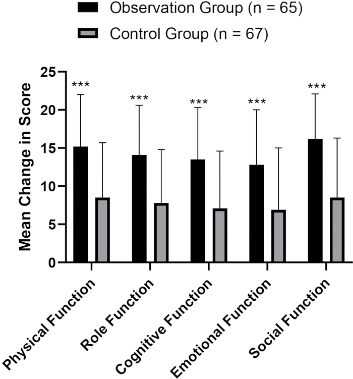
Figure 2. Changes in EORTC QLQ-C30 Functional Domain Scores. Changes from baseline to post-treatment are presented as mean ± SD for the observation group (n = 65) and control group (n = 67). Change was calculated as the post-treatment score minus the baseline score. Error bars represent SD. *** P < 0.001 versus the control group for all five functional domains. EORTC QLQ-C30, European Organization for Research and Treatment of Cancer Quality of Life Questionnaire-Core 30; SD, standard deviation. Please click here to view a larger version of this figure.
Immune Function
Changes from baseline to post-treatment in CD4⁺ T-cell count and CD4⁺/CD8⁺ ratio were significantly greater in the observation group than in the control group (Table 3). The mean change in CD4⁺ count was +283.5 ± 98.2 cells/µL in the observation group versus +80.4 ± 105.6 cells/µL in the control group (P < 0.001). The mean change in CD4⁺/CD8⁺ ratio was +0.60 ± 0.28 in the observation group versus +0.16 ± 0.25 in the control group (P < 0.001). There was no significant between-group difference in the change in CD8⁺ T-cell count (P = 0.81).
| Indicator | Observation Group (n = 65) | Control Group (n = 67) | Between-Group Difference in Change | 95% CI | P |
| CD4⁺ count (cells/µL) | +283.5 ± 98.2 | +80.4 ± 105.6 | +203.1 | 168.5–237.7 | < 0.001 |
| CD8⁺ count (cells/µL) | −24.9 ± 35.6 | −26.4 ± 38.1 | 1.5 | −10.9–13.9 | 0.81 |
| CD4⁺/CD8⁺ ratio | +0.60 ± 0.28 | +0.16 ± 0.25 | +0.44 | 0.35–0.53 | < 0.001 |
Table 3: Changes in Immune Function Indicators from Baseline to Post-Treatment. Data are presented as mean change ± SD for the observation group (n = 65) and control group (n = 67). Change was calculated as the post-treatment value minus the baseline value. Between-group differences represent the difference in change between the observation and control groups. P values were calculated using independent-samples t-tests comparing change scores between groups. CI, confidence interval; SD, standard deviation.
Exploratory Serum Biomarkers
Changes from baseline to post-treatment in SCC-Ag and CYFRA21-1 levels were significantly greater in the observation group than in the control group (Table 4). The mean change in SCC-Ag was −1.40 ± 0.85 ng/mL in the observation group versus −0.92 ± 0.95 ng/mL in the control group (P = 0.003). The mean change in CYFRA21-1 was −1.66 ± 1.10 ng/mL in the observation group versus −0.88 ± 1.05 ng/mL in the control group (P < 0.001).
| Marker | Observation Group (n = 65) | Control Group (n = 67) | Between-Group Difference in Change | 95% CI | P |
| SCC-Ag (ng/mL) | −1.40 ± 0.85 | −0.92 ± 0.95 | −0.48 | −0.79 to −0.17 | 0.003 |
| CYFRA21-1 (ng/mL) | −1.66 ± 1.10 | −0.88 ± 1.05 | −0.78 | −1.15 to −0.41 | < 0.001 |
Table 4: Changes in Exploratory Serum Biomarker Levels from Baseline to Post-Treatment. Data are presented as mean change ± SD for the observation group (n = 65) and control group (n = 67). Change was calculated as the post-treatment value minus the baseline value. Between-group differences represent the difference in change between the observation and control groups. P values were calculated using independent-samples t-tests comparing change scores between groups. SCC-Ag, squamous cell carcinoma antigen; CYFRA21-1, cytokeratin 19 fragment; CI, confidence interval; SD, standard deviation.
Adverse Reactions
All 136 patients who received at least one cycle of treatment were included in the safety analysis. The incidence of grade ≥ 2 treatment-related adverse events was significantly lower in the observation group than in the control group (Table 5; Figure 3). Gastrointestinal reactions occurred in 10 patients (14.71%) in the observation group versus 22 patients (32.35%) in the control group (χ2 = 5.89, P = 0.015). Bone marrow suppression occurred in 8 patients (11.76%) versus 19 patients (27.94%) (χ2 = 5.45, P = 0.019). Hepatic and renal injury occurred in 7 patients (10.29%) versus 17 patients (25.00%) (χ2 = 4.92, P = 0.027). Peripheral neuropathy occurred in 6 patients (8.82%) versus 15 patients (22.06%) (χ2 = 4.52, P = 0.034).
| Adverse Event | Observation Group (n = 68) | Control Group (n = 68) | χ² | P |
| Gastrointestinal reaction | 10 (14.71%) | 22 (32.35%) | 5.89 | 0.015 |
| Bone marrow suppression | 8 (11.76%) | 19 (27.94%) | 5.45 | 0.019 |
| Hepatic and renal injury | 7 (10.29%) | 17 (25.00%) | 4.92 | 0.027 |
| Peripheral neuropathy | 6 (8.82%) | 15 (22.06%) | 4.52 | 0.034 |
Table 5: Incidence of Grade ≥2 Treatment-Related Adverse Events. Data are presented as n (%). All 136 patients who received at least one treatment cycle were included in the safety analysis (observation group, n = 68; control group, n = 68). Adverse events were graded according to the National Cancer Institute Common Terminology Criteria for Adverse Events (CTCAE), version 5.0. P values were calculated using chi-square tests. CTCAE, Common Terminology Criteria for Adverse Events.
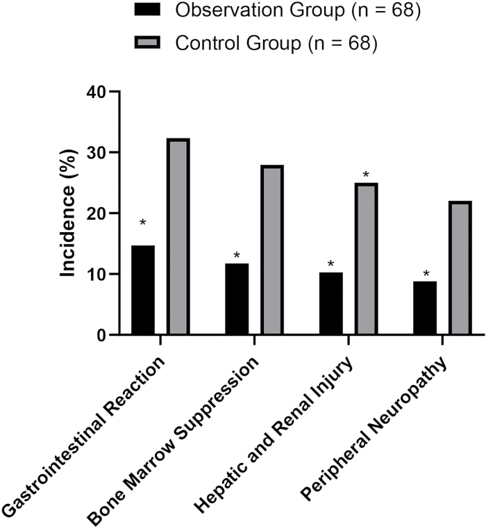
Figure 3. Incidence of Grade ≥2 Treatment-Related Adverse Events. The grouped bar chart shows the incidence of gastrointestinal reactions, bone marrow suppression, hepatic and renal injury, and peripheral neuropathy in the observation group (n = 68) and control group (n = 68). Data are presented as percentages of patients experiencing each adverse event. Adverse events were graded according to the National Cancer Institute Common Terminology Criteria for Adverse Events (CTCAE), version 5.0. * P < 0.05 versus the control group. CTCAE, Common Terminology Criteria for Adverse Events. Please click here to view a larger version of this figure.
Treatment Completion and Karnofsky Performance Status
Completion of all six chemotherapy cycles occurred in 65/68 patients (95.6%) in the observation group and 67/68 patients (98.5%) in the control group (P = 0.31). Changes from baseline to post-treatment in KPS scores were significantly greater in the observation group than in the control group (Table 6). The mean change in KPS was +15.25 ± 7.25 in the observation group versus +10.37 ± 8.10 in the control group (P < 0.001).
| Group | n | Baseline KPS | Post-Treatment KPS | Change in KPS | 95% CI of Change | P |
| Observation | 65 | 70.42 ± 8.56 | 85.67 ± 7.49 | +15.25 ± 7.25 | 13.45–17.05 | < 0.001 |
| Control | 67 | 69.98 ± 8.62 | 80.35 ± 7.95 | +10.37 ± 8.10 | 8.39–12.35 | < 0.001 |
| Between-group | — | P = 0.769 | P < 0.001 | Difference: +4.88 | 2.28–7.48 | < 0.001 |
Table 6: Changes in KPS Scores from Baseline to Post-Treatment. Data are presented as mean ± SD for the complete-case analysis populations (observation group, n = 65; control group, n = 67). Change was calculated as the post-treatment score minus the baseline score. The 95% CIs shown for each treatment group refer to the within-group change from baseline, whereas the between-group 95% CI refers to the difference in change between groups. Within-group P values were calculated using paired t-tests, and between-group P values were calculated using independent-samples t-tests. KPS, Karnofsky Performance Status; CI, confidence interval; SD, standard deviation.
Overall, the observation group showed greater changes in quality-of-life functional domain scores, CD4⁺ T-cell count, CD4⁺/CD8⁺ ratio, KPS score, and exploratory serum biomarkers, together with lower incidences of the reported grade ≥ 2 treatment-related adverse events, compared with the control group. These findings describe associations observed in this retrospective cohort and do not establish causality.
Data Availability:
Due to institutional privacy requirements, the de-identified patient-level data are not publicly available. The data may be made available by the corresponding author upon reasonable request, subject to institutional review board approval and applicable institutional requirements.
Discussion
This retrospective cohort study evaluated the clinical effects of adding Astragalus Injection to standard paclitaxel-carboplatin adjuvant chemotherapy in 136 postoperative patients with endometrial cancer. Astragalus Injection adjunctive therapy was associated with significantly greater improvements in quality of life, immune function, and performance status, as well as greater reductions in exploratory serum biomarker levels and lower incidences of selected chemotherapy-related adverse reactions compared with chemotherapy alone. These findings provide preliminary clinical evidence supporting the potential role of Astragalus Injection as an adjunctive intervention during endometrial cancer chemotherapy and warrant further investigation in prospective studies. The observed improvement in immune function, specifically the significantly greater increase in CD4⁺ T-cell count and CD4⁺/CD8⁺ ratio in the Astragalus Injection group, is consistent with preclinical evidence suggesting immunomodulatory properties of Astragalus membranaceus preparations. Astragalus polysaccharides have been shown to modulate lymphocyte activity and other immune responses in experimental models11,12. In the context of chemotherapy-induced immunosuppression, the ability of Astragalus components to influence T-cell homeostasis may be clinically relevant. The greater increase in the CD4⁺/CD8⁺ ratio observed in this study suggests an association with altered T-cell immune status; however, the present retrospective clinical data do not establish the underlying mechanism.
The greater reductions in the exploratory serum biomarkers SCC-Ag and CYFRA21-1 in the Astragalus Injection group warrant cautious interpretation. These markers have been investigated as potential biomarkers in endometrial cancer monitoring13,14, but they are not standard efficacy endpoints in endometrial cancer. The observed changes in these exploratory biomarkers should therefore be interpreted as hypothesis-generating findings rather than evidence of antitumor efficacy. Definitive efficacy endpoints, such as objective response rate, progression-free survival, and overall survival, are required to establish clinical benefit. The reduced incidence of selected chemotherapy-related adverse reactions in the Astragalus Injection group was another notable finding. Patients receiving Astragalus Injection had significantly lower rates of gastrointestinal reactions, bone marrow suppression, hepatic and renal injury, and peripheral neuropathy. This toxicity-sparing association may be related to the hematopoietic protective, anti-inflammatory, and antioxidant properties reported for Astragalus components. Astragalus polysaccharides have been shown to protect hematopoietic stem cells from chemotherapy-induced damage in preclinical models15, and Astragalus preparations have demonstrated hepatoprotective effects in various experimental studies16. The lower incidence of selected adverse reactions was accompanied by greater improvements in quality-of-life and KPS scores in the Astragalus Injection group; however, the retrospective design does not establish a causal relationship among these observations.
Several limitations of this study should be acknowledged. First, this was a single-center retrospective study with inherent potential for selection bias, and multicenter external validation was not performed. The non-randomized design also means that unmeasured confounders may have influenced the observed outcomes. Second, the retrospective nature of the study precludes definitive causal inference; therefore, the findings should be considered hypothesis-generating rather than confirmatory. Third, the relatively small sample size (n = 136) limits the generalizability of the findings. Fourth, the short follow-up period, encompassing six cycles of chemotherapy over approximately 4 months, precluded assessment of long-term outcomes, including DFS and OS. Survival data are not yet mature and will be the subject of future follow-up studies. Fifth, SCC-Ag and CYFRA21-1 are not standard efficacy endpoints in endometrial cancer; their use as exploratory biomarkers represents a limitation, and these findings should be confirmed using definitive efficacy measures in future prospective trials. Sixth, this study evaluated clinical phenotypes without investigating the molecular mechanisms by which Astragalus Injection may modulate immune function or influence biomarker levels. Although preclinical studies have identified several potential mechanisms involving Astragalus polysaccharides and astragalosides, the specific pathways operative in this clinical context remain to be elucidated. Seventh, the study population predominantly consisted of endometrioid adenocarcinoma, and the findings may therefore not be generalizable to rarer histological subtypes. Finally, the use of SCC-Ag and CYFRA21-1 reflected the biomarkers routinely measured in clinical practice at the study institution during the study period; other markers, such as CA125 and HE4, were not consistently available for all patients and were therefore not included in the analysis.
The standard of care for adjuvant treatment of high-risk endometrial cancer includes paclitaxel-carboplatin chemotherapy, which has established efficacy but can be associated with substantial toxicity. Various strategies have been explored to improve the therapeutic index of this regimen, including dose modification, growth factor support, and the addition of targeted agents. The present findings suggest that Astragalus Injection may warrant further investigation as an adjunctive supportive intervention during chemotherapy, given its association with changes in exploratory biomarker profiles, selected immune parameters, quality of life, performance status, and lower incidences of selected treatment-related adverse events. These associations may be particularly relevant in the adjuvant setting, where maintaining quality of life and treatment tolerability are important considerations. Several clinical studies conducted in China have reported that Astragalus Injection used as an adjunct to chemotherapy in endometrial cancer may be associated with reduced tumor marker levels and improved immune function14,17. However, high-quality prospective evidence from international settings remains limited, and the present findings should be interpreted with this context in mind. Alternative approaches to evaluating the present hypothesis include prospective controlled studies, randomized comparisons, and mechanistic investigations designed to distinguish treatment-associated clinical observations from potential confounding effects.
The drug evaluated in this study was Astragalus Injection, National Drug Approval Z19993281, a preparation of Astragalus membranaceus containing multiple active components, including astragalosides and Astragalus polysaccharides. Accordingly, the observed clinical associations cannot be attributed to a single compound but instead relate to the multi-component preparation administered in this cohort. This distinction is important when interpreting the present findings and considering their potential application in future research. Further characterization of the preparation and its individual components may help determine which constituents contribute to the observed clinical and immunological associations. In addition, the present retrospective method provides a means of generating clinical hypotheses from routinely collected treatment, quality-of-life, immune-function, biomarker, adverse-event, and performance-status data, but prospective approaches will be required to determine whether the observed associations are reproducible and clinically meaningful.
Future research should address the limitations of the present study through large-scale, multicenter, prospective randomized controlled trials with long-term follow-up to confirm these findings and assess DFS and OS outcomes. Characterization of the specific active components of Astragalus Injection, including astragalosides, polysaccharides, and flavonoids, and their individual contributions to the observed effects would help identify the optimal formulation for further clinical investigation. Mechanistic studies exploring the molecular pathways through which Astragalus-derived compounds modulate immune function are also warranted. Additionally, future studies should include adequately characterized histological subgroups, including endometrioid adenocarcinoma, to assess the generalizability of the findings across relevant patient populations. Finally, the optimal dosing and timing of Astragalus Injection administration in relation to chemotherapy cycles should be established through appropriately designed phase I/II dose-finding studies. Together, these approaches would help determine whether the associations observed in this retrospective cohort can be reproduced prospectively and translated into clinically meaningful benefits.
Disclosures
Conflict of Interest:
The authors declare that they have no competing interests.
Acknowledgements
This work was supported by the Hubei Provincial Health Commission Scientific Research Project (Grant No. WJ2023M044), “Effects of LDHB-mediated lactic acid metabolism abnormalities on type I endometrial carcinoma.”
Materials
| Name | Company | Catalog Number | Comments |
|---|---|---|---|
| 5% Glucose Solution | Solarbio | Cat. No. IG9001 | 250 mL used as the diluent for Astragalus Injection. |
| Antiemetic, 5-HT3 Receptor Antagonist | Qilu Pharmaceutical Co., Ltd. (Ondansetron Hydrochloride Injection) | NMPA Approval No. H10970065 | Used prophylactically during chemotherapy. Specifically, ondansetron 8 mg intravenously 30 min prior to chemotherapy on day 1, followed by oral ondansetron 4 mg twice daily on days 2–3. If granisetron was used instead, refer to NMPA Approval No. H20030161 (Taiji Group) or NMPA Approval No. H20083644. |
| Astragalus Injection | Shanghai No. 1 Biochemical & Pharmaceutical Co., Ltd. | NMPA Approval No. Z19993281 | 10 mL/ampoule; two ampoules (20 mL total) per administration; each ampoule is equivalent to 20 g of crude herb. |
| Biochemical Assay System/Reagents for ALT, AST, BUN, and Cr | Beckman Coulter, Inc. | AU5800 Automated Biochemical Analyzer | Used for assessment of hepatic and renal function. Liver function was assessed using serum ALT, AST, and total bilirubin; renal function was assessed using serum creatinine, BUN, and eGFR. |
| Blood Collection Tubes | BD Biosciences (Becton Dickinson) | BD Vacutainer K2EDTA Tubes (e.g., Cat. Nos. 367841, 367844, 367856, 366643) | Used for collection of peripheral blood samples. EDTA-coated tubes were used for flow-cytometry analyses. Common catalog numbers include 367841 (2 mL, 13 × 75 mm), 367844 (4 mL, 13 × 75 mm), 367856 (K2EDTA), and 366643 (BD Vacutainer Plastic K2EDTA). |
| Carboplatin Injection | Shanghai Chuanno Pharmaceutical Co., Ltd. | NMPA Approval No. H20203353 | 10 mL:100 mg. Administered at a target AUC of 5–6 using the Calvert formula. |
| CD4+ T-Cell Antibody/Reagent | BD Biosciences | Cat. No. 555346 (FITC anti-human CD4, clone RPA-T4); RRID: AB_395751 | Used for flow-cytometric measurement of CD4+ T cells. |
| CD8+ T-Cell Antibody/Reagent | BD Biosciences | Cat. No. 555367 (PE anti-human CD8, clone RPA-T8); RRID: AB_395770 | Used for flow-cytometric measurement of CD8+ T cells. |
| Centrifuge | Eppendorf AG | Models: 5424/5424 R, 5702, 5804/5804 R, or 5810/5810 R | Used for serum separation at 800 × g for 10 min at 4 °C. Common Eppendorf models for clinical laboratory use include 5424 (benchtop microcentrifuge), 5702 (cell culture centrifuge), and 5810/5810 R (refrigerated benchtop centrifuge). Please verify the actual model from institutional records. |
| CYFRA21-1 ELISA Kit | Cloud-Clone Corp. | Cat. No. SEB246Hu | Used to measure serum CYFRA21-1. Double-antibody sandwich ELISA; intra-assay CV <8%, inter-assay CV <10%. |
| ELISA Microplate Reader | Agilent Technologies (BioTek) | ELx808 Absorbance Microplate Reader | Used for measurement of SCC-Ag and CYFRA21-1 ELISA assays. Absorbance was read at 450 nm. |
| Flow Cytometer | BD Biosciences | BD FACSCanto II Flow Cytometer; RRID: SCR_018056 | Used for measurement of peripheral blood CD4+ and CD8+ T-cell counts. Equipped with three lasers (405 nm, 488 nm, and 633 nm) and eight fluorescence channels. |
| Granulocyte Colony-Stimulating Factor | Qilu Pharmaceutical Co., Ltd. (Filgrastim) | NMPA Approval No. S19990049 | Administered for febrile neutropenia according to institutional guidelines. Specifically, filgrastim 5 μg/kg/day subcutaneously for severe neutropenia (ANC <0.1 × 10⁹/L) or prolonged neutropenia (>7 days). |
| Hepatoprotective Agent | Shanghai Fudan Forward Pharmaceutical Co., Ltd. (Reduced Glutathione for Injection) | NMPA Approval No. H20031265 | Used as supportive care during chemotherapy. Specifically, reduced glutathione 1.2 g intravenously once daily on days 1–3 of each cycle. If a glycyrrhizic acid preparation was used instead, please verify the actual product. |
| Karnofsky Performance Status Scale | Not applicable | Not applicable | Used to assess performance status at baseline and after six chemotherapy cycles. |
| Paclitaxel | Hainan Tongyao Kangli Pharmaceutical Co., Ltd. | NMPA Approval No. H20065071 | 5 mL:30 mg. Administered at 175 mg/m² by intravenous infusion over 3 h on day 1 of each cycle. |
| Quality of Life Questionnaire-Core 30, Version 3.0 (EORTC QLQ-C30) | European Organisation for Research and Treatment of Cancer | Version 3.0 | Used for quality-of-life assessment at baseline and after six chemotherapy cycles. Copyright © 1995 EORTC Quality of Life Group. All rights reserved. Used under license from the EORTC. |
| SCC-Ag ELISA Kit | Cloud-Clone Corp. | Cat. No. SEC893Hu | Used to measure serum SCC-Ag. Sandwich ELISA; intra-assay CV <8%, inter-assay CV <10%. |
| SPSS Statistics, Version 26.0 | IBM Corp. | Version 26.0; RRID: SCR_019096 | Used for statistical analyses. |
References
- Colombo N, et al. ESMO-ESGO-ESTRO consensus conference on endometrial cancer: diagnosis, treatment and follow-up. Ann Oncol. 2016;27(1):16-41.
- Morice P, et al. Endometrial cancer. Lancet. 2016;387(10023):1094-1108.
- Ramírez SP, et al. SEOM-GEICO clinical guidelines on endometrial cancer (2025). Clin Transl Oncol. 2025;27(12):4368-4380.
- Miller DS, et al. Carboplatin and paclitaxel for advanced endometrial cancer: final overall survival and adverse event analysis of a phase III trial (NRG Oncology/GOG0209). J Clin Oncol. 2020;38(33):3841-3850.
- Pignata S, et al. Carboplatin plus paclitaxel versus carboplatin plus pegylated liposomal doxorubicin as first-line treatment for patients with ovarian cancer: the MITO-2 randomized phase III trial. J Clin Oncol. 2011;29(27):3628-3635.
- Lorusso D, et al. Quality of life in patients with ovarian cancer receiving first-line chemotherapy: a MITO-2 study. Int J Gynecol Cancer. 2014;24(3):527-534.
- Li X, et al. A review of recent research progress on the Astragalus genus. Molecules. 2014;19(11):18850-18880.
- Guo Y, et al. Astragalus polysaccharides inhibit ovarian cancer cell growth via microRNA-27a/FBXW7 signaling pathway. Biosci Rep. 2020;40(3):BSR20193396.
- Bao WR, et al. Astragalus polysaccharide RAP selectively attenuates paclitaxel-induced cytotoxicity toward RAW 264.7 cells by reversing cell cycle arrest and apoptosis. Front Pharmacol. 2019;9:1580.
- Chang CC, et al. Efficacy of astragalus polysaccharides (PG2) in alleviating chemotherapy-induced fatigue in gynecologic cancer: a retrospective cohort study. Discov Oncol. 2025;16(1):1322.
- Salehi B, et al. Astragalus species: insights on its chemical composition toward pharmacological applications. Phytother Res. 2021;35(5):2445-2476.
- Zhang Q, Huang X. The modulatory properties of Astragalus membranaceus treatment on endometrial cancer: an integrated pharmacological method. PeerJ. 2021;9:e11995.
- Bedkowska GE, Lawicki S, Szmitkowski M. Tumor markers useful in the diagnostics and monitoring of endometrial and cervical cancer. Postepy Hig Med Dosw (Online). 2007;61:122-128.
- Gao X, et al. Effect of combination of traditional Chinese medicine with Western medicine on endometrial carcinoma and its influence on ultrasound, MRI, tumor markers HE4 and CA125. Evid Based Complement Alternat Med. 2021;2021:6053406.
- Bao W, et al. Radix Astragali polysaccharide RAP directly protects hematopoietic stem cells from chemotherapy-induced myelosuppression by increasing FOS expression. Int J Biol Macromol. 2021;183:1715-1722.
- Cheng M, et al. Efficacy and safety of Astragalus-containing traditional Chinese medicine combined with platinum-based chemotherapy in advanced gastric cancer: a systematic review and meta-analysis. Front Oncol. 2021;11:632168.
- Li Q, Chen Q, Zhang Z, Yang S. Comparative efficacy of Chinese herbal injections for treating endometrial carcinoma: a Bayesian network meta-analysis. Medicine (Baltimore). 2023;102(41):e34676.