This study describes the microsurgical dissection of fascial and ligamentous support structures in breast tissues removed during prophylactic mastectomy. The findings provide a valuable resource for surgeons and anatomists studying human female breast anatomy.
Method Article
Microsurgical Dissection of Fresh Human Mastectomy Specimens for Visualization of Breast Fascial and Ligamentous Support Structures
63 views
⸱
September 11th, 2026
In This Article
Summary
Abstract
Ligamentous and fascial support structures, such as Cooper’s ligaments and the superficial fascial system, are important structural components of the female breast, contributing to breast shape, mechanical support, and surgical plane definition during breast surgery. Although previous studies have investigated these structures through cadaveric dissection, intraoperative observation, and biomechanical analysis, there have been no reports describing their relationships using microsurgical dissection of fresh human breast tissue. In this study, fresh nonfixed breast tissue specimens were obtained from patients undergoing prophylactic mastectomy and sectioned into 2-cm-thick sagittal slices by a breast pathologist. Microsurgical dissection of each slice was performed under approximately 5×–10× magnification to visualize and examine Cooper’s ligaments and the superficial layer of the superficial fascia (SLSF) and deep layer of the superficial fascia (DLSF). Using this protocol, microsurgical dissections were completed in 24 breast tissue specimens, enabling visualization of the observed structural continuity between Cooper’s ligaments, the SLSF, the dermis, the DLSF, and the pectoral fascia. Specifically, Cooper’s ligaments were observed extending through the SLSF into the dermis and posteriorly through the DLSF toward the fascia overlying the pectoralis major muscle. This protocol enables direct visualization of the fascial-ligamentous support network of the breast and facilitates anatomical understanding of the relationships among these structures. This study and its protocol may serve as valuable educational and research resources for breast surgeons, plastic surgeons, and cross-disciplinary researchers seeking a better understanding of the fascial-ligamentous support structures of the breast.
Introduction
The anatomy of the breast has long been a subject of interest in both clinical practice and anatomical research, especially owing to its implications for breast aesthetic surgery and breast cancer treatment. One of the most important, yet underexplored, components of breast anatomy is Cooper’s ligaments, a network of fibrous connective tissue that runs through the breast, providing structural support. Initially described by Sir Astley Cooper in his 19th-century book, On the Anatomy of the Breast, Cooper’s ligaments play a critical role in maintaining the shape and position of the breast by anchoring the skin to the underlying pectoral fascia1. Their relevance in both surgical and aesthetic procedures has increased, particularly as breast surgery continues to evolve. In the 1990s, Ted Lockwood highlighted the significance of the superficial fascial system, especially in the context of breast and body contouring surgeries. His work emphasized the importance of this fascial layer for maintaining breast shape, distributing mechanical forces during surgery, and providing a surgical plane for procedures, such as breast lifts (mastopexy) and breast reconstructions2,3. Since then, the focus on breast anatomy has intensified, with various studies investigating Cooper’s ligaments and the superficial fascial system through cadaveric dissection4,5,6, histological characterization7, imaging8, endoscopy9 intraoperative findings4, and biomechanical studies7,10,11. Collectively, these investigations have broadened current understanding of the structural organization of the breast fascial system and highlighted the diversity of anatomical observations reported in the literature.
These investigations have enriched our understanding of the structural dynamics of the breast and how they change under physiological and pathological conditions. However, they lack the detailed, fine-scale observations necessary to fully understand the relationships between tissues as seen during microsurgical dissection. Despite the wealth of information from macroscopic dissections and clinical procedures, there have been no reports of microsurgical dissection of these structures in human breast tissue. Such observations are crucial for gaining a deeper understanding of how these anatomical systems work together to maintain the breast’s architecture. Therefore, there remains a significant knowledge gap regarding the features of Cooper’s ligaments and the superficial fascial system. For surgeons, especially those in breast oncology and plastic surgery, having a detailed understanding of these systems is important for surgical planning and anatomical orientation during procedures, such as breast reconstruction, mastectomy, and augmentation12.
The present study aims to fill this gap by providing detailed microsurgical observations of Cooper’s ligaments and the superficial fascial system in breast tissue obtained from prophylactic mastectomy specimens. Using tissue slices, we performed dissections of these structures using the microsurgical dissection techniques described in the protocol below. Unlike previous cadaveric or macroscopic studies, this protocol enables microsurgical visualization of Cooper’s ligaments and the superficial fascial system in fresh human breast tissue.
This study provides clear, visually documented evidence of the relationships between Cooper’s ligaments, the superficial fascial system, and surrounding breast tissues at the microsurgical level. These anatomical observations may facilitate future anatomical and surgical investigations. The potential applications of these findings in oncologic breast surgery require further validation using specimens from breast cancer patients. For junior breast and plastic surgeons, as well as cross-disciplinary researchers, the findings presented here will serve as an educational tool, enhancing their understanding of the intricate details of the breast’s fascial and ligamentous anatomy.
Protocol
This protocol was approved by The University of Texas MD Anderson Cancer Center’s Institutional Review Board (IRB) on 05/06/2016 with the record number PA16-0364. As per institutional guidelines, written informed consent was obtained from all patients who provided prophylactic mastectomy breast tissue samples.
NOTE: This protocol provides detailed steps for microsurgical dissection of breast tissue to visualize and study the ligaments and fascia of the breast structural support system. The breast surgeons at the institution where this study was conducted routinely use electrocautery to dissect the pectoralis major fascia (pectoral fascia) to remove the mastectomy specimen from the chest wall during a total skin-sparing or nipple-sparing mastectomy. Specimen preparation, orientation, and microsurgical dissection are described in detail in the following sections.
1. Preparation of tissue slices from mastectomy specimens
- Collect mastectomy specimens.
- Obtain the breast tissue from patients undergoing prophylactic mastectomy following approval of the protocol by the institutional IRB. Obtain informed consent and collect tissues in accordance with ethical and clinical guidelines.
- Section the mastectomy specimens.
- Immediately after the mastectomy, examine grossly and begin processing the mastectomy specimen. This is done by a breast pathologist. A schematic illustrating the specimen sectioning strategy is shown in Figure 1.
- Before sectioning, orient and ink the intact mastectomy specimen according to the standardized pathology color scheme: red (superior), orange (inferior), green (anterior), and black (posterior).
- Use a #20 pathology scalpel blade to section the specimen into approximately 2-cm-thick sagittal slices while maintaining its native anatomical orientation. Subsequently, use the retained ink markings to preserve the anatomical orientation of the tissue slice during subsequent handling and microdissection.
- Use a single representative peripheral tissue slice provided by the breast pathologist for microsurgical dissection, as the central slices containing the nipple or a portion of the nipple are usually retained for routine clinical pathologic evaluation.
- Transfer and store the breast tissue.
- Wrap each tissue slice in a saline-moistened paper towel and seal the tissue slice in a plastic biohazard bag. Transfer the wrapped slice to the institutional tissue bank (ITB), where they are refrigerated at 4 °C until microsurgical dissection.
NOTE: Specimens are not frozen or rewarmed prior to dissection. The time from mastectomy to refrigerated storage typically ranges from 2–3 h. Microsurgical dissection is typically performed within 6–24 h after mastectomy. The effect of storage duration on tissue quality was not formally evaluated in this study.
- Wrap each tissue slice in a saline-moistened paper towel and seal the tissue slice in a plastic biohazard bag. Transfer the wrapped slice to the institutional tissue bank (ITB), where they are refrigerated at 4 °C until microsurgical dissection.
2. Microsurgical setup
- Position the microscope.
- Position the surgical microscope and set it to 5×–10× magnification to ensure optimal visualization of tissue structures for precise microsurgical dissection. Use an adapter to connect a smartphone to the microscope (Figure 2) and ensure that the field of view is properly aligned.
NOTE: The smartphone setup provides sufficient image quality for visualization, documentation, and video recording of the dissection. Alternatively, an operating microscope equipped with an integrated camera may be used when available.
- Position the surgical microscope and set it to 5×–10× magnification to ensure optimal visualization of tissue structures for precise microsurgical dissection. Use an adapter to connect a smartphone to the microscope (Figure 2) and ensure that the field of view is properly aligned.
- Prepare dissection tools.
- Arrange all dissection tools, including forceps and scissors, on the dissection platform for easy access during the procedure.
- Set up the vacuum and solution.
- Use a vacuum system equipped with a 3-mm Frazier suction tip to assist with tissue removal during dissection. Set the vacuum pressure between 180 mmHg and 200 mmHg.
- Use normal saline to keep the tissue slices hydrated and prevent tissue desiccation throughout the procedure.
NOTE: When greater suction is needed, temporarily cover the vent hole of the Frazier suction tip with the thumb to increase suction. Apply suction gently to the adipose tissue on either side of the ligament, removing only enough adipose tissue to expose the ligament. Avoid direct suction of the ligament or fascial structures. Keep additional saline available to flush the suction tip if it becomes blocked or the suction strength weakens. This ensures continuous suction and prevents interruptions during dissection.
3. Dissection process
NOTE: Handle fresh human breast tissue in accordance with institutional biosafety procedures. Wear disposable gloves, a laboratory coat, and a surgical mask throughout specimen handling and microsurgical dissection. Collect suctioned material through the institutional wall suction system using a disposable suction collection canister equipped with an overflow protection mechanism to prevent accidental entry of fluids and suction debris into the wall vacuum system. Dispose of sharps, tissue specimens, suctioned material, and other biohazardous materials according to institutional biosafety procedures.
- Confirm the tissue orientation.
- Follow the orientation, labeled by the pathologist (Figure 3).
- Before microscopic dissection, routinely orient the tissue slice with the posterior (black-inked) surface facing the dissector and the skin surface facing the assistant.
NOTE: This standardized orientation places the pectoral fascia closest to the dissector and allows systematic identification of the DLSF, Cooper’s ligaments, and SLSF in a consistent anatomical sequence during dissection. - Double-check the orientation under low power magnification by identifying key anatomical landmarks, such as the epidermis, dermis, subcutaneous tissue, and pectoral fascia.
- Identify the superficial fascial system.
- Locate and observe the superficial fascial system, paying attention to how it connects with the skin and integrates with subcutaneous adipose tissue and deeper layers.
- Locate the superficial layer of the superficial fascia (SLSF):
- Identify the SLSF layer, which typically presents as a distinct, thin, somewhat translucent fascial layer beneath the skin and subcutaneous adipose tissue.
- Under 5×–10× magnification, it appears as a somewhat continuous linear membrane running parallel to the dermis, distinguishing it from the surrounding adipose tissue. The fascia often has an adipose lobule “traversing” the fascia and creating a “hole” in the fascia. Follow this plane carefully during dissection to confirm the fascia's identity and visualize its attachments to surrounding structures (Figure 4A,A’).
- Locate the deep layer of the superficial fascia (DLSF):
- Identify the DLSF as a somewhat translucent fascial layer located immediately superficial to the pectoral fascia and extending transversely across the chest wall. It runs parallel to the pectoral fascia and forms a continuous fascial plane during gentle dissection.
- Trace this structure carefully during dissection to confirm the fascia’s identity and visualize the adjacent anatomical structures (Figure 5A,A’).
NOTE: The DLSF lies close to the pectoral fascia and may not be easily distinguishable in all specimens. Compared to the superficial layer, the deep layer is more challenging to identify and usually requires additional time and careful dissection to locate and confirm. If the expected continuous fascial plane cannot be confidently identified, return to low magnification to relocate the fascial plane before continuing the dissection.
- Trace Cooper's ligaments and dissect adipose pockets.
- Recognize Cooper's ligaments as smooth, translucent fiber bundles, typically less than 1 mm thick, extending approximately perpendicular to the dermis, SLSF, DLSF, and pectoral fascia. They remain intimately adherent to the surrounding adipose lobules and breast ducts. Follow the ligaments continuously during gentle traction.
- (Exception) Dissection of Cooper's ligaments may not be feasible in specimens with dense glandular tissue. Carefully identify a candidate ligamentous structure and trace it in both the superficial and deep directions while preserving its relationships with the surrounding tissues.
- Document its observed anatomical course, including whether it terminates, branches, merges with adjacent fascial structures, or becomes disrupted during dissection.
- Manipulate ligamentous tissues gently with micro-forceps to prevent damage to the fascia. Cooper's ligaments are composed of collagen fibers that are predominantly oriented in a uniform direction, whereas ordinary fibrous septa contain collagen fiber bundles arranged in multiple directions.
- As dissection proceeds, incise the ligamentous cover of adjacent adipose pockets to expose the adipose tissue for dissection.
- For cut specimens with exposed adipose tissue, apply suction gently to the adipose tissue on both sides of the target ligament to remove only enough adipose tissue to expose the edge of the ligament to facilitate its microsurgical dissection (by scissor dissection). Avoid direct suction of the ligament or fascial structures. The adipose tissue is organized into lobules that remain adherent to the surrounding Cooper's ligaments.
- Carefully remove the adipose tissue to expose the ligamentous framework and facilitate visualization of the anatomical relationships between Cooper's ligaments, the superficial fascial system, and the surrounding adipose tissue (Figure 6).
- Maintain hydration with saline irrigation.
- Use normal saline irrigation throughout the procedure to prevent tissue desiccation.
- Perform irrigation as needed, guided by the tissue’s appearance rather than a predetermined flow rate or total volume.
- Adjust the suction evacuation as needed to keep the dissection field clear of excess fluid and debris, ensuring uninterrupted observation and precise dissection of each ligament.
4. Video documentation
- Record the entire dissection.
- Use the microscope’s built-in camera system to capture the complete dissection process in real time for reference and review, ensuring no steps are missed. Perform the dissection under 5×–10× optical magnification using the operating microscope.
- Focus on key anatomical structures.
- During dissection and recording, zoom in from low to high power on critical structures, including Cooper’s ligaments and their structural relationships with the superficial fascial system, to provide detailed views that highlight their relationships with surrounding tissues.
- Annotate findings.
- During the dissection, record verbal narration. After acquiring the video, review the recorded video and audio to add text annotations during postproduction.
NOTE: These annotations highlight key anatomical structures, procedural steps, and important observations, thereby improving the educational value and clarity of the final video.
- During the dissection, record verbal narration. After acquiring the video, review the recorded video and audio to add text annotations during postproduction.
Results
Over the course of 1 year, microsurgical dissections were performed on 24 unilateral prophylactic mastectomy specimens obtained from 24 patients. The visibility of individual fascial and ligamentous structures varied among specimens; therefore, the representative images presented here were selected to illustrate the protocol, the anatomical appearance of the fascial and ligamentous structures, and the observed anatomical relationships among Cooper's ligaments, the DLSF, SLSF, and the surrounding adipose tissue. A summary of the representative anatomical observations documented using this protocol is provided in Table 2. Representative examples are described below.
Cooper’s ligaments extend to the SLSF and dermis:
We observed that Cooper’s ligaments appear to traverse the SLSF and extend into the dermis, reinforcing the structural integrity of the superficial breast architecture. These extensions suggest a more complex network than previously described13, which may warrant further exploration and potential nomenclature updates to differentiate these extended segments (Figure 7A).
Cooper’s ligaments connect to the DLSF and Pectoralis Major fascia:
We observed that Cooper’s ligaments appear to extend posteriorly through the DLSF and continue into the fascia overlying the pectoralis major muscle. This observation highlights a previously underappreciated structural relationship that may have implications for surgical techniques involving deep fascia manipulation (Figure 7B). The relationship between Cooper's ligaments and the SLSF is further demonstrated in Figure 8. Gentle elevation of the SLSF provides an additional representative view of its continuity with the Cooper's ligament, facilitating visualization of their anatomical relationships.
Representative figures and Table 2 summarize these anatomical observations and illustrate the continuity and anatomical relationships among Cooper's ligaments, the fascial layers, and the surrounding adipose tissue.
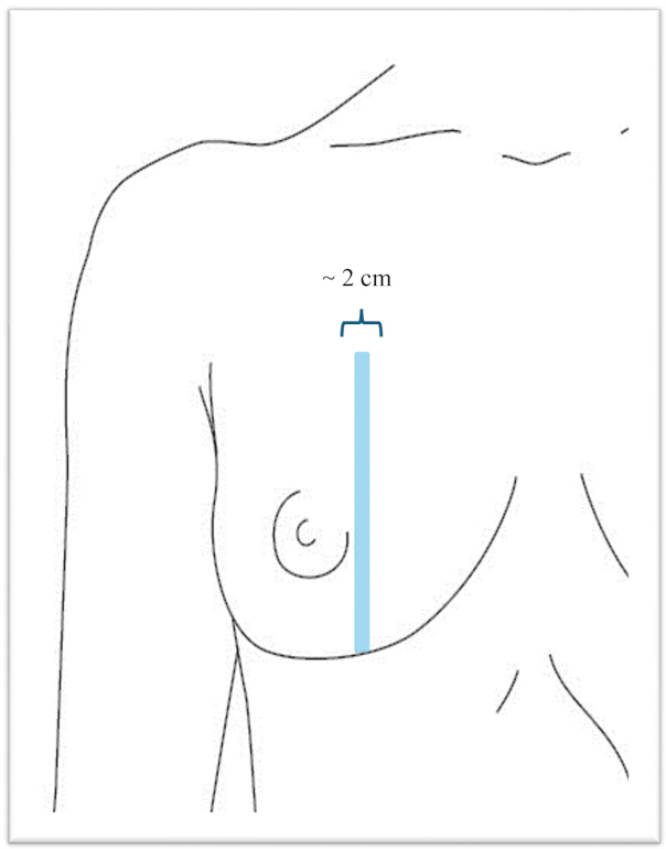
Figure 1: Schematic illustration of specimen sagittal sectioning. Please click here to view a larger version of this figure.
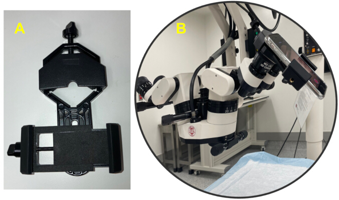
Figure 2: Adapter to connect a smartphone to the microscope. (A) Adapter. (B) Smartphone attached to the microscope. Please click here to view a larger version of this figure.
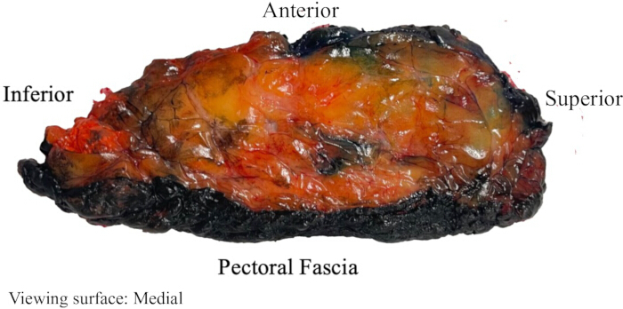
Figure 3: Representative sagittal breast tissue slice before microsurgical dissection. The specimen orientation is indicated by the superior, inferior, anterior, and pectoral fascial surfaces. The surface shown represents the medial aspect of the sagittal tissue slice; the opposite surface corresponds to the lateral aspect of the breast. Please click here to view a larger version of this figure.
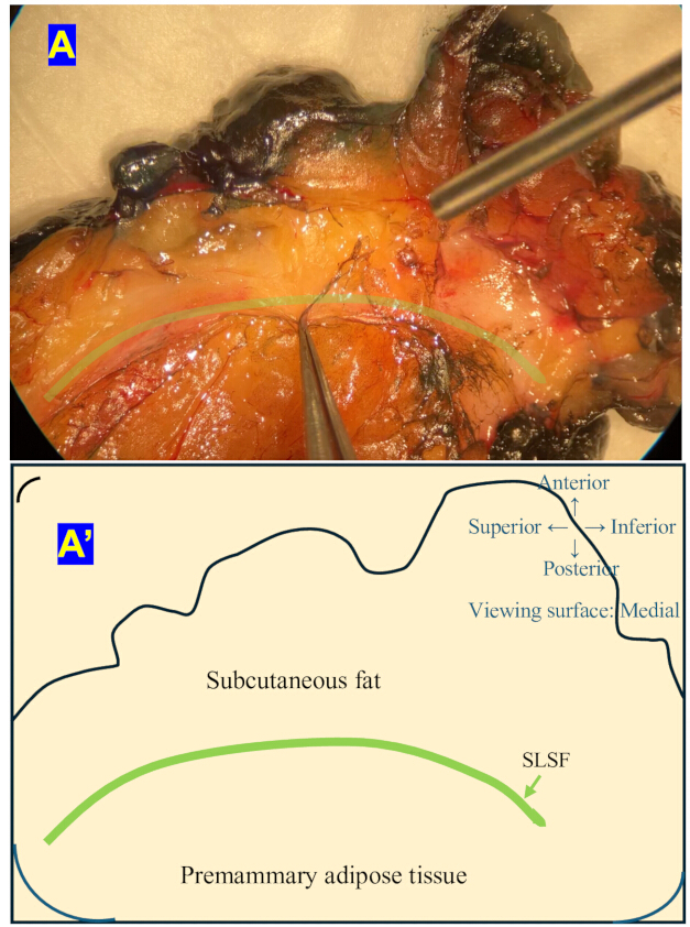
Figure 4: Identification of the superficial layer of the superficial fascia (SLSF). (A) Representative microsurgical dissection demonstrating the SLSF beneath the subcutaneous fat. (A’) Corresponding schematic illustrating the anatomical location of the SLSF. Anatomical orientation is indicated. Please click here to view a larger version of this figure.
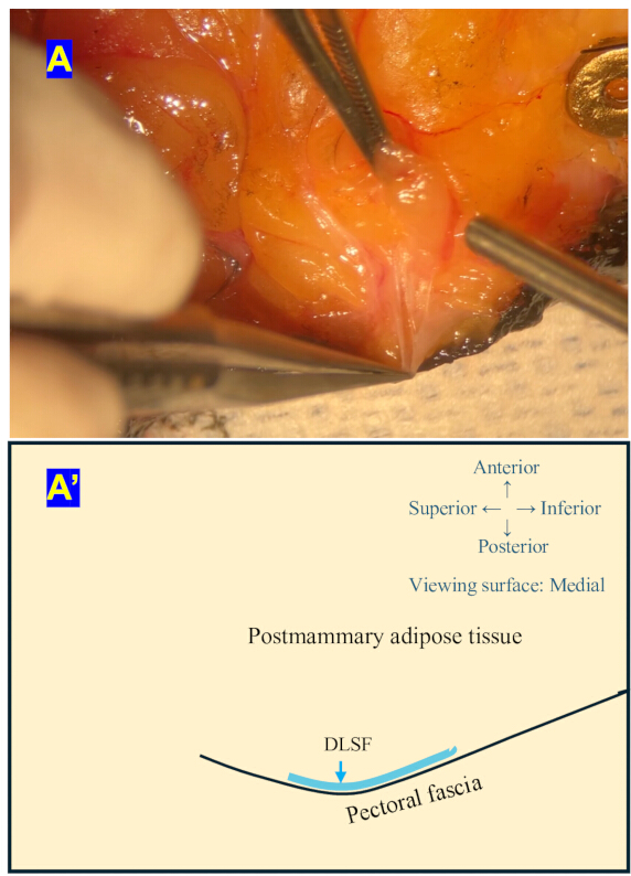
Figure 5: Identification of the deep layer of the superficial fascia (DLSF). (A) Representative microsurgical dissection demonstrating the DLSF immediately superficial to the pectoral fascia. (A’) Corresponding schematic illustrating the anatomical location of the DLSF. Anatomical orientation is indicated. Please click here to view a larger version of this figure.
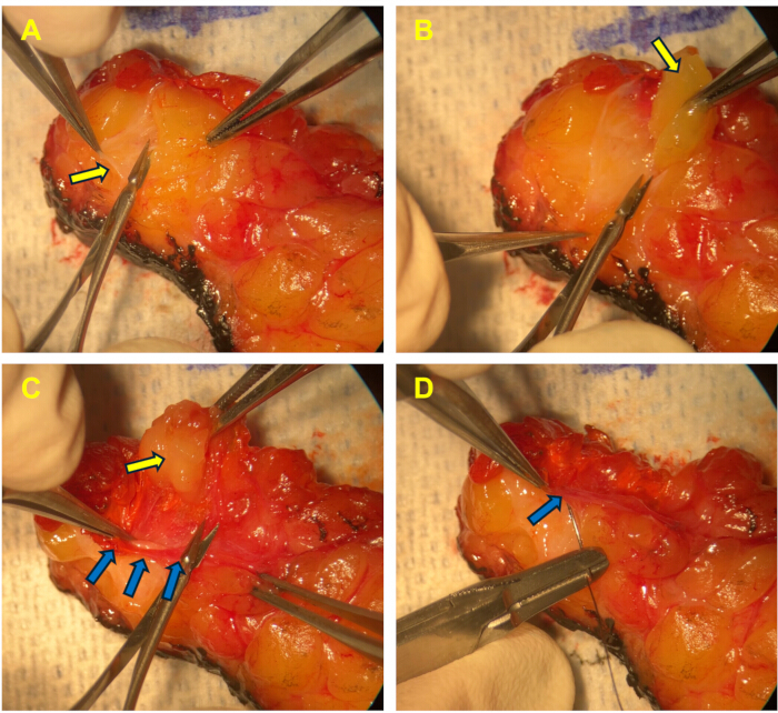
Figure 6: Stepwise dissection demonstrating progressive exposure of the superficial layer of the superficial fascia (SLSF) and an associated Cooper's ligament. Orientation: Top = anterior; bottom = posterior; left = superior; right = inferior. The specimen is viewed from the medial aspect. (A) A Cooper’s ligament is lifted with forceps and separated from the surrounding adipose tissue using scissors. The yellow arrow indicates the Cooper’s ligament. (B) Fat lobules are nearly completely separated (highlighted by the yellow arrow) from the ligament. (C) The SLSF (indicated by the blue arrows) is surrounded by subcutaneous fat tissue (highlighted by the yellow arrow). (D) The SLSF is isolated after removing the inner and outer adipose tissues. The blue arrow indicates the SLSF. Please click here to view a larger version of this figure.
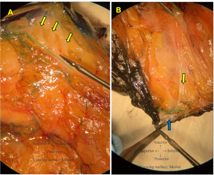
Figure 7: Representative visualization of the anatomical relationships between Cooper's ligaments and the superficial fascial system. (A) Cooper’s ligaments appear to extend through the superficial layer of the superficial fascia (green line) to the dermis, as indicated by the yellow arrows. (B) A Cooper’s ligament (yellow arrow) appears to extend through the deep layer of the superficial fascia (green line) to the pectoral fascia, as indicated by the blue arrow. Please click here to view a larger version of this figure.
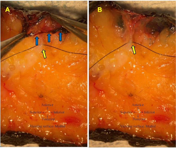
Figure 8: Relationship between a Cooper's ligament and the superficial layer of the superficial fascia (SLSF). (A) A Cooper's ligament was selectively marked with a suture after anatomical identification for documentation purposes and is indicated by the yellow arrow. The SLSF is indicated by the blue arrows. (B) The SLSF was gently elevated to better demonstrate its anatomical relationship with the suture-marked Cooper's ligament (yellow arrow). Please click here to view a larger version of this figure.
| Characteristic | Value |
| Patients, n | 24 |
| Mastectomy specimens, n | 24 (all unilateral) |
| Age, years | 52.5 ± 12.2 (33–86) |
| BMI, kg/m² | 29.49 ± 5.62 (20.55–42.38) |
| Prophylactic mastectomy | 24 (100%) |
| Prior breast surgery | 0 (0%) |
| Prior radiotherapy | 0 (0%) |
| BRCA1 mutation | 1 (4.2%) |
| BRCA2 mutation | 2 (8.3%) |
| No documented BRCA1/2 mutation | 21 (87.5%) |
Table 1: Patient demographics and specimen characteristics.
| Representative anatomical finding | Supporting figure |
| The superficial layer of the superficial fascia (SLSF) can be identified as a continuous fascial layer beneath the dermis. | Figure 4, Figure 6, Figure 8 |
| The deep layer of the superficial fascia (DLSF) can be identified anterior and running parallel to the pectoral fascia. | Figure 5 |
| Cooper's ligaments extend through the SLSF into the dermis. | Figure 7A |
| Cooper's ligaments continue posteriorly through the DLSF toward the fascia overlying the pectoralis major muscle. | Figure 7B |
Table 2: Representative anatomical findings observed using the microsurgical dissection protocol.
Discussion
The dissection process outlined in this study emphasizes several critical steps necessary to achieve optimal visualization of Cooper’s ligaments and the superficial fascial system. Proper sagittal sectioning of the breast tissue into 2-cm-thick slices under the guidance of a pathologist is fundamental to preserving the anatomical orientation and integrity of the structures of interest. Additionally, the use of a surgical microscope with 5×–10× magnification ensures precise dissection and observation of delicate structures. The irrigation of tissue slices with saline and the use of a suction system are indispensable for maintaining tissue hydration and ensuring visual clarity during dissection.
Several procedural refinements were introduced to address the technical challenges encountered during dissection and to enhance the consistency and clarity of anatomical visualization. Increased optical magnification using an operative microscope and fine-tipped microsurgical instruments to improve tissue handling and facilitate identification of fascial and ligamentous structures. A smartphone adapter connected to the surgical microscope was used to facilitate live monitoring and high-resolution recording. A microscope with an integrated video output may also be used as an alternative. Periodic saline flushing of the suction tip and intermittent saline irrigation were used throughout the procedure to prevent clogging and maintain adequate suction. Keep tissues adequately hydrated. Hydration preserves the native appearance of fascial and ligamentous structures for more accurate identification. Remove only enough adipose tissue on either side of the target ligament by gentle suction to improve visualization of the edge of the ligamentous and fascial structures while preserving the target ligament or fascial structure. Collectively, these refinements improve procedural consistency, enhance visualization of fascial and ligamentous structures, and increase the protocol's educational value.
Despite the methodological advancements listed above, certain limitations persist. The anatomical identity of the SLSF, DLSF, Cooper's ligaments, and pectoral fascia was determined during microsurgical dissection based on the gross anatomical characteristics and tissue relationships. The identification of fascial and ligamentous structures was not independently confirmed by a breast pathologist or an anatomist, and histological or microscopic correlation was not performed because it was not the aim of this study. However, identification of these structures was based on the senior author’s 40-year experience performing breast reconstruction and other types of breast surgery. Therefore, the absence of independent anatomical and histological validation represents a small but definite limitation of this study.
Electrocautery is routinely used during mastectomy as part of the standard surgical practice at our institution. Although all specimens underwent the same tissue procurement process, thermal injury from electrocautery may have altered the gross appearance of the pectoral fascial structure. Because thermal artifacts were not formally evaluated, their influence on the anatomical identification of the pectoral fascia was not fully evaluated in this study. However, the pectoral fascia was identified based on its location relative to the mastectomy specimen, itself i.e., it is the last structural layer included with the mastectomy specimen as the breast surgeon cuts the specimen from the pectoral muscles. The identification of the DLSF remains challenging due to its variable proximity to the pectoral fascia and its variable presentation across specimens. Additionally, the reliance on tissue from patients undergoing a prophylactic mastectomy may introduce sampling bias, as these specimens may not fully represent the anatomical variations found in the general population. Furthermore, the use of 5×–10× magnification, while sufficient for this protocol, may not capture ultra-fine details that higher magnifications provide. Another potential limitation of this study is the age range of the patients. All samples were obtained from women approximately 33 to 86 years old and thus may not fully represent the anatomical characteristics of women outside this range. Finally, this study was designed to present a microsurgical dissection protocol rather than to prospectively validate the study’s performance and quantitative metrics, such as identification rates, dissection time, technical failure rates, and operator-related variability. Thus, quantitative metrics were not systematically recorded.
This protocol enables microsurgical visualization of Cooper's ligaments and the superficial fascial system in fresh human breast tissue, providing a suggested approach for observing their anatomical relationships. The observations here support the general concept that the breast parenchyma is situated between superficial and deep layers of the superficial fascial system and is traversed by Cooper’s ligaments. However, differences in scope and nomenclature exist among previous anatomical descriptions. Rehnke et al. proposed a broader three-dimensional model in which the anterior and posterior lamellae surround the corpus mammae and fuse peripherally with the deep fascia of the chest wall to form the circummammary ligament4. Instead, the present protocol focused on reproducible identification of SLSF, DLSF, Cooper's ligaments, and pectoral fascia. By comparison, the terminology summarized by Duncan et al., particularly the superficial and deep layers of the superficial fascial system and Cooper’s ligaments, more closely aligns with the structures identified in our dissections13. Building on these previous approaches, this protocol provides a complementary microsurgical dissection technique for direct visualization of these structures and their three-dimensional relationships in fresh human breast tissue. By providing a clearer understanding of these relationships, the technique expands the anatomical knowledge base and provides a foundation for future anatomical and surgical investigations. Compared to cadaveric studies, the protocol's use of fresh non-fixed tissue specimens may better preserve the native ligamentous architecture of the breast than cadaveric specimens14.
The findings from this study may have implications for future anatomical research and surgical education. For breast and plastic surgeons, detailed visualization of Cooper's ligaments, the superficial fascial system, and their continuity with the pectoral fascia provides valuable anatomical guidance for identifying tissue structures visualized and used during breast reconstruction, implant pocket preparation, mastopexy, and mastectomy. Preservation of the superficial fascial layer and its ligamentous attachments may help maintain the native supporting framework in selected breast procedures where the superficial fascial system is preserved, such as reconstruction following segmental mastectomy. However, these potential applications require further clinical validation. This dissection protocol has potential applications in biomechanical studies, where a detailed anatomical understanding is crucial for modeling the mechanical behavior of breast tissues. Beyond anatomical characterization, this protocol may facilitate future studies correlating imaging findings with gross anatomy, support finite-element modeling and surgical simulation, and contribute to the development of anatomical training resources. The protocol may serve as a useful resource for further investigations of breast anatomy and changes under pathological conditions, such as cancer or trauma15.
In conclusion, this study provides a standardized microsurgical dissection protocol for visualization of Cooper's ligaments and the superficial fascial system in fresh human breast tissue. The protocol provides a useful resource for future anatomical, translational, and surgical investigations.
Disclosures
We hereby declare that there are no conflicts of interest regarding this scientific article. AI-assisted tools (ChatGPT and Grammarly) were used for language editing only. All scientific content was reviewed and approved by the authors.
Acknowledgements
The authors thank the NIH for their financial support through the project Enhanced Biomechanical Modeling of the Breast for Women’s Health (R01EB032533). Special thanks to Esther Sey, Sara Hull, and Tina Alvarado for their contributions to this study, including patient screening, obtaining consent, specimen collection, and equipment preparation. The authors also sincerely thank Dawn Chalaire, Associate Director of Editing Services at the Research Medical Library, The University of Texas MD Anderson Cancer Center, for her thoughtful editing and valuable suggestions, which greatly improved the clarity and readability of the manuscript. This would not have been possible without their efforts.
Materials
| Name | Company | Catalog Number | Comments |
|---|---|---|---|
| 0.9% NaCl solution | General use | N/A | Used for tissue irrigation and periodic flushing of the Frazier suction tip during dissection. |
| Adapter (Smartphone and Microscope) | GOSKY | Standard | Smartphone adapter used to attach the camera to the operating microscope. |
| Camera | Apple | iPhone SE (2nd generation) | Used for video recording through the operating microscope. |
| CDI tissue marking dye | Cancer Diagnostics, Inc. | #0727-1, #0727-3, #0727-4, #0727-6 | Used to ink the intact mastectomy specimen before sectioning for orientation (Red = superior, Orange = inferior, Green = anterior, Black = posterior). |
| Forceps | Precision Medical Devices | N/A | Used for microsurgical tissue manipulation. |
| Frazier suction tip | Integra Miltex | Model not specified | 3-mm Frazier suction tip instrument used for tissue suction during dissection. |
| Hospital wall suction | N/A | N/A | Hospital wall suction system connected to the operating room vacuum source. |
| Microscissors | Precision Medical Devices | S1059 1575 | Used for fine microsurgical tissue dissection. |
| No.20 scalpel blade | Swann-Morton | Example | Used for gross specimen trimming. |
| Non-absorbable suture (Silk) | Ethicon | Mersilk 4-0 | Used to mark anatomical structures for photography and video documentation. |
| Scalpel handle | General surgical supply | N/A | Compatible with No.20 scalpel blade. |
| Surgical microscope | Med Link | Z880 | Operating microscope used for microsurgical dissection |
| Tissue culture dishes | Corning | 430167 | Used to hold saline for periodic flushing of the Frazier suction tip. |
References
- Cooper AP. On the Anatomy of the breast. Longman, Orme, Green, Brown, and Longmans; London; 1840.
- Lockwood T. Reduction mammaplasty and mastopexy with superficial fascial system suspension. Plast Reconstr Surg. 1999;103(5):1411-20.
- Lockwood TE. Superficial fascial system (SFS) of the trunk and extremities: a new concept. Plast Reconstr Surg. 1991;87(6):1009-18.
- Rehnke RD, Groening RM, Van Buskirk ER, Clarke JM. Anatomy of the superficial fascia system of the breast: A comprehensive theory of breast fascial anatomy. Plast Reconstr Surg. 2018;142(5):1135-44.
- Hall MI, et al. A reinterpretation of human breast anatomy includes all the layers of the anterior body wall. Anat Rec (Hoboken). 2024;307(11):3564-73.
- Gaskin KM, Peoples GE, McGhee DE. The attachments of the breast to the chest wall: A dissection study. Plast Reconstr Surg. 2020;146(1):11e-22e.
- Briot N, Chagnon G, Burlet L, Gil H, Girard E, Payan Y. Experimental characterisation and modelling of breast Cooper's ligaments. Biomech Model Mechanobiol. 2022;21(4):1157-68.
- Duraes M, et al. New insights on breast anatomy based on magnetic resonance imaging and surgical observations. Clin Anat. 2026;doi: 10.1002/ca.70071.
- Liu Q, et al. Endoscopic anatomy of the breast fascia system: Peripheral-to-central approach. Plast Reconstr Surg Glob Open. 2025;13(11):e7315.
- Teixeira AM, Martins P. A review of bioengineering techniques applied to breast tissue: Mechanical properties, tissue engineering and finite element analysis. Front Bioeng Biotechnol. 2023;11:1161815.
- Bhattarai A, Reece GP, Brock KK, Ravi-Chandar K. A comprehensive biomechanical material characterization of the human breast fibro-structural support system. J Mech Behav Biomed Mater. 2026;175:107283.
- Matousek SA, Corlett RJ, Ashton MW. Understanding the fascial supporting network of the breast: key ligamentous structures in breast augmentation and a proposed system of nomenclature. Plast Reconstr Surg. 2014;133(2):273-81.
- Duncan AM, Al Youha S, Joukhadar N, Konder R, Stecco C, Wheelock ME. Anatomy of the breast fascial system: A systematic review of the literature. Plast Reconstr Surg. 2022;149(1):28-40.
- Hubbell DS, Dwornik JJ, Alway SE, Eliason R, Norenberg RE. Teaching gross anatomy using living tissue. Clin Anat. 2002;15(2):157-9.
- Vidya R, Ghulam H, Wild J. Breast anatomy: The importance of understanding the superficial fascial system for oncoplastic dissection. Plast Reconstr Surg. 2019;144(2):320e.
Reprints and Permissions
This article has been published
Video Coming Soon