Alcohol-associated liver disease progresses through steatosis, inflammation, and fibrosis via microRNA regulatory networks targeting signaling pathways. High-throughput studies reveal dual microRNA roles, yet network-level cooperation and clinical translation remain underexplored. This review synthesizes mechanisms to support early diagnostic biomarkers and targeted therapies.
Review Article
MicroRNA Regulatory Networks in Alcohol-Associated Liver Disease: Mechanisms from Steatosis and Inflammation to Fibrosis
In This Article
Summary
Abstract
Alcohol-associated liver disease (ALD) is a leading cause of liver-related morbidity and mortality worldwide and progresses through steatosis, alcohol-associated hepatitis, fibrosis, and cirrhosis. MicroRNAs (miRNAs) are established post-transcriptional regulators whose roles in ALD-associated signaling networks have been increasingly characterized. Recent high-throughput sequencing and functional studies have revealed dual regulatory roles of miRNA families, such as miR-122, miR-155, and miR-21, at different disease stages. However, most studies have focused on individual miRNAs, and network-level cooperative regulation and clinical translation remain underexplored. This narrative review examines the molecular mechanisms of miRNAs in alcohol-induced steatosis, inflammation, and fibrosis, with a particular focus on network-level cooperative regulation. Key findings indicate that miRNAs function as stage-specific and cell-type-specific regulators, with miR-122, miR-155, miR-21, miR-29, and miR-34a acting as central network nodes. These insights provide a framework for the identification of early diagnostic biomarkers and potential therapeutic targets in ALD.
Introduction
Alcohol-associated liver disease (ALD) is a leading cause of liver-related morbidity and mortality worldwide. It has various clinical features, such as simple steatosis and fatty liver, alcohol-associated hepatitis, fibrosis, cirrhosis and hepatocellular carcinoma1. ALD progression is the result of the combined action of parenchymal cells (such as hepatocytes) and non-parenchymal cells such as Kupffer cells, hepatic stellate cells (HSCs) and sinusoidal endothelial cells, along with abnormal regulation of multiple molecular pathways2. Alcohol exposure directly damages hepatocytes through its metabolites, acetaldehyde and reactive oxygen species (ROS), while also causing gut dysbiosis and intestinal barrier dysfunction. As a result, pathogen-associated molecular patterns (PAMPs), such as lipopolysaccharide (LPS), are translocated to the liver, activating the innate immune system and initiating an inflammatory response3,4; thus, alcohol-associated hepatitis contributes to a vicious cycle of steatosis, inflammation, and fibrosis. Although alcohol exposure is the established etiological driver of ALD, the molecular mechanisms underlying inter-individual susceptibility and disease progression remain incompletely understood, and effective treatment options remain limited; therefore, further investigation of the molecular regulatory networks of ALD is needed to facilitate clinical translation5.
miRNAs are relatively short non-coding RNAs, 21-23 nucleotides in length. They generally regulate gene expression at the post-transcriptional level by binding to the 3' untranslated region (3'UTR) of their target mRNAs and thus cause mRNA degradation or inhibit translation6. miRNAs are implicated in multiple pathological processes underlying liver disease, such as altered lipid metabolism, oxidative stress, inflammation, apoptosis, and fibrosis5. Alcohol exposure alters the expression pattern of liver-specific miRNAs in ALD and thus modifies a 'miRNA regulatory network'7. miR-122 is a hepatocyte-specific miRNA that shows reduced expression in the livers of ALD patients and alcohol-fed mice, and its downregulation is closely linked to the progression of liver injury8. In addition, miR-155, miR-223, and miR-34a are frequently involved in the regulation of inflammation and fibrosis in ALD5. The abnormal expression of these miRNAs directly alters the process of hepatic lipid metabolism and inflammation in hepatocytes and also promotes fibrosis by regulating the activation state of HSCs9.
In this review, a miRNA regulatory network is defined as a set of miRNAs that jointly regulate a signaling pathway or pathological process through experimentally validated target interactions, including feed-forward loops, feedback loops, competing endogenous RNA interactions, and cross-stage bridging miRNAs. Predicted or inferred interactions are clearly distinguished from experimentally validated interactions. At present, research on miRNAs in ALD has shifted from functional analyses of individual miRNAs to network-level integration, and their interactions and feedback regulatory mechanisms have been uncovered. For instance, in the livers of patients with alcohol-associated hepatitis, miR-182 is one of the most significantly upregulated miRNAs; its abundance is associated with the extent of cholangiopathy, disease severity, and short-term mortality, and miR-182 mimics can induce an increase in inflammatory mediators in cholangiocytes10. Members of the miRNA family also work together to regulate liver fibrosis; for example, the miR-29 family mainly promotes apoptosis and regulates extracellular matrix (ECM) accumulation by modulating the PI3K/AKT signaling pathway, while the miR-34 family accelerates the fibrotic process by promoting HSC activation11. Therefore, it can be inferred that miRNAs do not act independently but rather form complex regulatory networks to synergistically regulate multiple signaling pathways in the pathogenesis of ALD12. Compared with protein biomarkers and genetic variants, miRNAs offer advantages including stability in circulation, tissue-specific expression patterns, and amenability to therapeutic targeting. However, practical limitations remain, including the lack of standardized detection methods and inter-study variability in miRNA expression profiles.
This review examines the molecular mechanisms of miRNA regulatory networks across three stages of ALD: steatosis, inflammation, and fibrosis. Oxidative stress and lipid peroxidation caused by alcohol metabolism in the steatosis stage can lead to the downregulation of miRNAs, such as miR-122 in hepatocytes, and thus alter the expression of genes involved in fatty acid oxidation and lipid synthesis8. Alcohol-induced gut dysbiosis and LPS translocation during the inflammatory stage can activate the Toll-like receptor 4 (TLR4) signaling pathway in Kupffer cells, upregulate pro-inflammatory miRNAs such as miR-155, and thus worsen the inflammatory response of the liver5,13. At the time of fibrosis, activated HSCs are the main producers of ECM, and miRNAs such as miR-29, miR-34a, and miR-21 are crucial regulators of HSC activation, proliferation, and apoptosis by modulating the TGF-β/Smad and Wnt/β-catenin signaling pathways9,14. By integrating the functions of these miRNAs at different stages of disease, this review aims to provide insights into potential diagnostic biomarkers and therapeutic targets for ALD.
Review and Perspective
1. Regulatory networks of miRNAs in hepatic steatosis of ALD
- Alcohol-induced lipid metabolic dysregulation and altered miRNA expression
An early feature of ALD is hepatic steatosis, and at its base, alcohol and its products disrupt the regulation of lipid metabolism in the liver. Aldehydes and ROS produced by alcohol metabolism can inhibit the activity of essential transcription factors, such as peroxisome proliferator-activated receptor alpha (PPARα) and sterol regulatory element-binding protein-1c (SREBP-1c), thus reducing fatty acid β-oxidation and promoting de novo lipogenesis15. This metabolic disturbance is closely associated with altered expression of several miRNAs. For example, miR-122, which is highly enriched or predominantly expressed in the liver, shows a reduction after alcohol consumption. In ALD, miR-122 downregulation has been associated with impaired lipid homeostasis. In non-alcoholic fatty liver models, miR-122 has been shown to regulate lipid metabolism through the YY1/ACE2-MAS1 pathway16. In NAFLD and type 2 diabetes, miR-122 and miR-34a exhibit dual roles in metabolic disorders17. In ALD, reduced hepatic miR‑122 expression has been validated in human patients and alcohol‑fed animal models, and its downregulation correlates with disturbed hepatic lipid homeostasis8. Its downstream regulatory effects on SIRT1 and PGC‑1α that impair fatty acid β‑oxidation are inferred mainly from non‑ALD metabolic liver‑disease models16,17. Although miR‑122 down‑regulation is thought to promote hepatic triglyceride accumulation in ALD, these specific molecular interactions still require direct experimental verification under ALD conditions. Additionally, miR-34a is elevated in ALD; it directly binds to the mRNA of PPARα to suppress its expression and thus reduce the level of fatty acid oxidase CPT1A18. All these alterations in miRNA expression work together to form a regulatory network that promotes alcohol-induced dysregulation of lipid metabolism. In alcohol-associated steatosis, the miRNA network is characterized by coordinated downregulation of miR-122 and upregulation of miR-34a, miR-26a/30c, miR-33, and miR-182, which together impair fatty acid oxidation, promote de novo lipogenesis, and disrupt lipid droplet homeostasis. The core mechanisms of this network are visualized in Figure 1, and the key miRNAs involved in steatosis are summarized in Table 1. - Lipid droplet formation and miRNA regulatory mechanisms
Lipid droplets are dynamic cellular organelles that store neutral lipids, such as triglycerides, and their formation and breakdown are tightly regulated. Many miRNAs are dysregulated in alcohol-associated fatty liver disease and affect lipid droplet homeostasis by targeting lipid droplet-associated proteins and necessary enzymes for lipid metabolism. In NAFLD models, miR-26a and miR-30c have been reported to target PLIN2/ADFP (perilipin-2), thereby altering lipid droplet coat protein distribution and modulating lipid storage and release19. Whether these mechanisms are directly operative in alcohol-induced steatosis requires further validation. In addition, members of the miR-33 family (miR-33a and miR-33b) regulate cholesterol and fatty acid synthesis by targeting genes such as ABCA1 and ACAT2, thereby promoting lipid accumulation20. Chronic alcohol feeding has been shown to upregulate hepatic miR-33 expression and decrease VLDL secretion, contributing to alcohol-induced steatosis. Prolonged alcohol consumption is linked to an increase in miR-182-5p; this miRNA directly targets FOXO1 and promotes hepatic lipid accumulation, thereby contributing to alcohol-induced steatosis20,21. Together, these miRNAs regulate the formation, growth, and turnover of lipid droplets to some extent and are associated with the development of alcohol-associated fatty liver disease. - Cross-regulation between endoplasmic reticulum (ER) stress and miRNAs
ER stress is a feature of alcohol-associated liver injury, and ER stress and miRNAs form a reciprocal regulatory network. Alcohol-induced ER stress regulates the expression of certain miRNAs by activating signaling pathways such as PERK and IRE1α. ER stress can induce the expression of miR-204 and miR-211, and these miRNAs regulate the balance between steatosis and apoptosis by targeting CHOP and XBP119. At the same time, miR-199a-5p is downregulated under conditions of ER stress, leading to an increase in the expression of its target gene ATF6, and thus promoting the harmful effects of the unfolded protein response (UPR) in steatosis19. Recently, studies have shown that miRNA-451 can reduce alcohol-induced hepatic steatosis by regulating the AMPK signaling pathway under ER stress22. These findings suggest a feedback loop between ER stress and miRNAs that influences hepatocyte adaptation or apoptosis and the progression of steatosis.
The activation of the NLRP3 inflammasome is a typical path for the alcohol-induced inflammatory response in the liver; it recruits ASC and Caspase-1 to promote the maturation and release of IL-1β and IL-18, thereby strengthening the inflammatory cycle28. miRNAs are involved in the regulation of this process. Among them, miR-223 has been shown to directly bind to the 3'-UTR of NLRP3 mRNA, suppress its expression, inhibit IL-1β secretion, and thus exhibit anti-inflammatory properties29. In ALD, downregulation of the SIRT1-C/EBPα-miR-223 axis in neutrophils worsens liver injury, and therefore the protective effect of miR-223 is reduced30. In NASH models, miR-142-5p and miR-150 suppress TSLP and inhibit JAK-STAT signaling, thereby ameliorating steatohepatitis30,31. Whether miR-142-5p exerts similar effects in ALD remains unknown. In contrast, miR-142-3p has been implicated in inflammasome regulation, but direct evidence in alcohol-associated hepatitis is still limited31. Within the inflammasome network, miR-223 in neutrophils and miR-27a in hepatocytes may suppress NLRP3 activation, while miR-142-5p in macrophages regulates inflammasome-related signaling. However, most evidence comes from NASH models or cell culture, and ALD-specific validation is still limited.
2. Regulatory mechanisms of miRNAs in the inflammatory response of ALD
- Synergistic interaction between oxidative stress and miRNA regulation
ROS produced in the metabolism of alcohol are the main reasons for inflammation in ALD. Alcohol is mainly degraded by the cytochrome P450 2E1 (CYP2E1) pathway, and this process produces a large amount of ROS that causes oxidative stress32. Oxidative stress directly damages hepatocytes and also activates antioxidant pathways, such as the nuclear factor erythroid 2-related factor 2 (Nrf2) pathway33. In a Western diet-induced NASH model, miR-144-3p is elevated and promotes microvesicular steatosis by targeting ALDH1A334. Whether miR-144-3p is similarly regulated in ALD requires further study. In NASH models, PPARγ attenuates hepatic inflammation and oxidative stress via modulating the miR-21-5p/SFRP5 pathway33. These studies show that miRNAs can regulate oxidative stress by modulating the expression of antioxidant and ROS-generating enzymes, thereby forming a regulatory network that influences ALD pathogenesis. The detailed relevant miRNAs are listed in Table 2. Together, these findings suggest that oxidative stress and miRNAs form a feedback network in which ROS regulate miRNA expression, and miRNAs in turn modulate antioxidant enzymes, NOX4, and NLRP3 inflammasome activation. However, most evidence comes from NASH models or alcohol-exposed hepatocytes, and human ALD validation is still needed.
3. Regulatory networks of miRNAs in the fibrotic process of ALD
- miRNA regulation in hepatic stellate cell activation
An early event in alcohol-associated liver fibrosis is the activation of HSCs into myofibroblasts. Many miRNAs regulate this process, and miR-21 has been found to be upregulated in the livers of patients with alcohol-associated hepatitis and fibrosis. In CCl4-induced liver fibrosis models, miR-21 has been shown to target PTEN and SMAD7, thereby increasing TGF-β/SMAD signaling activity and promoting HSC activation and collagen production35. These findings suggest that miR-21 may similarly promote HSC activation in ALD, but direct ALD-specific validation is still needed. Additionally, in chronic-binge ethanol feeding models, members of the miR-29 family (miR-29a/b/c) are downregulated in the fibrotic state, and their targets include several collagens (such as COL1A1 and COL3A1) and matrix metalloproteinases (MMPs)36. Downregulation of miR-29 promotes collagen synthesis and reduces ECM degradation, leading to ECM accumulation. These findings support a central role for miR-29 in alcohol-associated liver fibrosis. In non-alcohol-associated liver fibrosis models, miR-455-3p is downregulated in fibrotic liver tissue and activated HSCs. In CCl4-induced liver fibrosis models, upregulation of miR-455-3p suppresses PAK2 expression, thereby inhibiting HSC activation and collagen deposition35. In a separate model of liver fibrosis, miR-455-3p promotes apoptosis in activated HSCs by targeting HDAC2, and its downregulation is linked to DNA methylation-mediated epigenetic regulation37. These findings suggest that miR-455-3p may function as a fibrosis suppressor across different etiologies, but its specific role in alcohol-induced liver fibrosis remains to be directly validated. Together, miR-21, miR-29, and miR-455-3p form a regulatory network in HSCs, in which miR-21 promotes activation, while miR-29 and miR-455-3p suppress collagen deposition and ECM accumulation. However, most evidence comes from non-alcohol-associated liver fibrosis models, and ALD-specific validation is still limited. - The feedback loop between transforming growth factor-β signaling and miRNAs
Transforming growth factor-β1 (TGF-β1) is a primary cytokine in the process of liver fibrosis, and it regulates the expression levels of various miRNAs by altering transcription via SMAD proteins. In liver fibrosis models, activation of the TGF-β1 signaling pathway enhances the expression of pro-fibrotic miRNAs, such as miR-21 and miR-199a, and reduces the expression of anti-fibrotic miRNAs, such as miR-29 and miR-122; thus, a positive feedback loop is formed that promotes the progression of fibrosis38. In ALD, this feedback loop may be particularly relevant because alcohol-induced inflammation increases TGF-β1 levels, but direct ALD-specific validation of each miRNA node is still limited. In liver fibrosis models, members of the miR-200 family are upregulated after TGF-β stimulation, and by targeting the ZEB1 and ZEB2 transcription factors, they promote the process of epithelial-mesenchymal transition (EMT) and thus accelerate liver fibrosis39. Whether this mechanism operates in ALD requires further investigation. In CCl4-induced liver fibrosis models, miR-199a-5p is also highly expressed in activated HSCs, targets DDX3X, suppresses apoptosis of HSCs, prolongs the life of activated cells, and thus promotes the progression of chronic fibrosis35. Its role in alcohol-induced HSC activation remains to be determined. TGF-β and miRNAs exhibit reciprocal regulation; TGF-β directly regulates miRNA transcription through SMAD proteins, and at the same time, miRNAs also regulate the strength of the TGF-β signal by targeting key components in the TGF-β pathway, such as SMAD738. For example, miR-21 enhances the effect of TGF-β by reducing the expression of SMAD7 and thus forms a positive feedback loop. This feedback loop may sustain fibrosis progression, providing a rationale for miRNA-targeted strategies aimed at disrupting this cycle. Together, TGF-β1 and miRNAs form a positive feedback loop in which TGF-β1 upregulates miR-21 and miR-199a while downregulating miR-29 and miR-122. This loop may sustain HSC activation and fibrosis progression in ALD, but most evidence comes from non-alcohol-associated liver fibrosis models. - miRNA regulation in ECM remodeling
ALD leads to fibrosis, accompanied by an imbalance between ECM synthesis and degradation. miRNAs regulate these processes. In liver fibrosis models, miR-9 and miR-132 directly regulate MMP2 and MMP9 expression, thereby affecting ECM degradation and promoting fibrosis39. However, the specific contribution of these miRNAs to alcohol-associated liver fibrosis requires further validation. In liver fibrosis models, miR-221 and miR-222 target TIMP3, thereby promoting ECM degradation, but overexpression may also promote angiogenesis and fibrosis progression38. Their role in ALD remains to be established. The core regulatory mechanisms of miRNAs in hepatic fibrosis are illustrated in Figure 2, and the relevant miRNAs are listed in Table 3. Disruptions in miRNA expression can lead to dysregulation of ECM remodeling in ALD and promote the development and progression of fibrosis. Together, these miRNAs form an ECM remodeling network in which miR-9, miR-132, and miR-221/222 regulate MMPs and TIMP3, while miR-455-3p suppresses ECM deposition. However, most evidence comes from non-alcohol-associated liver fibrosis models, and ALD-specific validation is urgently needed.
4. Integration of miRNA regulatory networks and cross-stage crosstalk
- miRNA bridging mechanism between steatosis and inflammation
Chronic inflammation contributes to the progression of ALD to liver fibrosis, and miRNAs participate in this transition. A prolonged inflammatory state can lead to an increase in the concentration of pro-inflammatory factors, such as TNF-α and IL-1β, which promote the transcription of miR-21 and repress the transcription of the miR-29 family. At the same time, miR-21 promotes the activation and proliferation of HSCs by suppressing its target genes (such as PTEN and SMAD7), and miR-29, which has anti-fibrogenic functions, suppresses genes for various collagens and ECM components through downregulation. Thus, together, they promote the start of fibrosis by establishing a positive feedback loop between inflammation and fibrosis43. In the prefrontal cortex of individuals with alcohol use disorder and cirrhosis, miR-146a-5p expression is altered, but its increase does not correlate with decreased IRAK1 or TRAF6 expression, suggesting a loss of regulatory control of the TLR4 pathway44. Whether a similar dysregulation occurs in the liver during ALD remains to be established. In addition, through the construction of an integrated analysis network for mRNA-miRNA interactions, miR-222, miR-24, and miR-424 have been identified as the core regulatory module of inflammation-to-fibrosis conversion. These miRNAs work together to regulate HSC activation, proliferation, and matrix synthesis, and provide additional insight into the mechanisms of ALD fibrosis45. - Multistage miRNA co-regulation and signaling pathway integration
The progression of ALD is a multi-step process of steatosis, inflammation, and fibrosis; at the same time, certain miRNAs can regulate all these stages through multiple signaling pathways by targeting various genes. miR-148a is consistently downregulated in alcohol-associated liver fibrosis models and human alcohol-associated hepatitis. miR-148a-3p mimics reduce liver injury markers and inhibit HSC activation by targeting ERBB346. In hepatocytes, alcohol decreases miR-148a via FoxO1, facilitating TXNIP overexpression and NLRP3 inflammasome activation. These findings position miR-148a as a multistage regulator linking steatosis, inflammation, and fibrosis in ALD. The PI3K/mTOR signaling pathway is another necessary regulator of cell metabolism and proliferation; miR-125b and miR-99a are known to belong to this pathway and have numerous functions in the development of ALD. They can adjust lipid metabolism and the survival of hepatic cells, as well as the activation of immune cells and the proliferation of HSCs, thus affecting the development of ALD at different levels, such as metabolism, inflammation, and fibrosis47. In addition, genome-wide ontology analysis shows that the differentially expressed miRNAs in ALD are not randomly distributed but tend to cluster in specific chromosomal regions. This clustered distribution indicates that these miRNAs are likely regulated by the same transcriptional regulatory factors and thus form a co-regulatory unit. As a result, they often co-express and can function together to regulate multiple pathological processes during the progression of ALD, exhibiting a complex and integrated miRNA regulatory network48. The inter-stage bridging and multistage co-regulatory mechanisms are comprehensively demonstrated in Figure 3.
5. Circulating miRNAs as diagnostic, prognostic, and severity biomarkers in ALD
- The potential of miRNAs as diagnostic biomarkers for ALD
Circulating miRNAs are relatively stable in body fluids and have emerged as candidates for non-invasive diagnosis of ALD. However, tissue and circulating miRNA levels should be distinguished, as circulating miRNAs reflect liver-derived injury signals rather than disease-specific signatures. A recent systematic review and meta-analysis of 40 studies showed that serum miR-122 and miR-155 were upregulated, and miR-146a was downregulated, in individuals with alcohol use and in patients with ALD compared with controls. In patients with alcohol-associated hepatitis, only serum miR-122 was significantly upregulated. Circulating miR-122 emerged as the most promising single biomarker for ALD, although its diagnostic accuracy requires further refinement. In HCV/HIV-infected liver transplant recipients, miR-122 is dynamically regulated; however, this represents non-ALD evidence and should not be interpreted as direct ALD biomarker validation49. Additionally, the long non-coding RNA UCA1 regulates KLF5 expression in the serum of patients with alcohol-associated fatty liver disease by sponging miR-214, indicating that lncRNA–miRNA interactions may alter miRNA networks early in the disease course50. Exosomal miRNAs serve as carriers for intercellular communication and are altered under different conditions of ALD. Chronic alcohol consumption changes the composition of the gut microbiota and alters the expression pattern of miRNAs in exosomes; together, these changes promote ALD development51. Exosomal miR-122 is upregulated in the serum of patients with alcohol-associated hepatitis and may serve as a diagnostic biomarker. Exosomal let-7f, miR-29a, and miR-340 are elevated in ALD patients but not in other liver disease models, suggesting potential ALD-specific diagnostic value. These findings remain exploratory and require validation in larger cohorts. Because a single miRNA biomarker has limited diagnostic performance, multi-miRNA panels are being explored to improve specificity and sensitivity. For prognosis, serum miR-30b-5p, miR-20a-5p, miR-146a-5p, and miR-26b-5p are reduced in alcohol-associated hepatitis patients, and higher expression of these miRNAs predicts short-term mortality. Non-miRNA biomarkers such as serum histone H4 may complement miRNA panels, but their added value requires direct comparative studies52. Combined miRNA panels or ratios, such as miR-122/miR-29, may improve diagnostic performance by integrating multiple injury signals, but quantitative prospective validation is currently lacking. - miRNA-targeted therapeutic strategies: current evidence and translation gaps
Preclinical studies in alcohol-fed mouse models suggest that miRNA-based therapy may reduce hepatic steatosis and fibrosis. Inhibition of miR-21 and miR-155 has reduced hepatic steatosis and fibrosis in these models; however, human validation is still lacking. In alcohol-associated fatty liver models, knockdown of the long non-coding RNA UCA1 counteracts the inhibitory effect of ethanol on hepatocyte proliferation, migration, and apoptosis by sequestering miR-214, suggesting a potential lncRNA–miRNA therapeutic axis in ALD50. miRNA mimics are being explored to restore the expression of protective miRNAs that are downregulated in ALD. In animal models, miR-29 and miR-146a mimics have been reported to increase the levels of these miRNAs, inhibit HSC activation, and reduce ECM accumulation. However, these findings remain preclinical. Lipid nanoparticles functionalized with GalNAc specifically target receptors on liver cell surfaces, increasing specificity and reducing systemic side effects. However, no clinical trials of miRNA-targeted therapy specifically in ALD have been conducted to date. Clinical trials involving miRNAs relevant to ALD have been conducted in other disease contexts, including miravirsen (anti-miR-122) for HCV, MRX34 for advanced melanoma, and cobomarsen (anti-miR-155) for cutaneous T-cell lymphoma. MRX34 was halted because of off-target delivery. Key barriers include inefficient liver targeting, off-target effects, long-term safety, immune activation, and interference with normal miRNA homeostasis. - From network mechanisms to clinical tools: priority directions for miRNA-based diagnosis and therapy in ALD
Circulating miRNAs are attractive non-invasive biomarkers because they are relatively stable in body fluids. However, tissue and circulating miRNA levels should be distinguished, and diagnostic, prognostic, and severity biomarkers should be evaluated separately. Circulating miR-122 and miR-192 exemplify liver-derived injury signals rather than disease-specific signatures, emphasizing the need for multi-miRNA panels integrated with clinical parameters to improve diagnostic specificity. Prospective, cross-center validation with standardized sample preparation, internal controls, and detection platforms is required. The therapeutic potential of miRNAs depends on cell-type-specific delivery. In hepatocytes, restoring miR-122 or inhibiting miR-34a may improve lipid metabolism. In Kupffer cells, inhibiting miR-155 or restoring miR-146a-5p may reduce innate immune activation. In HSCs, inhibiting miR-21 or restoring miR-29 and miR-455-3p may suppress activation and ECM deposition. However, most evidence comes from animal alcohol models or cell culture, and human ALD validation is still lacking53. Key barriers include inefficient liver targeting, off-target effects, long-term safety, immune activation, and interference with normal miRNA homeostasis. LNP and GalNAc delivery systems have improved liver specificity in broader RNA therapeutics, but their application in ALD remains preclinical. Future studies should integrate single-cell sequencing, spatial transcriptomics, and multi-omics data to build dynamic miRNA network models and identify cell-type-specific therapeutic targets.
Conclusions
ALD progresses through steatosis, inflammation, and fibrosis, with miRNAs acting as central nodes in a complex regulatory network rather than isolated regulators. Evidence from human ALD, animal alcohol models, and cell culture indicates that miRNAs such as miR-122, miR-155, miR-21, miR-29, and miR-34a coordinate multiple signaling pathways across disease stages. In steatosis, miR-122 downregulation and miR-34a upregulation impair lipid metabolism and promote lipid droplet accumulation. In inflammation, miR-155 amplifies TLR4/NF-κB signaling in Kupffer cells, while miR-146a-5p and miR-223 provide negative feedback that is lost during chronic alcohol exposure. In fibrosis, miR-21 promotes HSC activation, whereas miR-29 and miR-455-3p suppress collagen deposition and ECM accumulation. Cross-stage bridge miRNAs, including miR-122, miR-155, and miR-34a-5p, link steatosis to inflammation, while miR-21, miR-29, and miR-146a-5p mediate the inflammation-fibrosis transition. However, most mechanistic evidence derives from non-ALD models or cell culture, and direct validation in human ALD remains limited. The same miRNA can exert opposing effects depending on cell type and disease stage, underscoring the need to specify experimental models and cellular context. Circulating miRNAs, particularly miR-122, show promise as non-invasive diagnostic and prognostic biomarkers, but standardization of detection methods and prospective cohort validation are required. Therapeutic strategies targeting miR-21, miR-155, miR-29, and miR-146a have shown preclinical efficacy in alcohol-fed animal models, yet no clinical trials specifically in ALD have been conducted. Key barriers include inefficient liver targeting, off-target effects, and long-term safety. Future research should move from point discoveries to systemic integration, using single-cell sequencing, spatial transcriptomics, and multi-omics to build dynamic miRNA network models. Such models could identify cell-type-specific therapeutic targets and guide precision medicine in ALD. Only through standardized validation and interdisciplinary collaboration will miRNAs be effectively translated into clinical tools for ALD diagnosis and therapy.
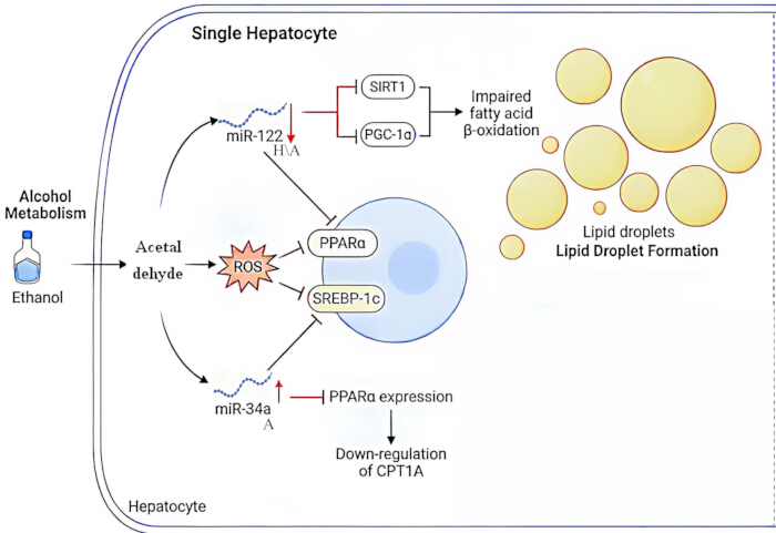
Figure 1: Regulatory networks of miRNAs in the steatosis stage of ALD. Alcohol metabolites (acetaldehyde and reactive oxygen species) disturb hepatic lipid homeostasis. Directly validated regulatory interactions are supported by human alcohol-associated liver disease samples (H) or alcohol-exposed animal models (A). Please click here to view a larger version of this figure.
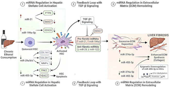
Figure 2: Regulatory networks of miRNAs in the fibrotic process of ALD. The diagram illustrates three core modules: miRNA-driven hepatic stellate cell activation, transforming growth factor beta (TGF-β)/Smad positive feedback loops, and extracellular matrix remodeling. Some regulatory relationships are directly validated in ALD-relevant experimental systems, whereas others are inferred from non-ALD liver disease studies (N) and have not yet been experimentally confirmed under ALD conditions. Please click here to view a larger version of this figure.
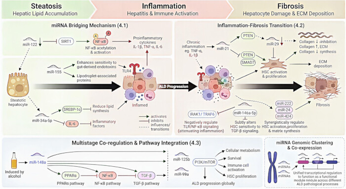
Figure 3: Integration of miRNA regulatory networks and cross-stage crosstalk in ALD. The schematic summarizes bridging hub miRNAs that mediate pathological transitions across steatosis, inflammation, and fibrosis, including the steatosis-inflammation transition, inflammation-fibrosis transition, and multistage miRNA co-regulation and signaling pathway integration. Certain hub miRNA-mediated effects are supported by direct evidence from human ALD samples or alcohol-treated animal models, whereas additional regulatory crosstalk is extrapolated from non-ALD studies. Please click here to view a larger version of this figure.
| Target Gene / Pathway | Cell Type | Model / Evidence Source | Biological Effect | Primary Reference |
| SIRT1, PGC-1α | Hepatocyte | Human ALD patients + alcohol‑fed mice (Ref8, ALD); mechanistic inference from non‑ALD models (Ref16,17, N) | Impairs fatty acid β-oxidation and promotes lipid accumulation; downstream effects involving SIRT1/PGC-1α are inferred mainly from non-ALD models and require direct validation in ALD | 8,16,17 |
| PPARα (↓ CPT1A) | Hepatocyte | Alcohol / miR-34a-5p–PPARα (Ref 18) | Suppresses PPARα → lowers CPT1A, reduces fatty acid oxidation, exacerbates lipid accumulation | 18 |
| FoxO1 (lipogenic genes) | Cholangiocyte / hepatocyte | Human alcohol-associated hepatitis (Ref 10, ALD) | Upregulates lipogenic genes; associated with cholangiopathy, disease severity and short-term mortality | 10 |
Table 1: Regulatory mechanisms of key miRNAs in the steatosis stage of ALD. The table summarizes miRNAs with experimental evidence from human ALD samples, alcohol-exposed animal models, in vitro cell culture, or non-ALD models. H = human ALD samples; A = alcohol-exposed animal models; C = in vitro cell-culture evidence; N = evidence extrapolated from non-ALD models and not directly validated in ALD; ↑ = increased expression; ↓ = decreased expression.
| miRNA | Expression Change | Target Gene / Pathway | Cell Type | Model / Evidence Source | Biological Effect | Primary Reference | |
| miR-155 | Up (↑) | SHIP1, SOCS1 | Kupffer cell / macrophage | A/C | Enhances NF-κB / MAPK signaling, amplifying inflammatory response (pro-inflammatory) | 24,25 | |
| miR-146a-5p | Early ↑, chronic ↓ | IRAK1, TRAF6 | Kupffer cell / macrophage | N (inferred from non‑ALD studies) | Negatively regulates TLR4 signaling; chronic decline weakens inhibition and sustains inflammation | 27,44 | |
| miR-223 | Down (↓) via SIRT1–C/EBPα axis | NLRP3 mRNA (3′UTR) | Neutrophil | A | Directly suppresses NLRP3, reduces IL-1β secretion; anti-inflammatory protection | 29,30 | |
| miR-144-3p | Up (↑) | ALDH1A3 | Hepatocyte | N (Western diet-induced NASH model) | Promotes microvesicular steatosis; direct relevance to ALD requires validation | 34 | |
Table 2: Regulatory mechanisms of key miRNAs in the inflammatory response stage of ALD. The table summarizes core miRNAs participating in alcohol-provoked hepatic inflammation, including TLR4/NF-κB signaling, NLRP3 inflammasome activation, and oxidative stress responses. H = human ALD samples; A = alcohol-exposed animal models; C = in vitro cell-culture evidence; N = evidence extrapolated from non-ALD models and not directly validated in ALD; ↑ = increased expression; ↓ = decreased expression.
| Target Gene / Pathway | Cell Type | Model / Evidence Source | Biological Effect | Primary Reference |
| PTEN, SMAD7 | Hepatic stellate cell (HSC) | N | Enhances TGF-β/SMAD signaling, promotes HSC activation and collagen deposition | 35,38 |
| COL1A1, COL3A1, MMPs | HSC | A | Anti-fibrotic; loss disrupts the ECM synthesis/degradation balance and promotes excess matrix deposition | 36 |
| PAK2, HDAC2 | HSC | N | Inhibits HSC activation and promotes apoptosis of activated HSCs | 35,37 |
| DDX3X | HSC | N | Suppresses HSC apoptosis, prolongs activated-HSC survival, drives chronic fibrosis | 35 |
| ZEB1, ZEB2 | HSC / EMT | N | Promotes epithelial-mesenchymal transition (EMT), accelerates fibrosis | 39 |
| TGF-β/SMAD feedback loop | HSC / hepatocyte | N | Anti-fibrotic; TGF-β lowers miR-122, completing a pro-fibrotic positive feedback loop | 38 |
| TIMP3 | HSC | N | Suppresses the MMP inhibitor TIMP3, thereby increasing MMP activity | 38 |
Table 3: Regulatory mechanisms of key miRNAs in the fibrotic stage of ALD. The table summarizes miRNAs involved in alcohol-associated liver fibrosis, including hepatic stellate cell activation/apoptosis, transforming growth factor beta (TGF-β)/Smad feedback regulation, and extracellular matrix remodeling. H = human ALD samples; A = alcohol-exposed animal models; C = in vitro cell-culture evidence; N = evidence extrapolated from non-ALD models and not directly validated in ALD; ↑ = increased expression; ↓ = decreased expression.
Disclosures
The authors have no conflicts of interest to disclose. All three figures were generated with AI assistance from Google Gemini and manually revised and scientifically verified by the authors.
Acknowledgements
The authors acknowledge Mr. Sun Qiang for his assistance.
References
- Seitz HK, et al. Alcoholic liver disease. Nat Rev Dis Primers. 2018;4(1):16.
- Seo W, Jeong WI. Hepatic non-parenchymal cells: Master regulators of alcoholic liver disease? World J Gastroenterol. 2016;22(4):1348-1356.
- Dukić M, et al. Alcohol, inflammation, and microbiota in alcoholic liver disease. Int J Mol Sci. 2023;24(4):3735.
- Jung JH, Kim SE, Suk KT, Kim DJ. Gut microbiota-modulating agents in alcoholic liver disease: Links between host metabolism and gut microbiota. Front Med. 2022;9:913842.
- Zhang L, Wang R, Nan Y, Kong L. Molecular regulators of alcoholic liver disease: a comprehensive analysis of microRNAs and long non-coding RNAs. Front Med. 2025;12:1482089.
- Wang X, He Y, Mackowiak B, Gao B. MicroRNAs as regulators, biomarkers and therapeutic targets in liver diseases. Gut. 2021;70(4):784-795.
- Li HD, et al. Noncoding RNAs in alcoholic liver disease. J Cell Physiol. 2019;234(9):14709-14720.
- Satishchandran A, et al. MicroRNA 122, regulated by GRLH2, protects livers of mice and patients from ethanol-induced liver disease. Gastroenterology. 2018;154(1):238-252.e237.
- Ezhilarasan D. MicroRNA interplay between hepatic stellate cell quiescence and activation. Eur J Pharmacol. 2020;885:173507.
- Blaya D, et al. Integrative microRNA profiling in alcoholic hepatitis reveals a role for microRNA-182 in liver injury and inflammation. Gut. 2016;65(9):1535-1545.
- Jiang XP, et al. The roles of microRNA families in hepatic fibrosis. Cell Biosci. 2017;7(1):34.
- Yang C, Zheng SD, Wu HJ, Chen SJ. Regulatory mechanisms of the molecular pathways in fibrosis induced by microRNAs. Chin Med J (Engl). 2016;129(19):2365-2372.
- Tilg H, Moschen AR, Szabo G. Interleukin-1 and inflammasomes in alcoholic liver disease/acute alcoholic hepatitis and nonalcoholic fatty liver disease/nonalcoholic steatohepatitis. Hepatology. 2016;64(3):955-965.
- Shen Y, et al. SGLT2 inhibitor empagliflozin downregulates miRNA-34a-5p and targets GREM2 to inactivate hepatic stellate cells and ameliorate non-alcoholic fatty liver disease-associated fibrosis. Metabolism. 2023;146:155657.
- Yang Z, et al. The role of SHP/REV-ERBα/CYP4A axis in the pathogenesis of alcohol-associated liver disease. JCI Insight. 2021;6(16):e140687.
- Liu H, et al. Prenatal dexamethasone exposure induces nonalcoholic fatty liver disease in male rat offspring via the miR-122/YY1/ACE2-MAS1 pathway. Biochem Pharmacol. 2021;185:114420.
- Thakur S, Munshi A, Kaur P. MicroRNA crosstalk in metabolic disorders: The dual role of miR-122 and miR-34a in NAFLD and Type 2 diabetes. Mol Biotechnol. 2026;68(8):3323-3343.
- Chen L, et al. Therapeutic role of Artemether in the prevention of hepatic steatosis through miR-34a-5p/PPARα pathway. Drug Dev Res. 2023;84(2):156-171.
- Xu H, et al. An endoplasmic reticulum stress-microRNA-26a feedback circuit in NAFLD. Hepatology. 2021;73(4):1327-1345.
- Pan JH, et al. The role of microRNA-33 as a key regulator in hepatic lipogenesis signaling and a potential serological biomarker for NAFLD with excessive dietary fructose consumption in C57BL/6N mice. Food Funct. 2021;12(2):656-667.
- Krause C, et al. Liver microRNA transcriptome reveals miR-182 as link between type 2 diabetes and fatty liver disease in obesity. Elife. 2024;12:RP92075.
- Li B, et al. Schisanhenol ameliorates non-alcoholic fatty liver disease via inhibiting miR-802 activation of AMPK-mediated modulation of hepatic lipid metabolism. Acta Pharm Sin B. 2024;14(9):3949-3963.
- Scarlata GGM, et al. The role of cytokines in the pathogenesis and treatment of alcoholic liver disease. Diseases. 2024;12(4):69.
- Zhang L, et al. Butyrate alleviates alcoholic liver disease-associated inflammation through macrophage regulation and polarization via the HDAC1/miR-155 axis. Int Immunopharmacol. 2024;131:111852.
- Salama RM, et al. Regulation of NOX/p38 MAPK/PPARα pathways and miR-155 expression by boswellic acids reduces hepatic injury in experimentally-induced alcoholic liver disease mouse model: novel mechanistic insight. Arch Pharm Res. 2023;46(4):323-338.
- Bala S, et al. Steatosis, inflammasome upregulation, and fibrosis are attenuated in miR-155 deficient mice in a high fat-cholesterol-sugar diet-induced model of NASH. Lab Invest. 2021;101(12):1540-1549.
- Ravanidis S, et al. Fasting-mediated metabolic and toxicity reprogramming impacts circulating microRNA levels in humans. Food Chem Toxicol. 2021;152:112187.
- Wang S, Qing D. Progress in inflammasome activation and pyrolysis in alcoholic liver disease. J Cent South Univ Med Sci. 2020;45(8):999-1004.
- Ren R, et al. Aging exaggerates acute-on-chronic alcohol-induced liver injury in mice and humans by inhibiting neutrophilic sirtuin 1-C/EBPα-miRNA-223 axis. Hepatology. 2022;75(3):646-660.
- Orr C, et al. Longitudinal analysis of serum microRNAs as predictors of cirrhosis regression during treatment of hepatitis B virus infection. Liver Int. 2020;40(7):1693-1700.
- Zhou C, et al. Overexpression of miR-142-5p inhibits the progression of nonalcoholic steatohepatitis by targeting TSLP and inhibiting JAK-STAT signaling pathway. Aging. 2020;12(10):9066-9084.
- Michalak A, Lach T, Cichoż-Lach H. Oxidative stress-a key player in the course of alcohol-related liver disease. J Clin Med. 2021;10(14):3011.
- Zhang X, et al. PPARγ attenuates hepatic inflammation and oxidative stress of non-alcoholic steatohepatitis via modulating the miR-21-5p/SFRP5 pathway. Mol Med Rep. 2021;24(5):823.
- Kennedy L, et al. Mast cells promote nonalcoholic fatty liver disease phenotypes and microvesicular steatosis in mice fed a western diet. Hepatology. 2021;74(1):164-182.
- Zhou Q, et al. Human umbilical cord mesenchymal stem cells ameliorate hepatic stellate cell activation and liver fibrosis by upregulating microRNA-455-3p through suppression of p21-activated kinase-2. Biomed Res Int. 2021;2021(1):6685605.
- Hoebinger C, Rajcic D, Silva B, Hendrikx T. Chronic-binge ethanol feeding aggravates systemic dyslipidemia in Ldlr-/- mice, thereby accelerating hepatic fibrosis. Front Endocrinol. 2023;14:1148827.
- You H, et al. The miR-455-3p/HDAC2 axis plays a pivotal role in the progression and reversal of liver fibrosis and is regulated by epigenetics. FASEB J. 2021;35(7):e21700.
- Namachivayam A, Gopalakrishnan AV. A review on molecular mechanism of alcoholic liver disease. Life Sci. 2021;274:119328.
- Shan L, et al. Purine signaling regulating HSCs inflammatory cytokines secretion, activation, and proliferation plays a critical role in alcoholic liver disease. Mol Cell Biochem. 2020;466(1):91-102.
- Abdelgawwad El-Sehrawy AAM, et al. MicroRNA-SIRT1 crosstalk in liver diseases: molecular regulation of metabolism, inflammation, and cell survival. Mol Biol Rep. 2026;53(1):899.
- Feng X, et al. Functional role of miR-155 in physiological and pathological processes of liver injury. Mol Med Rep. 2021;24(4):714.
- Wan Y, et al. miR-34a regulates macrophage-associated inflammation and angiogenesis in alcohol-induced liver injury. Hepatol Commun. 2023;7(4):e0089.
- Hochreuter MY, Dall M, Treebak JT, Barrès R. MicroRNAs in non-alcoholic fatty liver disease: Progress and perspectives. Mol Metab. 2022;65:101581.
- Naidu C, Cox AJ, Lewohl JM. Influence of sex and liver cirrhosis on the expression of miR-146a-5p and its target genes, IRAK1 and TRAF6. Brain Res. 2024;1827:148763.
- Sun SY, et al. Identifying competing endogenous RNA regulatory networks and hub genes in alcoholic liver disease for early diagnosis and potential therapeutic target insights. Aging. 2024;16(10):9147.
- Xiong J, Ni J, Chen C, Wang K. miR-148a-3p regulates alcoholic liver fibrosis through targeting ERBB3. Int J Mol Med. 2020;46(3):1003-1012.
- Shen L, et al. Metabolic Dysfunction-associated Steatotic Liver Disease Diminishes the Efficacy of miR-125b-5p-engineered MSC Therapy for Hepatocellular Carcinoma via HK2 Upregulation: A Multimodal Theranostic Study. J Clin Transl Hepatol. 2026;14(5):511-524.
- Choudhary NS, Duseja A. Genetic and epigenetic disease modifiers: non-alcoholic fatty liver disease (NAFLD) and alcoholic liver disease (ALD). Transl Gastroenterol Hepatol. 2021;6:2.
- Bulfoni M, et al. miRNA expression profiles in liver grafts of HCV and HIV/HCV-infected recipients, 6 months after liver transplantation. J Med Virol. 2021;93(8):4992-5000.
- Xiang H, et al. Knockdown of UCA1 attenuated the progression of alcoholic fatty disease by sponging miR-214. Mamm Genome. 2022;33(3):534-542.
- Cheng Z, Yang L, Chu H. The role of gut microbiota, exosomes, and their interaction in the pathogenesis of ALD. J Adv Res. 2025;72:353-367.
- Küçük İ, et al. Circulating histone H4 values can relate to disease severity in patients with alcoholic hepatitis and cirrhosis. Cir Cir. 2025;93(4):411-418.
- Zhang G, Chen H, Ren W, Huang J. Efficacy of bile acid profiles in diagnosing and staging of alcoholic liver disease. Scand J Clin Lab Invest. 2023;83(1):8-17.