This study evaluates the protective effects of Yangjing Capsule on cyclophosphamide-induced spermatogenic dysfunction in mice and examines the involvement of the lncRNA NONMMUT031883.2/QKI-5/p38 MAPK signaling pathway.
A subscription to JoVE is required to view this content. Sign in or start your free trial.
Research Article
Yangjing Capsule Alleviates Spermatogenic Dysfunction in Mice by Inhibiting the QKI-5/p38 MAPK Pathway Through Upregulation of IncRNA NONMMUT031883.2
In This Article
Summary
Abstract
Spermatogenic dysfunction is an important cause of male infertility, and cyclophosphamide (CP) can impair spermatogenesis by inducing abnormal spermatogonial apoptosis. Yangjing Capsule (YC) has been used to improve reproductive function, but its underlying molecular mechanism remains unclear. This study investigated the protective effects of YC in a CP-induced mouse model of spermatogenic dysfunction and in GC-1 spermatogonial cells. Sperm count and motility, male fertility, serum sex hormone levels, and histopathological changes in the testes and epididymides were evaluated. Cell viability, lactate dehydrogenase release, apoptosis, and apoptosis-related proteins were assessed in GC-1 cells. Quantitative reverse transcription polymerase chain reaction, RNA pull-down assays, immunofluorescence, and Western blotting were used to examine lncRNA NONMMUT031883.2, Quaking-5 (QKI-5), and p38 mitogen-activated protein kinase (MAPK) signaling. YC dose-dependently improved sperm quality, serum sex hormone levels, reproductive organ morphology, testicular histopathology, and fertility in CP-treated mice. In GC-1 cells, YC increased cell viability and reduced cellular damage and apoptosis. LncRNA NONMMUT031883.2 physically interacted with QKI-5, and YC treatment increased lncRNA NONMMUT031883.2 expression while reducing QKI-5 expression and p38 phosphorylation. YC treatment inhibited apoptosis induced by lncRNA NONMMUT031883.2 knockdown in GC-1 cells, whereas QKI-5 overexpression reduced the anti-apoptotic effects of YC. These findings suggest that YC alleviates CP-induced spermatogenic dysfunction and that its protective effects are mediated by regulation of the lncRNA NONMMUT031883.2/QKI-5/p38 MAPK pathway.
Introduction
Spermatogenic dysfunction refers to reproductive impairment caused by abnormalities in sperm production, maturation, or function in the male reproductive system. It is characterized by reduced sperm count and motility and an increased rate of sperm deformity rate1,2. The pathogenesis of spermatogenic dysfunction is complex and is associated with genetic factors, autoimmune disorders, environmental toxins, chemotherapy-induced damage, and reproductive organ lesions3-5. Chemotherapeutic drugs such as cyclophosphamide (CP) can directly damage the testicular seminiferous tubules and induce spermatogonial apoptosis, thereby contributing to male spermatogenic dysfunction6,7. Hormone replacement therapy, antioxidant supplementation, and assisted reproductive technologies are currently used as clinical interventions. However, these approaches inconsistent efficacy and substantial adverse effects8,9,10. Therefore, identifying safe and effective interventions and clarifying their molecular targets are clinically important for improving fertility outcomes in patients with spermatogenic dysfunction.
Chinese herbal medicine has shown potential in protecting male reproductive function and treating spermatogenic dysfunction because of its multitarget properties and relatively low toxicity11,12. Yangjing Capsule (YC) is a traditional Chinese patent medicine composed of Rehmannia glutinosa, Epimedium, placenta, Polygonatum sibiricum, Angelica sinensis, and other Chinese herbal medicines. It is used to tonify the kidney and essence, nourish the blood and marrow, and regulate reproductive function13. Previous studies have shown that YC can regulate sex hormone levels in male animals, improve sperm quality, repair testicular tissue damage, and protect against reproductive injury induced by chemical agents14,15. Spermatogonial cells form the basis of spermatogenesis, and abnormal spermatogonial apoptosis is a central event in the development of spermatogenic dysfunction16. YC has also been reported to improve spermatogonial viability, increase the proportion of cells in the S phase, and inhibit apoptosis17. However, its protective effects and specific molecular mechanisms in CP-induced spermatogenic dysfunction remain unclear.
Long noncoding RNAs (lncRNAs) are RNA molecules longer than 200 nucleotides that lack protein-coding capacity18. LncRNAs can regulate gene expression through mechanisms including chromosomal silencing, chromatin modification, genomic imprinting, and transcriptional activation19,20. The RNA-binding protein Quaking-5 (QKI-5) participates in physiological and pathological processes, including cell proliferation and apoptosis, by regulating the alternative splicing of downstream genes21. The p38 mitogen-activated protein kinase (MAPK) pathway is involved in apoptosis regulation and also plays an important role in male fertility and testicular development22,23. A previous study showed that QKI-5 overexpression activates the p38 MAPK pathway in hepatocellular carcinoma cells24. In addition, lncRNA NONMMUT074098.2 was shown to bind QKI-5 in spermatogonia, whereas QKI-5 knockdown inhibited the p38 MAPK pathway and reduced spermatogonial apoptosis25. This study used a CP-induced mouse model of spermatogenic dysfunction to investigate whether YC regulates lncRNA NONMMUT031883.2 expression and the QKI-5/p38 MAPK signaling axis, thereby reducing abnormal spermatogonial apoptosis and alleviating CP-induced spermatogenic dysfunction (Figure 1). The objective was to clarify the molecular mechanism underlying YC's protective effects on spermatogenesis and to expand understanding of its pharmacological role in protecting male reproductive function.
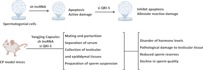
Figure 1: Schematic diagram of the experimental design. The upper panel summarizes the in vitro experiments in GC-1 spermatogonial cells. Knockdown of lncRNA NONMMUT031883.2 was used to induce apoptosis and cellular damage, followed by QKI-5 knockdown to evaluate its effects on apoptosis and cell injury. The lower panel summarizes the in vivo experiments in cyclophosphamide (CP)-induced mouse models of spermatogenic dysfunction. Mice received Yangjing Capsule treatment and/or lncRNA NONMMUT031883.2 or QKI-5 intervention. Fertility, serum hormone levels, testicular and epididymal tissues, sperm suspension, sperm reserves, sperm quality, and testicular histopathological damage were subsequently evaluated. Please click here to view a larger version of this figure.
Access restricted. Please log in or start a trial to view this content.
Protocol
All animal experiments were performed in accordance with the ARRIVE guidelines and the National Institutes of Health Guide for the Care and Use of Laboratory Animals. All experimental protocols were reviewed and approved by the Animal Ethics Committee of Southeast University (approval no. 20230216022).
1. Preparation of YC-containing serum
Two Yangjing Capsules (YC; 0.5 g/capsule) were opened, and 0.8 g of the capsule contents was weighed. Normal saline was added to a final volume of 10 mL, and the mixture was vortexed thoroughly to prepare an 80 mg/mL suspension. Specific pathogen-free male BALB/c mice weighing 22–25 g and aged 8 weeks were maintained at 22 °C and 55%–60% humidity under a 12 h light/dark cycle, with food and water provided ad libitum. After 1 week of acclimatization, the mice received YC by oral gavage at 800 mg/kg body weight in a volume of 10 mL/kg once daily for 5 consecutive days. Two hours after the final administration, the mice were anesthetized with intraperitoneal 1% sodium pentobarbital, and blood samples were collected to prepare YC-containing serum. The serum was inactivated at 56 °C for 30 min, filtered through a 0.22 µm membrane, and stored at −80 °C until use.
2. Cell culture and transfection
Mouse GC-1 spermatogonial cells were maintained in DMEM containing 1% penicillin-streptomycin and 10% fetal bovine serum. All cultures were maintained at 37 °C in an atmosphere containing 5% CO₂ (v/v).
LncRNA-specific short hairpin RNA (sh-lncRNA), a QKI-5 overexpression plasmid (OE-QKI-5), QKI-5 small interfering RNA (si-QKI-5), and their corresponding negative controls (sh-NC, OE-NC, and si-NC) were synthesized and constructed. GC-1 cells were transfected with sh-lncRNA or sh-NC, si-QKI-5 or si-NC, or OE-QKI-5 or OE-NC according to the transfection reagent instructions. After incubation for 48 h, transfection efficiency was assessed by measuring lncRNA NONMMUT031883.2 or QKI-5 expression.
For the sh-lncRNA + si-QKI-5 + etoposide group, GC-1 cells were transfected with sh-lncRNA and si-QKI-5 and subsequently treated with 100 µM etoposide for 12 h26. For subsequent cell viability assays, GC-1 cells were cultured in medium supplemented with 5%, 10%, or 20% YC-containing serum and designated as the low-dose YC (L-YC), medium-dose YC (M-YC), and high-dose YC (H-YC) groups, respectively. The concentrations of YC-containing serum (5%, 10%, and 20% YC) were determined based on the results of preliminary concentration-gradient experiments. For the sh-lncRNA + H-YC + OE-NC and sh-lncRNA + H-YC + OE-QKI-5 groups, GC-1 cells were transfected with sh-lncRNA plus OE-NC or sh-lncRNA plus OE-QKI-5 for 48 h and then exposed to 20% YC-containing serum for 24 h.
3. Cell Counting Kit-8 assay
GC-1 cells were seeded into 96-well plates at 5 × 10³ cells/well and cultured for 24 h. After cell attachment, the original medium was replaced with 100 µL of complete medium supplemented with YC-containing serum, and the cells were incubated for 6, 12, or 24 h. After incubation, 10 µL of Cell Counting Kit-8 reagent was added to each well, and the cells were incubated at 37 °C in darkness for 2 h. Absorbance at 450 nm was measured using a microplate reader.
4. Lactate dehydrogenase assay
Following transfection with sh-NC or sh-lncRNA for 48 h, the GC-1 cell culture medium was collected, and the supernatant was obtained by centrifugation. According to the lactate dehydrogenase assay kit instructions, the supernatant was gently mixed with the detection working solution and transferred to a 96-well plate. The plate was incubated at 25 °C in darkness for 30 min. Absorbance at 490 nm was measured using a microplate reader, and lactate dehydrogenase release was calculated to assess cell damage.
5. Calcein AM/propidium iodide staining
After transfection with sh-NC or sh-lncRNA for 48 h, GC-1 cell survival was assessed using Calcein AM/propidium iodide (PI) staining. Cells were seeded into 12-well plates at 5 × 10⁴ cells/well and cultured until they reached 60%–70% confluence. The cells were gently rinsed with phosphate-buffered saline, and 500 µL of Calcein AM/PI staining solution was added to each well. The cells were incubated at 37 °C in darkness for 30 min and observed under a fluorescence microscope. Live and dead cells were counted using image-analysis software, and the proportion of surviving cells was calculated.
6. Flow cytometry
GC-1 cells were collected by centrifugation and gently rinsed with phosphate-buffered saline. The cell pellet was resuspended in 500 µL of binding buffer at a density of 5 × 10⁵ cells/mL. Annexin V-APC and PI were then added at 5 µL each, and the suspension was mixed thoroughly and incubated at 25 °C in darkness for 10 min. Apoptosis was analyzed by flow cytometry using excitation/emission wavelengths of 633/660 nm for APC and 488/630 nm for PI. The apoptosis rate was evaluated using flow cytometry analysis software.
7. Caspase-3 activity assay
After the indicated treatments, approximately 1 × 10⁶ GC-1 cells were collected, rinsed, and resuspended in phosphate-buffered saline. The cells were mixed with 100 µL of reagent II from the caspase-3 activity assay kit and lysed on ice for 15 min. Following centrifugation, 50 µL of the lysate supernatant was collected and mixed with 40 µL of reagent I and 10 µL of reagent III. The mixture was incubated at 37 °C for 2 h. Absorbance at 405 nm was measured using a microplate reader, and caspase-3 activity was evaluated according to the kit instructions.
8. RNA pull-down assay
The sense and antisense strands of lncRNA NONMMUT031883.2 were synthesized according to the corresponding sequence and labeled with biotin. Cells were lysed in immunoprecipitation cell lysis buffer on ice for 30 min, and the protein-containing supernatant was collected by centrifugation. Streptavidin magnetic beads were washed three times with RNA-binding buffer and incubated in binding buffer containing 1% bovine serum albumin for 30 min.
The pretreated beads were combined with the biotin-labeled probes and incubated at 25 °C for 30 min. Unbound probes were removed by magnetic separation. The cell lysate supernatant was then added, and the mixture was incubated at 4 °C for 4 h to form magnetic bead-RNA-protein complexes. The magnetic beads were washed three times with washing buffer to remove nonspecifically bound proteins. The washing buffer was removed by magnetic separation, and 2× SDS sample-loading buffer was added. The samples were heated at 95 °C for 10 min, and the supernatant was collected by magnetic separation for subsequent Western blot analysis.
9. Construction of a mouse model of spermatogenic dysfunction
After 5 days of acclimatization, specific pathogen-free male BALB/c mice weighing 22–25 g and aged 8 weeks received cyclophosphamide (CP) intraperitoneally at 50 mg/kg once daily for 7 days to establish a spermatogenic dysfunction model14,27. Successfully modeled mice were randomly assigned using a random-number table to the CP, 200 mg/kg YC, 400 mg/kg YC, 800 mg/kg YC, 800 mg/kg YC + sh-NC, and 800 mg/kg YC + sh-lncRNA groups, with six mice in each group.
Mice in the YC-treated groups received the corresponding dose of YC by oral gavage once daily for 30 days13. For the 800 mg/kg YC + sh-NC and 800 mg/kg YC + sh-lncRNA groups, after successful model establishment, the mice were anesthetized by inhalation of 2% isoflurane. After confirming that each mouse showed no foot withdrawal reflex and that the respiratory rate was stable, the surgical site was shaved and disinfected with povidone-iodine. Under aseptic conditions, a ventral midline incision was made through the skin and body wall anterior to the genitalia. The testis was exposed by carefully manipulating the epididymal fat pad, with care taken to avoid vascular damage. Using a micromanipulator, the seminiferous tubules were visualized, and 1 µL of the adenoviral suspension (1 × 10¹⁰ TU/mL) was slowly injected using a microinjection needle28. The needle was left in place for 5 min after injection before being slowly withdrawn, and hemostasis was achieved with a sterile cotton swab. The same procedure was subsequently performed on the contralateral testis. The incision was then sutured, and the mice were allowed to recover on a warming pad postoperatively. Control mice received an equal volume of saline solution by gavage and injection daily.
10. Assessment of fertility
After completion of drug administration, male mice in each group were mated freely with female BALB/c mice at a ratio of 1:2 for 7 days. Vaginal plug formation in the female mice was monitored and recorded daily. After successful mating was confirmed, the male mice were removed, and the female mice were housed individually until natural delivery. Nine pregnant female mice per group were obtained and included in the analysis. The average litter size was recorded to evaluate male fertility in each group.
At the end of the experimental period, the mice were euthanized by intraperitoneal administration of pentobarbital sodium at 100 mg/kg. This euthanasia method was approved by the Animal Ethics Committee of Southeast University (approval no. 20230216022). The testes and epididymides were carefully separated, photographed, and weighed. The testis and epididymis indices were calculated as organ weight/body weight. One epididymis from each mouse was collected, cut into pieces, placed in 1 mL of prewarmed phosphate-buffered saline (37 °C), and incubated for 20 min to allow sperm to swim out29. The sperm suspension was gently mixed, and 10 µL of the sperm suspension was placed on a preheated blood cell counting plate. Sperm were counted manually under a light microscope at 200× magnification, and the results were expressed as 106/L. Furthermore, the percentage of progressively motile spermatozoa relative to the total number of spermatozoa was calculated as sperm motility13.
11. Determination of sex hormone levels
Blood was collected from the orbital vein of mice in each group and allowed to stand at 25 °C for 30 min. Serum was collected by centrifugation at 3,000 × g for 10 min30. Serum testosterone, luteinizing hormone (LH), and follicle-stimulating hormone (FSH) levels were measured using the corresponding enzyme-linked immunosorbent assay kits.
12. RNA stability assay
GC-1 cells were seeded into 96-well culture plates, transfected with si-QKI-5 or si-NC, and cultured for 48 h. Actinomycin D was then added at 5 µg/mL, and the cells were collected at 0, 3, 6, and 9 h. Total RNA was isolated, and the relative expression level of lncRNA NONMMUT031883.2 was determined by quantitative reverse transcription polymerase chain reaction following reverse transcription. The relative RNA level at each time point was calculated using the expression level at 0 h as 100%.
13. Quantitative reverse transcription polymerase chain reaction
GC-1 cells and mouse testicular tissues were rinsed with phosphate-buffered saline and lysed for total RNA isolation. Complementary DNA was generated by reverse transcription using AMV reverse transcriptase. Polymerase chain reaction amplification was performed using a real-time PCR system and a quantitative PCR kit, with complementary DNA used as the amplification template. U6 was used as the internal reference for lncRNA NONMMUT031883.2, and GAPDH was used as the internal reference for QKI-5. Relative target-gene expression was calculated using the 2−ΔΔCt method25. RNA was also isolated separately from the cytoplasmic and nuclear fractions to determine the subcellular localization of lncRNA NONMMUT031883.2. The primer sequences are provided in Table 1.
| Gene | Forward primer (5' to 3') | Reverse primer (5' to 3') |
| QKI-5 | CTGTCATGCCAAACGGAAC | GATGGACACGCATATCGTG |
| GAPDH | AGGTCGGTGTGAACGGATTTG | TGTAGACCATGTAGTTGAGGTCA |
| lncRNA NONMMUT031883.2 | CCACTGTGGCACATTGAAGTA | TTAAGTTAATGGGTGCTGTGTT |
| U6 RNA | CTCGCTTCGGCAGCACA | AACGCTTCACGAATTTGCGT |
Table 1: Primer sequences used for quantitative reverse transcription polymerase chain reaction. Forward and reverse primer sequences are listed in the 5′–3′ direction for lncRNA NONMMUT031883.2, QKI-5, U6, and GAPDH.
14. Hematoxylin and eosin staining
Mouse testicular and epididymal tissues were immersed in 4% paraformaldehyde and fixed at 4 °C for 24 h. After rinsing with phosphate-buffered saline, the tissues were dehydrated through a graded ethanol series, embedded in paraffin, and sectioned at 5 µm. The sections were heated at 60 °C for 60 min and dewaxed in xylene for 30 min.
After hydration through a graded ethanol series, the sections were stained with hematoxylin for 5 min, treated with hydrochloric acid-ethanol differentiation solution for 10 s, washed with tap water for bluing, and stained with 1% eosin for 2 min. The sections were then dehydrated in 95% ethanol and absolute ethanol and cleared in xylene for 30 min. After air-drying, the sections were mounted with neutral gum. Morphological changes in the testes and epididymides were observed under a microscope.
15. Immunofluorescence
Mouse testicular paraffin sections were dewaxed and hydrated through a graded ethanol series, followed by microwave-mediated antigen retrieval in citrate buffer at pH 6.0. After cooling naturally to room temperature, the sections were incubated with 0.3% Triton X-100 for 10 min. The sections were blocked with 5% bovine serum albumin at 25 °C for 60 min, rinsed with phosphate-buffered saline, and incubated overnight at 4 °C with a primary antibody against Ki67. The following day, a fluorochrome-labeled secondary immunoglobulin G antibody was applied, and the sections were incubated at 37 °C in darkness for 60 min. After rinsing with phosphate-buffered saline, the sections were counterstained with DAPI for 10 min and observed under a fluorescence microscope. Fluorescence intensity was analyzed using image-analysis software.
For immunofluorescence staining of GC-1 cells, the cells were seeded into 12-well plates at 1 × 10⁵ cells/well and incubated for 24 h. The cells were fixed with 4% paraformaldehyde for 30 min, permeabilized with 0.3% Triton X-100 for 10 min, blocked with 5% bovine serum albumin, and incubated overnight at 4 °C with a primary antibody against QKI-5. The remaining procedure was performed as described for the testicular tissue sections.
16. TUNEL staining
Mouse testicular paraffin sections were dewaxed, hydrated through a graded ethanol series, and incubated with 0.1% Triton X-100 for 5 min to increase cell membrane permeability. Proteinase K working solution at 20 µg/mL without DNase was applied uniformly to the sections, which were then incubated at 37 °C for 15 min. TUNEL detection solution was added, and the sections were incubated at 37 °C in darkness for 90 min. After incubation with DAPI for 10 min, antifade mounting medium was applied, and the testicular tissue was observed under a fluorescence microscope.
GC-1 cells were seeded into 12-well plates and fixed with 4% paraformaldehyde for 30 min. After permeabilization with 0.1% Triton X-100 for 5 min, TUNEL detection solution was applied to the cells, followed by incubation in darkness for 60 min. The cells were then stained with DAPI for 10 min and observed under a fluorescence microscope.
17. Western blotting
Mouse testicular tissues and GC-1 cells were lysed in immunoprecipitation cell lysis buffer on ice for 30 min and disrupted by ultrasonication. After centrifugation, the protein-containing supernatant was collected, and protein concentration was determined using a bicinchoninic acid assay kit. The protein supernatant was mixed with SDS loading buffer and heated in boiling water for 10 min. After separation by gel electrophoresis, the proteins were transferred to polyvinylidene fluoride membranes and blocked with 5% bovine serum albumin for 2 h31.
The membranes were incubated overnight at 4 °C with primary antibodies against cleaved caspase-3 (1:500), Bcl-2 (1:1000), caspase-3 (1:2000), Bax (1:500), QKI-5 (1:1000), phosphorylated p38 (1:1000), or p38 (1:500). On the following day, the membranes were rinsed with Tris-buffered saline containing Tween 20 and incubated with goat anti-rabbit immunoglobulin G at 1:10000 for 2 h at 25 °C. The enhanced chemiluminescence working solution was applied to the membrane surface, and the protein bands were imaged with a gel imaging system. Band intensity was analyzed using image-analysis software, and relative protein expression was normalized to GAPDH (1:3000).
Cycloheximide at 50 µg/mL was used to inhibit protein synthesis. To determine the half-life of QKI-5, cells were collected at 0, 3, 6, and 9 h, and QKI-5 protein levels were subsequently determined by Western blotting25.
18. Statistical analysis
All data were statistically analyzed and plotted using statistical, graphing, and image-analysis software. The Shapiro-Wilk test was used to assess data normality. Experimental data are presented as the mean ± standard deviation. One-way analysis of variance was used to evaluate differences among groups. The LSD-t test was used for post hoc pairwise comparisons when variances were homogeneous, whereas Dunnett’s T3 test was used when variances were heterogeneous. A value of p < 0.05 was considered statistically significant.
Access restricted. Please log in or start a trial to view this content.
Results
Knockdown of lncRNA NONMMUT031883.2 induces spermatogonial apoptosis
GC-1 cells were transfected with sh-lncRNA to knock down lncRNA NONMMUT031883.2 expression. Compared with sh-NC-transfected cells, the transcriptional level of lncRNA NONMMUT031883.2 was markedly reduced following sh-lncRNA transfection, confirming successful knockdown (Figure 2A). Following lncRNA NONMMUT031883.2 knockdown, GC-1 cell viability was significantly reduced (Figure 2B
Access restricted. Please log in or start a trial to view this content.
Discussion
Chemotherapy-induced acquired spermatogenic dysfunction is an important cause of male infertility32,33,34. However, the specific molecular mechanisms remain incompletely understood. The findings of this study demonstrate that Yangjing Capsule (YC) dose-dependently improves cyclophosphamide (CP)-induced sex hormone abnormalities, testicular histopathological damage, and fertility impairment in male mice. These improvements were a...
Access restricted. Please log in or start a trial to view this content.
Disclosures
The authors declare no financial or nonfinancial conflicts of interest.
Acknowledgements
This work was supported by the National Natural Science Foundation of China (grant no. 82374257), the Fourth Batch of Peak Academic Talent Training Program of Jiangsu Provincial Hospital of Chinese Medicine (grant no. k2026yrc74), and the Scientific Research Projects of Traditional Chinese Medicine and Integrated Traditional Chinese and Western Medicine of Jiangsu Province (grant no. CYTF2026019).
Access restricted. Please log in or start a trial to view this content.
Materials
| Name | Company | Catalog Number | Comments |
|---|---|---|---|
| Actinomycin D | MedChemExpress, Monmouth Junction, NJ, USA | HY-17559 | Transcription inhibitor used for RNA stability assays |
| AMV reverse transcriptase | Takara, Tokyo, Japan | 2621 | Enzyme for complementary DNA synthesis |
| Annexin V-APC/PI detection kit | BioLab Technology, Beijing, China | KFS191 | Kit for flow cytometric analysis of apoptosis |
| Antifade mounting medium | MedChemExpress, Monmouth Junction, NJ, USA | HY-K1042 | Mounting medium for fluorescence preservation |
| Bax primary antibody | Affinity Biosciences, OH, USA | AF0120 | Primary antibody for detection of Bax |
| BCA kit | Macklin, Shanghai, China | B917925 | Kit for protein concentration determination |
| Bcl-2 primary antibody | Affinity Biosciences, OH, USA | AF0769 | Primary antibody for detection of Bcl-2 |
| Bovine serum albumin | BioLab Technology, Beijing, China | BTN131070 | Blocking reagent |
| Calcein/PI detection kit | Beyotime, Shanghai, China | C2015M | Kit for live/dead cell staining |
| Caspase-3 activity assay kit | BioLab Technology, Beijing, China | QN2866 | Kit for measuring caspase-3 activity |
| Caspase-3 primary antibody | Abcam, MA, USA | ab184787 | Primary antibody for detection of caspase-3 |
| CCK-8 reagent | MedChemExpress, Monmouth Junction, NJ, USA | HY-K0301 | Reagent for cell viability assessment |
| Cleaved caspase-3 primary antibody | Abcam, MA, USA | ab32042 | Primary antibody for detection of cleaved caspase-3 |
| Cycloheximide | Macklin, Shanghai, China | H823111 | Protein synthesis inhibitor used for protein stability assays |
| Cyclophosphamide | MedChemExpress, Monmouth Junction, NJ, USA | HY-17420 | Used to induce spermatogenic dysfunction |
| DAPI reagent | Beyotime, Shanghai, China | C1006 | Nuclear counterstain |
| DMEM medium | Wuhan Sunncell Biotechnology, Hubei, China | SNLM-302 | Cell culture medium for GC-1 cells |
| ECL working solution | Solarbio, Beijing, China | SW2030 | Chemiluminescent substrate for protein-band detection |
| Etoposide | MedChemExpress, Monmouth Junction, NJ, USA | HY-13629 | Apoptosis-inducing agent |
| Flow cytometer | BD Biosciences, CA, USA | BD FACSCalibur | Instrument for apoptosis analysis |
| FlowJo software | BD Biosciences, CA, USA | v10.8 | Software for flow cytometry data analysis |
| Fluorescence microscope | Leica, Heidelberg, Germany | DM3000 | Instrument for fluorescence imaging |
| Fluorescence-labeled secondary antibody IgG | Servicebio, Wuhan, China | GB22303 | Secondary antibody for immunofluorescence detection |
| Follicle-stimulating hormone ELISA kit | Huamei Biotech, Wuhan, China | CSB-E06871m | Kit for measuring serum follicle-stimulating hormone |
| GAPDH primary antibody | Affinity Biosciences, OH, USA | T0004 | Loading-control antibody for Western blotting |
| GC-1 cells | Wuhan Sunncell Biotechnology, Hubei, China | SNL-302 | Mouse spermatogonial cell line |
| Gel imaging system | Jinpeng Analysis Instrument, Shanghai, China | JP-2880 | Imaging system for gel and blot visualization |
| Gel imaging system | Invitrogen, Carlsbad, CA, USA | iBright CL1500 | Chemiluminescence imaging system for Western blots |
| Goat anti-rabbit IgG | Servicebio, Wuhan, China | GB23303 | Secondary antibody for Western blotting |
| Hematoxylin solution | BioLab Technology, Beijing, China | YT165 | Nuclear stain for histological analysis |
| Hydrochloric acid ethanol differentiation solution | Beyotime, Shanghai, China | C0163S | Differentiation reagent for hematoxylin staining |
| ImageJ software | National Institutes of Health, USA | 1.54h | Software for image quantification |
| IP cell lysis buffer | Beyotime, Shanghai, China | P0013 | Lysis buffer for immunoprecipitation and protein extraction |
| Ki67 primary antibody | Affinity Biosciences, OH, USA | AF0198 | Primary antibody for detection of Ki67 |
| LDH Cytotoxicity Assay Kit | Beyotime, Shanghai, China | C0016 | Kit for measuring lactate dehydrogenase release and cellular damage |
| Light microscope | Olympus, Tokyo, Japan | CKX53 | Used for sperm counting and morphological observation |
| Lipofectamine 3000 | Invitrogen, Carlsbad, CA, USA | L3000001 | Transfection reagent |
| Microplate reader | Molecular Devices, CA, USA | SpectraMax Mini | Instrument for absorbance measurements |
| Mouse luteinizing hormone ELISA kit | Huamei Biotech, Wuhan, China | CSB-E12770m | Kit for measuring serum luteinizing hormone |
| Mouse testosterone ELISA kit | Huamei Biotech, Wuhan, China | CSB-E05101m | Kit for measuring serum testosterone |
| Neutral gum | BioLab Technology, Beijing, China | QN1253 | Mounting medium for histological sections |
| p38 primary antibody | Affinity Biosciences, OH, USA | AF6456 | Primary antibody for detection of total p38 |
| Paraformaldehyde | MedChemExpress, Monmouth Junction, NJ, USA | HY-Y0333 | Fixative for cells and tissues |
| PARIS kit | Invitrogen, Carlsbad, CA, USA | AM1921 | Kit for nuclear and cytoplasmic RNA fractionation |
| PCR system | ABI, CA, USA | ABI PRISM 7300 | Real-time PCR instrument |
| p-p38 primary antibody | Affinity Biosciences, OH, USA | AF3455 | Primary antibody for detection of phosphorylated p38 |
| Proteinase K solution | Beyotime, Shanghai, China | ST532 | Reagent for tissue permeabilization before TUNEL staining |
| PVDF membranes | Millipore, MA, USA | ISEQ00010 | Membranes for protein transfer in Western blotting |
| QKI-5 primary antibody | Abcam, MA, USA | ab126742 | Primary antibody for detection of QKI-5 |
| RNA binding buffer | Invitrogen, Carlsbad, CA, USA | 65040D | Buffer for RNA-protein binding reactions |
| SDS loading buffer | Beyotime, Shanghai, China | P0286 | Sample-loading buffer for SDS-PAGE |
| Streptavidin magnetic beads | MedChemExpress, Monmouth Junction, NJ, USA | HY-K0208 | Magnetic beads for capture of biotin-labeled RNA probes |
| TB Green FAST qPCR Kit | Takara, Tokyo, Japan | CN830S | Reagent kit for quantitative PCR |
| Triton X-100 | Macklin, Shanghai, China | T824275 | Permeabilization reagent |
| TRIzol reagent | Invitrogen, Carlsbad, CA, USA | 15596018CN | Reagent for total RNA extraction |
| TUNEL detection solution | Beyotime, Shanghai, China | C1086 | Reagent for detection of DNA fragmentation |
| Xylene | Macklin, Shanghai, China | X821391 | Reagent for paraffin removal and tissue clearing |
References
- Bhattacharya I, Sharma SS, Majumdar SS. Etiology of male infertility: an update. Reprod Sci. 2024;31(4):942-965.
- Dong S, et al. Testicular aging, male fertility and beyond. Front Endocrinol (Lausanne). 2022;13:1012119.
- Wang X, et al. Decoding the pathogenesis of spermatogenic failure in cryptorchidism through single-cell transcriptomic profiling. Cell Rep Med. 2024;5(9):101709.
- Stallmeyer B, et al. Inherited defects of piRNA biogenesis cause transposon de-repression, impaired spermatogenesis, and human male infertility. Nat Commun. 2024;15(1):6637.
- Szlachcikowska D, Koszła O, Sołek P, Tabęcka-Łonczyńska A. Non-canonical apoptosis mediates oxaliplatin-induced disruption of spermatogenic cells: implications for male fertility. Chem Biol Interact. 2026;431:112009.
- Chi YN, et al. Geniposide attenuates spermatogenic dysfunction via .inhibition of endoplasmic reticulum stress in male mice. Chem Biol Interact. 2022;366:110144.
- Guo XB, et al. Protective effect of bone marrow mesenchymal stem cell-derived exosomes against the reproductive toxicity of cyclophosphamide is associated with the p38 MAPK/ERK and AKT signaling pathways. Asian J Androl. 2021;23(4):386-391.
- Lamb DJ. Chromosome defects and male factor infertility. Fertil Steril. 2025;123(6):933-942.
- Kathrins M, Niederberger C. Diagnosis and treatment of infertility-related male hormonal dysfunction. Nat Rev Urol. 2016;13(6):309-323.
- Piechka A, et al. Molecular mechanisms of cellular dysfunction in testes from men with non-obstructive azoospermia. Nat Rev Urol. 2024;21(2):67-90.
- Li Z, et al. The role of Cistanches Herba and its ingredients in improving reproductive outcomes: a comprehensive review. Phytomedicine. 2024;129:155681.
- Jiang X, et al. Shenling Baizhu San improves spermatogenic dysfunction in hyperuricemia mice by regulating Sirt3/Nrf2 to inhibit testicular ferroptosis. J Ethnopharmacol. 2025;340:119310.
- Deng W, Sun D, Cai B, Jin B. Yangjing capsule improves oligoasthenozoospermia by promoting nitric oxide production through the PLCγ1/AKT/eNOS pathway. Front Pharmacol. 2023;14:1056091.
- Zhao H, et al. Yangjing Capsule ameliorates spermatogenesis in male mice exposed to cyclophosphamide. Evid Based Complement Alternat Med. 2015;2015:980583.
- Jin B, et al. Yangjing Capsule improves testicular angiogenesis by activating the VEGFA/eNOS signaling pathway. Evid Based Complement Alternat Med. 2020;2020:1957267.
- Guo W, et al. Chlorocholine chloride exposure induced spermatogenic dysfunction via .iron overload caused by AhR/PERK axis-dependent ferritinophagy activation. Ecotoxicol Environ Saf. 2024;274:116193.
- Wang Z, et al. Yangjing Capsule extract promotes proliferation of GC-1 Spg cells. Evid Based Complement Alternat Med. 2014;2014:640857.
- Zhang P, et al. LncRNA-mediated adipogenesis in different adipocytes. Int J Mol Sci. 2022;23(13).
- Núñez-Martínez HN, Recillas-Targa F. Emerging functions of lncRNA loci beyond the transcript itself. Int J Mol Sci. 2022;23(11).
- Herman AB, Tsitsipatis D, Gorospe M. Integrated lncRNA function upon genomic and epigenomic regulation. Mol Cell. 2022;82(12):2252-2266.
- Wang JZ, et al. QKI-5 regulates the alternative splicing of the cytoskeletal gene ADD3 in lung cancer. J Mol Cell Biol. 2021;13(5):347-360.
- Zeng Y, et al. Nickel induces blood-testis barrier damage through ROS-mediated p38 MAPK pathways in mice. Redox Biol. 2023;67:102886.
- Luo D, He Z, Yu C, Guan Q. Role of p38 MAPK signaling in testis development and male fertility. Oxid Med Cell Longev. 2022;2022:6891897.
- Xia C, et al. LncRNA CCAT1 enhances chemoresistance in hepatocellular carcinoma by targeting QKI-5. Sci Rep. 2022;12(1):7826.
- Li K, et al. A long noncoding RNA binding to QKI-5 regulates germ cell apoptosis via .the p38 MAPK signaling pathway. Cell Death Dis. 2019;10(10):699.
- Zhou X, et al. ANKRD49 inhibits etoposide-induced intrinsic apoptosis of GC-1 cells by modulating NF-κB signaling. Mol Cell Biochem. 2019;457(1-2):21-29.
- Fang C, et al. Integrative proteomics and metabolomics approach to identify the key roles of icariin-mediated protective effects against cyclophosphamide-induced spermatogenesis dysfunction in mice. Front Pharmacol. 2022;13:1040544.
- Li K, et al. Adeno-associated-virus-mediated delivery of CRISPR-CasRx induces efficient RNA knockdown in the mouse testis. Theranostics. 2024;14(10):3827-3842.
- Yi H, et al. Retinoic acid mitigates NSC319726-induced spermatogenesis dysfunction through cuproptosis-independent mechanisms. Cell Biol Toxicol. 2024;40(1):26.
- Guo Y, Wang A, Liu X, Li E. Effects of resveratrol on reducing spermatogenic dysfunction caused by high-intensity exercise. Reprod Biol Endocrinol. 2019;17(1):42.
- Beeraka NM, et al. Ginsenoside Rh4 suppresses Notch3 and PI3K/Akt signaling to inhibit the growth and metastasis of gastric cancer cells. J Cancer Biomol Ther. 2025;2(1):145-156.
- Li P, et al. Proteomic analysis of cisplatin-induced spermatogenesis defects in mice. Ann Med. 2026;58(1):2612816.
- Wang Z, et al. Melatonin ameliorates paclitaxel-induced spermatogenesis and fertility defects in mice. J Cell Mol Med. 2022;26(4):1219-1228.
- Abdi M, et al. Cyclophosphamide-induced infertility and the impact of antioxidants. Am J Reprod Immunol. 2024;92(6):e70014.
- Özbilgin MK, Demirören S, Üçöz M, Oztatlici M. Cyclophosphamide suppresses spermatogenesis in the testes of mice through downregulation of miR-34b and miR-34c. Andrologia. 2021;53(7):e14071.
- Poojary KK, et al. Curcumin nanocrystals attenuate cyclophosphamide-induced testicular toxicity in mice. Toxicol Appl Pharmacol. 2021;433:115772.
- Cao J, et al. Single-cell map of human azoospermic testes caused by cyclophosphamide chemotherapy. Sci Data. 2024;11(1):163.
- Dong Y, et al. New insights into polycyclic aromatic hydrocarbons: Phenanthrene targets ribosomes to induce neuronal apoptosis. Aquat. Toxicol. 2026;298:107884.
- Wang Y, et al. Interplay between elemental imbalance-related PI3K/Akt/mTOR-regulated apoptosis and autophagy in arsenic (III)-induced jejunum toxicity of chicken. Environ. Sci. Pollut. Res. Int. 2018;25(19):18662-18672.
- Liu H, et al. Integrating network toxicology, transcriptomics and experimental validation to elucidate the potential mechanisms of fluoride-induced neurotoxicity in zebrafish. Comp. Biochem. Physiol. C Toxicol. Pharmacol. 2026;306:110549.
- Saleh DO, et al. Cyclophosphamide-induced testicular injury: the role of chrysin in mitigating iron overload and ferroptosis. Naunyn Schmiedebergs Arch Pharmacol. 2025;398(5):5475-5489.
- Dong S, et al. Loss of lncRNAs 1700101O22Rik and 1700027A15Rik causes sperm malformation and subfertility. Andrology. 2025;13(5):1223-1235.
- Liu W, et al. A novel meiosis-related lncRNA, Rbakdn, contributes to spermatogenesis by stabilizing Ptbp2. Front Genet. 2021;12:752495.
- Sun TC, et al. LncRNAs induce oxidative stress and spermatogenesis by regulating endoplasmic reticulum genes and pathways. Aging (Albany NY). 2021;13(10):13764-13787.
- Hua R, et al. DNM3OS enhances the apoptosis and senescence of spermatogonia associated with nonobstructive azoospermia by providing miR-214-5p and decreasing E2F2 expression. Anal Cell Pathol (Amst). 2023;2023:1477658.
- Wang S, et al. Quaking 5 suppresses TGF-β-induced epithelial-mesenchymal transition and cell invasion in lung adenocarcinoma. EMBO Rep. 2021;22(6):e52079.
- Chen X, et al. The emerging roles of the RNA-binding protein QKI in cardiovascular development and function. Front Cell Dev Biol. 2021;9:668659.
- Li J, et al. Qiangjing tablets repair blood-testis barrier dysfunction in rats by regulating oxidative stress and the p38 MAPK pathway. BMC Complement Med Ther. 2022;22(1):133.
- Tang Z, et al. MRE11 is essential for the long-term viability of undifferentiated spermatogonia. Cell Prolif. 2024;57(9):e13685.
- Jiao Y, Feng Z, Zhang T, Chen X. Impact of Vitamin D(3) Supplementation on Androgens and Semen Parameters in Infertile Men: A Systematic Review and Meta-Analysis Based on Prospective Randomised Controlled Trials. Arch. Esp. 2025;78(6):772-781.
Access restricted. Please log in or start a trial to view this content.