Endogenous retrovirus-derived cis-regulatory elements regulate early embryonic gene expression. This protocol describes their CRISPR-Cas9-mediated deletion in human naive pluripotent stem cells to assess their roles in gene regulation and peri-implantation developmental processes.
Method Article
Genetic Deletion of Cis-Regulatory Elements to Dissect the Function of the Non-coding Genome in human Preimplantation Models
231 views
⸱
September 11th, 2026
* These authors contributed equally
In This Article
Summary
Abstract
Cis-regulatory elements coordinate gene expression in a spatially and temporally controlled manner and contribute to the establishment of distinct cellular states during development. A substantial proportion of transcriptionally active cis-regulatory elements in primate embryos originated from ancient retroviral integrations into the germline. These endogenous retroviruses, also known as long terminal repeat retrotransposons, retain intrinsic regulatory activity and are often species-specific, making them strong candidates for regulating species-divergent aspects of embryonic development. Ethical and legal restrictions on human embryo research have historically limited direct investigation of gene regulation during human embryogenesis. Human naive pluripotent stem cells and three-dimensional stem cell-based blastocyst models provide alternative systems for studying early developmental processes. This protocol describes the CRISPR-Cas9-mediated deletion of endogenous retrovirus-derived cis-regulatory elements in human naive pluripotent stem cells. Preassembled Cas9 and single-guide RNA ribonucleoprotein complexes are delivered by nucleofection, followed by single-cell cloning, PCR-based genotyping, Sanger sequencing, expansion, cryopreservation, and genomic stability assessment of the edited lines. The resulting wild-type, heterozygous, and homozygous or hemizygous deletion clones provide a platform for investigating the contribution of individual endogenous retrovirus-derived elements to gene regulation in human preimplantation models. This method enables direct functional interrogation of species-specific non-coding regulatory sequences and supports the study of transcriptional mechanisms involved in early human development.
Introduction
The precise regulation of gene expression during development is fundamental to the establishment of complex biological systems. Much of this regulatory information is encoded by cis-regulatory elements (CREs), which are non-coding DNA sequences containing transcription factor-binding sites that control transcriptional activity1. During primate embryogenesis, a substantial proportion of active CREs is derived from endogenous retroviruses (ERVs), also known as long terminal repeat (LTR) retrotransposons2,3. ERVs originated from ancient retroviral infections of the germline that became fixed in the population. Over evolutionary time, most ERV insertions accumulated mutations that disrupted the open reading frames encoding viral proteins, frequently resulting in the formation of solo LTRs4. Despite this degeneration, many ERV-derived sequences retain intrinsic regulatory activity and are often species-specific, making them strong candidates for driving species-divergent developmental processes. ERVs can function as enhancers, promoters, or chromatin-boundary elements and thereby regulate gene-expression programs and three-dimensional genome organization5,6.
Many evolutionarily young ERV families become transcriptionally active in a stage-specific manner during mammalian preimplantation development. This activation is facilitated by the widespread DNA hypomethylation characteristic of early embryogenesis, which exposes transcription factor-binding sites within LTRs7,8,9,10. Accumulating evidence indicates that the co-option of ERVs as CREs contributes substantially to mammalian embryogenesis. In mice, murine endogenous retrovirus-L elements are highly expressed at the two-cell stage and contribute to zygotic genome activation by functioning as gene promoters11. The mouse-specific retrotransposon MT2B2 also functions as a promoter that drives expression of a conserved CDK2AP1 isoform required for embryonic proliferation12. In humans, distinct ERV families can act as enhancers or promoters regulating trophoblast gene expression13. ERVs of the human endogenous retrovirus K (HML-2) family, particularly the LTR5_Hs subtypes, are transcriptionally active from the eight-cell to the blastocyst stage9. LTR5_Hs elements can function as enhancers14,15,16,17, and at least one human-specific insertion is essential for preimplantation development through activation of ZNF729, a transcription factor that regulates hundreds of housekeeping-gene promoters in human naive pluripotent stem cells (hnPSCs)14. Together, these findings demonstrate that ERVs have remodeled developmental gene-regulatory networks and may contribute to species-specific aspects of peri-implantation development.
Human naive pluripotent stem cells resemble the human preimplantation epiblast with respect to their transcriptome, DNA methylation status, and, in female cells, X-chromosome activation state18. Stable propagation of hnPSCs requires leukemia inhibitory factor, the mitogen-activated protein kinase inhibitor PD0325901, the atypical protein kinase C inhibitor Gö6983, and inhibition of the Wnt signaling pathway, collectively referred to as PXGL culture conditions19. Under these conditions, hnPSCs retain the capacity to differentiate into trophoblast and hypoblast lineages20,21,22. They can also generate blastoids, three-dimensional stem cell-based models that recapitulate key features of the human blastocyst23,24,25,26,27,28,29. Blastoids provide a scalable experimental system for investigating human-specific features of early embryogenesis while reducing the ethical and legal constraints associated with the use of human embryos. Consequently, hnPSCs and blastoids offer a valuable platform for functionally examining regulatory sequences involved in human preimplantation and peri-implantation development.
Clustered regularly interspaced short palindromic repeat-associated protein 9 (CRISPR-Cas9) technology enables targeted editing of genomic loci in mammalian cells and is widely used for functional genomic studies30. The system uses a single-guide RNA (sgRNA), typically 17–24 base pairs in length, to direct Cas9 to a complementary genomic sequence adjacent to a protospacer-adjacent motif. Cas9 then cleaves both DNA strands, generating a double-strand break. In the absence of a homologous repair template, repair via non-homologous end joining can introduce nucleotide insertions or deletions30. When two sgRNAs are designed to flank a CRE, simultaneous cleavage can remove the intervening sequence and generate a CRE loss-of-function allele. However, functional interrogation of ERV-derived CREs in hnPSCs remains technically challenging because plasmid-based delivery may produce low editing efficiency, unprotected sgRNAs are susceptible to degradation, and conventional hnPSC culture often depends on mouse embryonic fibroblast feeder cells.
The present protocol addresses these limitations by delivering preassembled Cas9-sgRNA ribonucleoprotein complexes into hnPSCs via nucleofection31,32. This approach avoids plasmid integration, limits the duration of Cas9 activity, and enables efficient deletion of ERV-derived CREs. Using this workflow, ERV complete deletion efficiencies of approximately 10%–78.9% were achieved, with homo- or hemizygous deletions obtained in an average of 43.6% of isolated clones. The resulting edited hnPSC lines can be used to investigate the contribution of individual ERV-derived regulatory elements to gene expression and early developmental phenotypes (Figure 1). The novelty of this method lies in combining ribonucleoprotein-based genome editing with human naive pluripotent stem cell models to permit direct functional interrogation of species-specific non-coding regulatory elements during human preimplantation development.
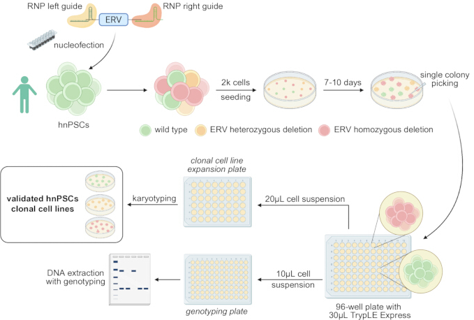
Figure 1: Overview of the ERV deletion workflow in human naive pluripotent stem cells. Endogenous retrovirus (ERV)-derived cis-regulatory elements are deleted using paired Cas9-single-guide RNA (sgRNA) ribonucleoprotein complexes delivered into human naive pluripotent stem cells (hnPSCs) by nucleofection. Following recovery and low-density plating, individual colonies are isolated, expanded, genotyped, and karyotyped to identify wild-type, heterozygous, and homozygous deletion clones. Please click here to view a larger version of this figure.
Protocol
The PB004 human naive pluripotent stem cell line was generated by reprogramming peripheral blood cells from volunteer donors who provided written informed consent, in accordance with applicable institutional and national guidelines, and with approval from the relevant institutional ethics committee (Institute of Medical Science of the University of Tokyo, approval number 21-68-0409)33. These cells were STR-authenticated and routinely tested for mycoplasma infection. The research tools used in this protocol are listed in the Table of Materials.
1. Culture human naive pluripotent stem cells
- Prepare the culture media
NOTE: Begin with human naive pluripotent stem cells (hnPSCs) maintained on inactivated mouse embryonic fibroblast (MEF) feeder layers. Passage the cells at least once after thawing before performing genome editing.
NOTE: Warm only the volume of PXGL medium required for immediate use to 37 °C. Do not repeatedly warm the complete stock.- Prepare N2B27 base medium and PXGL medium
- Combine 500 mL of high-glucose DMEM/F-12 with 500 mL of Neurobasal medium. Add L-glutamine to a final concentration of 2 mM and 2-mercaptoethanol to a final concentration of 100 µM.
CAUTION: Handle 2-mercaptoethanol according to institutional chemical-safety procedures. - Add 5 mL of N2 supplement, 10 mL of B27 supplement, and 10 mL of antibiotic-antimycotic solution, if required. Filter the medium through a 0.22 µm or 0.45 µm pore-size filter unit, if required.
- Store the resulting N2B27 base medium at 4 °C in the dark for up to 1 month.
- Estimate the volume of PXGL medium required for 1 week. Supplement the N2B27 base medium with 1 µM PD0325901, 2 µM XAV939, 2 µM Gö6983, and 10 ng/mL recombinant human leukemia inhibitory factor.
- Add 10 µM Y-27632 to the PXGL medium when thawing, passaging, nucleofecting, or plating hnPSCs as single cells. Store the PXGL medium at 4 °C in the dark for up to 1 week.
- Combine 500 mL of high-glucose DMEM/F-12 with 500 mL of Neurobasal medium. Add L-glutamine to a final concentration of 2 mM and 2-mercaptoethanol to a final concentration of 100 µM.
- Prepare the washing medium
NOTE: Use washing medium containing bovine serum albumin (BSA) during centrifugation to reduce the loss of hnPSCs through adherence to plastic consumables.- Add BSA to 500 mL of DMEM/F-12 to obtain a final concentration of 0.1%. Store the washing medium at 4 °C for up to 2 months.
- Prepare the MEF medium
- Add 50 mL of fetal bovine serum, 1 mM sodium pyruvate, and 5 mL of stabilized L-glutamine supplement to 500 mL of high-glucose Dulbecco’s modified Eagle medium.
- Add antibiotic-antimycotic solution, if required. Filter the medium through a 0.22 µm or 0.45 µm pore-size filter unit, if required, and store the MEF medium at 4 °C for up to 1 month.
- Prepare N2B27 base medium and PXGL medium
- Prepare the MEF feeder layers
NOTE: MEFs provide growth support for hnPSCs34. Use MEFs inactivated by gamma irradiation or mitomycin C treatment. Generate the MEFs in-house35 or obtain them from a commercial source. MEFs derived from CD1 or CF1 mice are suitable for this protocol. The experiments described here were performed using purchased CF1 MEFs.- Prepare the required number of MEF-coated wells 48 h before thawing or passaging the hnPSCs. Coat the tissue-culture surface with 0.05–0.1 mL/cm2 of 0.1% gelatin solution and incubate at 37 °C for 30 min.
- Aspirate the gelatin solution and plate 2–3 × 10⁴ inactivated MEFs/cm2 in MEF medium.
- Incubate the MEFs at 37 °C, 95% humidity, and 5% CO₂ for at least 48 h before use. Replace the MEF medium every 3 days when the feeder layers are not used immediately.
- Use the MEF feeder layers within 7 days of plating. Examine the feeder layers before plating hnPSCs and discard any wells showing extensive detachment or deterioration.
- Passage hnPSCs
- Aspirate the PXGL medium from the hnPSC culture. Add 0.05–0.1 mL/cm2 of cell-dissociation reagent and incubate the cells at 37 °C for 5 min.
- Pipette the colonies gently several times using a 1,000 µL pipette to generate a cell suspension. Transfer the suspension to a 15 mL conical tube containing 5–10 mL of washing medium and invert the tube three times.
- Centrifuge the cells at 250 × g for 3 min. During centrifugation, aspirate the medium from a prepared MEF feeder layer, wash the feeder layer once with phosphate-buffered saline, and aspirate the wash solution.
- Aspirate the washing medium from the hnPSC pellet without disturbing the pellet. Resuspend the cells in PXGL medium containing 10 µM Y-27632 and plate the cells at a split ratio of 1:3 or 1:4 onto the prepared MEF feeder layer.
- Passage the cells every 3–4 days and incubate them at 37 °C, 7% CO₂, and 5% O₂.
2. Identify and design the deletion of an endogenous retrovirus-derived cis-regulatory element
NOTE: The following procedure is optimized for endogenous retrovirus (ERV)-derived cis-regulatory elements (CREs), but it may be adapted for other defined CREs.
- Identify the genomic coordinates of the ERV
- Select an ERV candidate according to the biological question and the available evidence of promoter, enhancer, or chromatin-boundary activity.
- Open the UCSC Genome Browser (accessed 15 May 2026), select the appropriate species and genome assembly36, and navigate to the Repeats section.
- Set the RepeatMasker track to Full (track last updated at UCSC on 18 October 2022). Refresh the browser and locate the candidate ERV, which is displayed as an LTR retrotransposon.
NOTE: RepeatMasker identifies and classifies transposable elements using curated resources such as RepBase and Dfam37–39. - Select the candidate LTR to display its genomic coordinates. Record the ERV coordinates and the 100 bp regions immediately upstream and downstream of the element.
NOTE: For an LTR5_Hs element located at chr1:207635112–207636074 in the hg38 assembly, use chr1:207635012–207635112 to design the left guide and chr1:207636074–207636174 to design the right guide.
- Design the guide RNAs using Benchling
- Open a Benchling project (accessed 15 May 2026). Select +, navigate to CRISPR, and select CRISPR guides.
- Select Import from chromosomal coordinates, select the appropriate genome assembly, and enter the coordinates of the 100 bp region upstream of the ERV.
- Select a single guide as the design type and set the guide length to 20 bp. Select the protospacer-adjacent motif compatible with the Cas9 nuclease and use NGG when using Streptococcus pyogenes Cas9.
- Enable Include masked regions in off-target search under the advanced settings.
NOTE: Exclude guide RNAs predicted to target multicopy repetitive sequences. Cleavage at multiple genomic loci may reduce cell viability and confound interpretation. - Inspect the genomic region in the genome browser and confirm that the guide sequence does not overlap a nearby repetitive element. Select Finish.
- Navigate to the Sequence map and select the complete sequence. Select Design CRISPR, followed by +.
- Select a guide with a high predicted on-target score and a low predicted off-target score40. Design the selected upstream guide as the left guide and repeat the procedure using the 100 bp region downstream of the ERV to obtain the right guide.
- Perform an additional in silico off-target analysis using COSMID41 (accessed 15 May 2026). Exclude guides predicted to target coding genes, other CREs, or multicopy repetitive elements.
- Design the guide RNAs using CRISPOR
- Open the 100 bp region upstream of the ERV in the UCSC Genome Browser. Select View, followed by DNA sequence, and select Get DNA to obtain the upstream sequence in FASTA format.
- Open CRISPOR (version 5.2)42 and paste the FASTA-formatted upstream sequence into the Step 1 field.
- Select the appropriate genome assembly under Step 2 and select hg38 when working with the example human locus. Select the appropriate protospacer-adjacent motif under Step 3 and submit the sequence for analysis.
- Review the candidate guides ranked by specificity score and select a guide with a high specificity score and suitable predicted efficiency.
- Design the selected guide as the left guide. Repeat steps 2.3.1–2.3.4 using the 100 bp region downstream of the ERV to obtain the right guide.
- Obtain each guide as a single-guide RNA in which the CRISPR RNA is prehybridized to the trans-activating CRISPR RNA.
- Design PCR primers for genotyping
- Extend the ERV genomic interval by approximately 500 bp upstream and 500 bp downstream.
NOTE: For the example locus chr1:207635112–207636074, use the extended interval chr1:207634612–207636574. - Import the extended sequence into Primer3 (version 4.1.0)43.
- Set the minimum, optimal, and maximum primer lengths to 18, 20, and 25 nucleotides, respectively; the minimum, optimal, and maximum melting temperatures to 57 °C, 59 °C, and 62 °C, respectively; and the minimum, optimal, and maximum GC contents to 30%, 50%, and 70%, respectively.
- Set the product-size range to generate an amplicon that spans the complete ERV and both guide-RNA cleavage sites. Design one primer upstream and one primer downstream of the targeted ERV.
- Confirm that the expected wild-type and deletion products can be distinguished by agarose-gel electrophoresis. Label the primer pair as primers 1 and 2, as illustrated in Figure 2A.
- Extend the ERV genomic interval by approximately 500 bp upstream and 500 bp downstream.
3. Nucleofect the paired guide RNAs into hnPSCs
- Prepare a 12-well plate containing inactivated MEF feeder layers 48 h before nucleofection. Calculate the number of nucleofection reactions required and use one reaction for each left-guide and right-guide pair.
- For each reaction, combine 3.6 µL of nucleofection supplement with 16.4 µL of nucleofection solution and equilibrate the mixture to room temperature.
- Prepare the left-guide and right-guide Cas9 ribonucleoprotein complexes in separate tubes. For each guide, combine 2.9 µL of phosphate-buffered saline, 0.6 µL of 100 µM sgRNA, and 0.5 µL of 62 µM high-fidelity Cas9 nuclease, and incubate at room temperature for 10–20 min.
- During ribonucleoprotein incubation, aspirate the culture medium from the hnPSCs, add sufficient cell-dissociation reagent to cover the cells, and incubate at 37 °C for 5 min.
- Add 5 mL of washing medium and collect the dissociated cells. Centrifuge at 250 × g for 3 min, aspirate the supernatant, and gently resuspend the pellet in 2 mL of PXGL medium containing 10 µM Y-27632.
- Count 2–3 × 10⁵ cells for each nucleofection reaction. Centrifuge the required number of cells at 250 × g for 3 min, aspirate the supernatant completely, and remove residual liquid using a 10 µL pipette tip without disturbing the pellet.
NOTE: Do not perform MEF depletion before nucleofection. Count the dissociated mixture as hnPSCs together with residual MEFs. - Combine 2 µL of the left-guide Cas9 complex with 2 µL of the right-guide Cas9 complex and mix by pipetting five times. Add 4 µL of the combined ribonucleoprotein mixture to the 20 µL nucleofection solution.
- Resuspend the hnPSC pellet in the complete nucleofection mixture without introducing bubbles. Transfer the suspension to one well of a nucleofection microcuvette, then gently tap the cuvette to ensure the liquid covers the bottom.
NOTE: Proceed immediately after resuspending the cells. Process no more than two reactions simultaneously. - Nucleofect the cells using the P3 Primary Cell program DN-100. Immediately resuspend the nucleofected cells in prewarmed PXGL medium containing 10 µM Y-27632.
- Plate the cells into one well of the prepared 12-well MEF feeder plate and move the plate gently in a T-shaped pattern to distribute the cells evenly.
- Incubate the cells for at least 2 days before initiating clonal derivation. Maintain a separate stock of unedited hnPSCs and derive wild-type clonal lines in parallel.
4. Derive clonal hnPSC lines
- Prepare a 10 cm dish containing an inactivated MEF feeder layer 48 h before plating single cells.
- Aspirate the culture medium from the nucleofected and parallel wild-type hnPSC cultures. Add cell-dissociation reagent and incubate the cells at 37 °C for 5 min.
- Add 5 mL of washing medium and collect the dissociated cells. Centrifuge the cells at 250 × g for 3 min, aspirate the supernatant, and resuspend the cells thoroughly in 400 µL of PXGL medium containing 10 µM Y-27632.
NOTE: Generate a complete single-cell suspension. Incomplete dissociation may produce colonies containing mixed genotypes. - Count the cells and plate approximately 2,000 cells onto the prepared 10 cm MEF feeder dish in PXGL medium containing 10 µM Y-27632. Distribute the cells evenly to minimize contact between emerging colonies.
- Divide the remaining cell suspension into two portions. Cryopreserve one portion as a backup and isolate genomic DNA from the second portion for bulk-population genotyping.
- Culture the sparsely plated cells for 7–10 days. Do not replace the medium on the first day after plating; on days 2–4, replace 3 mL of medium daily with 3 mL of fresh PXGL medium, and from day 5 onward, replace half of the medium daily. Examine the culture daily and proceed to colony picking when spatially separated single-cell colonies are visible.
NOTE: Adjust the culture period according to the growth rate of the hnPSC line.
5. Perform bulk genotyping of the wild-type and edited populations
NOTE: Use a genomic DNA extraction kit to extract genomic DNA from the bulk population. Testing different DNA polymerases may be necessary to optimize these PCRs. A high-fidelity DNA polymerase master mix was used routinely for the experiments described here.
- Isolate genomic DNA from the wild-type and edited bulk-cell populations using a genomic DNA extraction kit.
- Amplify the targeted genomic region by PCR using primers 1 and 2 and a suitable DNA polymerase. Use a high-fidelity DNA polymerase master mix in a total reaction volume of 20 µL containing 10 µL of 2× master mix, 7 µL of H₂O, 0.5 µL of 10 µM primer 1, 0.5 µL of 10 µM primer 2, and 2 µL of genomic DNA at approximately 50 ng/µL. For the PCR shown in Figure 2 corresponding to an LTR5_Hs element located at coordinates chr1:207635112–207636074 (hg38), the primer sequences were CCCCTCCAAGATGACCAGTT (primer 1) and GGCCTCGCAAATCTTAACCC (primer 2). The cycling parameters were 98 °C (30 s), 30 cycles of 98 °C (10 s), 64 °C (30 s), and 72 °C (45 s), followed by a final extension at 72 °C for 10 min.
- Include genomic DNA from the wild-type population as a control.
- Separate the PCR products by electrophoresis using a 1% agarose gel stained with a safe DNA stain in TAE (Tris-acetate-EDTA) buffer. Run the gel at 80 V for approximately 45 min. Include a DNA ladder in the first lane to estimate the band sizes.
- Visualize the amplified DNA bands using a digital gel-imaging system.
- Compare the products obtained from the wild-type and edited bulk populations. In Figure 2B, primers 1 and 2 amplify a band of 1,468 bp from wild-type alleles and a band of 500 bp from edited alleles.
- Confirm that the primer pair produces distinguishable full-length and deletion products before proceeding to colony picking.
- Redesign the guide RNAs or optimize the nucleofection conditions when no deletion product is detected.
6. Pick and expand single-cell colonies
NOTE: Determine the number of colonies to pick according to the bulk-population PCR. Begin with at least 48 edited colonies when a clear deletion product is detected. Pick approximately eight colonies from the parallel wild-type population. Aim to obtain an excess of clones (for example, 4 wild-type, 5 homozygous, and 3 heterozygous clones).
- Prepare the plates and reagents
- Determine the number of colonies to be processed and prepare the corresponding number of MEF feeder-layer wells in a 48-well plate at least 48 h before colony picking. On the day of colony picking, aspirate the MEF medium, wash each well once with phosphate-buffered saline, aspirate the wash solution, and add PXGL medium containing 10 µM Y-27632.
- Add 30 µL of cell-dissociation reagent to the required number of wells in a round-bottom 96-well plate. Dilute the extracellular matrix reagent 1:4 in cold DMEM/F-12 and prepare the required number of wells in a second 96-well plate with PXGL medium containing 10 µM Y-27632. Add the diluted extracellular matrix reagent to each well to obtain a final concentration of 20 µL/mL.
NOTE: Use this feeder-free 96-well plate for genotyping to avoid amplification of mouse genomic DNA. - Dispense approximately 40 µL drops of washing medium onto the inner surface of a sterile 10 cm dish lid. Place the dissociation plate, expansion plate, genotyping plate, and dish lid inside the biological safety cabinet.
- Pick the colonies
NOTE: Perform colony picking under a microscope inside a biological safety cabinet. Process colonies in groups of eight when using an adjustable tip-spacing pipette.- Set a pipette to 20 µL and attach a sterile pipette tip. Prime the tip by dipping it into a washing-medium drop and pipetting twice.
- Identify a spatially isolated colony that appears to originate from a single cell. Avoid colonies that contact adjacent colonies or contain visibly separate subcolonies, and select colonies representing the range of sizes and morphologies present in the culture.
- Gently scratch beneath the colony with the primed pipette tip, aspirate the detached colony, and transfer it to one well of the round-bottom 96-well plate containing 30 µL of cell-dissociation reagent. Repeat until eight colonies have been transferred into one column of the plate.
- Incubate the colonies in the cell-dissociation reagent for 5–10 min at 37 °C. Set an adjustable tip-spacing pipette to the spacing for the 96-well plate and dissociate each colony by pipetting approximately 15 times.
- Examine the wells microscopically and confirm that the colonies have been dissociated. Aspirate 20 µL of each dissociated colony suspension, adjust the pipette spacing to the 48-well plate format, and dispense the suspension into the corresponding MEF-containing well of the clonal expansion plate.
- Aspirate the remaining approximately 10 µL of each colony suspension, adjust the pipette spacing to the 96-well plate format, and dispense the suspension into the corresponding well of the feeder-free genotyping plate.
- Repeat the procedure until the required number of edited and wild-type colonies has been picked. Return the expansion and genotyping plates to the incubator.
7. Genotype and preserve the clonal cell lines
NOTE: Use a rapid genomic DNA extraction reagent instead of a kit when processing numerous clones.
- Extract genomic DNA
- Incubate the genotyping plate for 24–48 h after colony picking and confirm that sufficient attached cells are visible in each well. Aspirate the medium and add 20 µL of rapid genomic DNA extraction solution to each well.
- Incubate the plate at room temperature for 5 min and transfer the complete volume from each well to the corresponding well of a PCR plate. Seal the PCR plate with foil.
- Incubate the plate at 65 °C for 6 min, followed by 98 °C for 2 min. Use 2–4 µL of the resulting lysate directly as the DNA template for PCR.
- Identify and validate the edited clones
- Amplify the targeted genomic region using primers 1 and 2 under the PCR and cycling conditions described previously. Include DNA from an unedited wild-type clone and the edited bulk population as controls.
- Separate the PCR products by agarose-gel electrophoresis using the conditions outlined in steps 5.4–5.5. Compare the PCR band sizes obtained from the clonal lines with the wild-type and edited bulk-population controls, as illustrated in Figure 2C.
- Excise the relevant PCR bands using a clean scalpel and purify the DNA from the excised gel fragments using a kit. Submit the purified PCR products for Sanger sequencing to a suitable sequencing provider, align the sequencing results with the reference sequence, and confirm the deletion junction and genotype of each clone. Discard or re-evaluate clones that produce unexpected PCR products or ambiguous sequencing results.
- Expand, cryopreserve, and quality-control the validated clones
- Identify the corresponding wells in the clonal expansion plate after confirming the genotype. Expand at least two independently derived edited clones and multiple wild-type clones derived in parallel through the same single-cell cloning procedure.
- Cryopreserve sufficient vials of each validated edited and wild-type clone before initiating downstream experiments. Retain the edited bulk population as a backup until the required clonal lines have been validated and cryopreserved.
- Perform karyotype analysis or another validated genomic-stability assessment on the selected hnPSC clones. Confirm that the selected clones retain the expected hnPSC morphology and growth characteristics before downstream use.
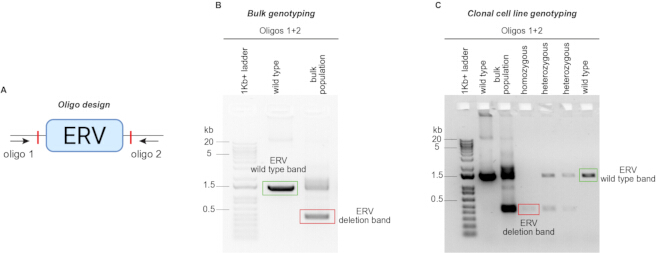
Figure 2: PCR-based assessment of ERV deletion. (A) Design of the genotyping primers positioned upstream and downstream of the targeted ERV. (B) Representative bulk-population PCR showing the full-length wild-type product and the smaller deletion product. Detection of the deletion product confirms successful editing and helps guide the number of colonies selected for clonal screening. (C) Representative PCR genotyping of individual clonal cell lines before Sanger sequencing. Wild-type clones produce the full-length product, heterozygous clones produce both full-length and deletion products, and homozygous deletion clones produce only the smaller deletion product. Please click here to view a larger version of this figure.
Results
The workflow enables the generation of clonal human naive pluripotent stem cell (hnPSC) lines carrying targeted deletions of endogenous retrovirus (ERV)-derived cis-regulatory elements (CREs). As summarized in Figure 1, paired single-guide RNAs (sgRNAs) are designed to flank the ERV of interest, assembled separately with Cas9, combined before nucleofection, and delivered into hnPSCs. Following recovery, the edited population is plated at low density to permit the formation and isolation of colonies derived from individual cells.
Bulk-population PCR provides an early indication of whether the intended deletion occurred. Primers positioned outside the two sgRNA cleavage sites amplify a larger product from the wild-type allele and a smaller product from the deletion allele (Figure 2A, B). Detection of both products in the edited bulk population indicates that the culture contains a mixture of unedited and edited cells. Failure to detect the deletion product suggests inefficient editing and indicates that guide design, ribonucleoprotein assembly, or nucleofection conditions should be reassessed before extensive colony picking. PCR genotyping of individual clones distinguishes wild-type, heterozygous, and homozygous deletion genotypes (Figure 2C). Wild-type clones produce only the full-length product, heterozygous clones produce both full-length and deletion products, and homozygous deletion clones produce only the smaller deletion product. Sanger sequencing of the PCR products confirms the expected deletion junction and the identity of the amplified allele.
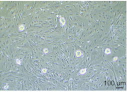
Single-cell colonies are typically visible after 7–10 days of culture and can be identified by their spatial separation and compact morphology (Figure 3). Colonies should be picked across the range of sizes and morphologies observed because deletion of a functional regulatory element may alter proliferation or differentiation behavior. Colonies arising from overlapping or incompletely separated cells should be avoided because they may contain mixed genotypes.
 0
0
Figure 3: Representative morphology of hnPSC colonies before colony picking. Bright-field microscopy image of spatially separated hnPSC colonies at the stage used for clonal isolation. Scale bar = 100 µm. Please click here to view a larger version of this figure.
Across tested ERV loci, complete deletion frequencies ranged from 10%–78.9% of isolated hnPSC clones, with mean and median homo- or hemizygous deletion frequencies of 43.6% and 45.8%, respectively (Table 1). The observed variation likely reflects locus-specific differences in guide activity, chromatin accessibility, cell-line background, and growth conditions. Parallel derivation of wild-type clones provides controls that have undergone the same single-cell cloning, expansion, and culture history as the edited lines.
| Left guide | Right guide | Forward PCR primer | Reverse PCR primer | Picked clones | Wild type | Hetero zygous deletion | Com- plete deletion | Un- desired re- arrange ments | Effective | Bands not clearly visible | % hetero zygous | % homo zygous | ||||
| TAATGACCG TGTAATTTATA | AACAGCCATG ATGATGATGG | CCCCTCCAAG ATGACCAGTT | GGCCTCGCAA ATCTTAACCC | 24 | 6 | 6 | 6 (Homo zygous) | 1 | 13 | 5 | 46.2 | 31.6 | ||||
| TTGATTTAGA AAGATCAGTG | TCTGACAGC ACTGATCTGAA | CCCCAACCC ACCATTACCTA | TTGGGAGCTG TCTTGCCTAA | 24 | 3 | 3 | 11 (Homo zygous) | 0 | 6 | 7 | 50.0 | 64.7 | ||||
| AGGCAGTCCT TGTCCCACAG | CTAGAGAAA AGCATCCACGT | CTACCTTGGA GCCCCTCTTC | TCTCCCTCCTT CTCTCTGCT | 24 | 1 | 4 | 12 (Homo zygous) | 3 | 8 | 4 | 50.0 | 60.0 | ||||
| TTACTAGTTAT TGAGTGAAG | TGTATGTGT CCTCAATTGTG | TTGGTCACT TGGCATTTGTG | TCCCCTTGTT CACCTTGACT | 48 | 1 | 2 | 15 (Homo zygous) | 1 | 4 | 29 | 50.0 | 78.9 | ||||
| CCAAGTTAGG TGGCAAGTCC | ATGGATGACT ATCAGTAATA | CCATCTGTGA GCGCTTTTGG | GGGTTTGAG TCCCTGCCATT | 24 | 15 | 0 | 2 (Homo zygous) | 3 | 18 | 4 | 0.0 | 10.0 | ||||
| TCAATGCAGT GTTAGATTCC | GGAGGGAGT CTGAGTATTCG | CTGAGTGACG CAGCTGGATA | CAAGACTGCC TCACACACAC | 48 | 26 | 13 | 6 (Homo zygous) | 1 | 40 | 2 | 32.5 | 13.0 | ||||
| TAACCAATAGA ATGTGGCAA | GAATGTGTCT CCTTGAAAGA | TGGCTCCCA ACATCCCAAT | AACGCTTGAG GGGAATGGAT | 48 | 14 | NA (Hemi zygous) | 4 (Hemi zygous) | 1 | 15 | 29 | NA | 21.1 | ||||
| CCTTTCCAATA TTTATGCCT | AAATATTAAC AAGTGCAATA | ACTTTGAGG AGAATGGGCATC | GGTGTTTATT CCAGGGACGC | 24 | 4 | NA (Hemi zygous) | 9 (Hemi zygous) | 0 | 4 | 11 | NA | 69.2 | ||||
Table 1: Genetic deletion efficiencies of ERVs. Data are shown for ERVs belonging to the LTR5_Hs family. Clonal cell lines were generated to test whether LTR5_Hs elements regulate the expression of the indicated genes. The table includes the genomic coordinates of each ERV, guide RNAs used for deletion, PCR primers used for genotyping, and clone-genotyping outcomes. “Effective” refers to clones with interpretable genotyping results. Undesired rearrangements refer to products that did not correspond to the expected wild-type or deletion product sizes. “Bands not clearly visible” refers to samples for which no interpretable PCR product was obtained. Deletion percentages were calculated using clones with interpretable genotyping results as the denominator. Heterozygous deletions refer to clones in which the ERV was deleted in one of the two alleles. Complete deletions refer to clones when the ERV has been deleted in the two copies of the chromosome (homozygous clones) or in the only chromosome copy in the case of sex chromosomes (hemizygous clones).
Validated wild-type, heterozygous, and homozygous or hemizygous deletion clones can subsequently be expanded, cryopreserved, and assessed for genomic stability. These lines provide a platform for investigating the effects of individual ERV-derived CREs on gene expression, cell proliferation, lineage differentiation, and stem cell-based models of early human development.
Discussion
This protocol describes a CRISPR-Cas9 ribonucleoprotein-based approach for deleting ERV-derived CREs in hnPSCs. The method combines paired sgRNA design, transient delivery of preassembled Cas9-sgRNA complexes, clonal derivation, PCR-based screening, Sanger sequencing, and genomic-stability assessment. Because hnPSCs are sensitive to dissociation and culture perturbation, several steps are critical for success, including maintaining healthy feeder-supported cultures, using a complete single-cell suspension, minimizing the time between cell resuspension and nucleofection, and supplementing the medium with Y-27632 during stressful manipulations. The method also requires careful guide selection because ERVs are repetitive sequences and poorly selected guides may cleave multiple genomic loci. Off-target assessment should therefore include repetitive regions and should exclude guides with predicted activity at coding genes, other CREs, or multicopy transposable elements.
Bulk-population genotyping is an important decision point in the workflow. Detection of the deletion product before colony picking confirms that cleavage and deletion have occurred and helps determine how many colonies should be screened. When no deletion product is detected, the most effective corrective action is usually to redesign one or both sgRNAs rather than proceeding directly to large-scale colony isolation. Low recovery after nucleofection may result from poor starting-cell quality, incomplete removal of residual medium from the cell pellet, delayed processing after resuspension, or excessive manipulation of the cells. Incomplete single-cell dissociation can generate colonies containing mixed genotypes, whereas dense plating can cause adjacent colonies to merge. These problems can be reduced by thorough but gentle dissociation, sparse plating, and selection of well-isolated colonies.
The efficiency of complete ERV deletion ranged from 10% – 78.9% across the tested loci, with an average of 43.6%. This range indicates that editing efficiency is strongly locus-dependent and may also vary among hnPSC lines and culture conditions. Cell-cycle activity can influence CRISPR-Cas9 editing efficiency, and differences in proliferation rate may therefore affect clone recovery30. In addition, deletion of an ERV with an essential regulatory role may reduce proliferation, alter colony morphology, or prevent recovery of homozygous clones. Some edited populations may also acquire compensatory genomic alterations that permit continued growth. For these reasons, multiple independently derived clones should be analyzed, and unexpected phenotypes should be confirmed after rederiving or thawing additional clones. Genotype confirmation, karyotype analysis, and comparison with parallel wild-type clones are essential before downstream interpretation.
A principal limitation of this method is its low throughput. Each locus requires individual guide design, nucleofection, colony isolation, genotyping, sequence confirmation, and expansion. Consequently, the method is best suited to focused functional testing of selected ERVs rather than genome-wide screening. Multiplex or family-wide manipulation of ERVs using dCas9-based epigenetic regulators can perturb many related elements simultaneously14, but this approach may produce broader off-target effects and cannot readily identify which individual insertion is responsible for a phenotype. In contrast, the present strategy enables locus-specific deletion and allows molecular or developmental effects to be attributed directly to a defined ERV-derived CRE.
The method is particularly relevant because ERVs have contributed extensively to the evolution of mammalian regulatory networks and can function as promoters, enhancers, and other regulatory elements during early development5,6,13,14,44,45,46,47. Some individual ERVs have already been shown to perform essential developmental functions12,13,14. The edited hnPSC lines generated through this protocol can be used for gene expression analysis, proliferation assays, two-dimensional lineage differentiation, and the generation of human blastoids or other stem cell-based embryo models14,23. These applications should be conducted within the applicable ethical and regulatory frameworks for human stem cell-based embryo model research48,49,50.
Future advances in genome editing, automated clone handling, and high-throughput genotyping may permit more systematic evaluation of individual ERV insertions51. Until such approaches become routinely available, the present method offers a reproducible framework for testing selected non-coding regulatory elements at single-locus resolution. Its main advantage is the ability to link a defined genomic deletion to molecular and developmental phenotypes in a human preimplantation model, thereby helping clarify how species-specific ERV-derived sequences have shaped gene-regulatory programs during human embryogenesis.
Disclosures
No conflicts of interest are declared.
Acknowledgements
Dr. Jaaved Mohammed is acknowledged for helpful discussions on genome editing. The Max Planck Foundation is acknowledged for supporting this study and the Fueyo lab. Merel Wentink received an Erasmus+ scholarship (ID 50377).
Materials
| Name | Company | Catalog Number | Comments |
|---|---|---|---|
| 2-Mercaptoethanol | Gibco | 21985023 | Added to N2B27 base medium. Handle according to the institutional chemical-safety procedures. |
| 4D Nucleofector X-Unit | Lonza | AAF-1003X | Device used for nucleofection. |
| Agarose Standard, 1 kg | Roth | 3810.4 | Used to prepare DNA electrophoresis gels. |
| Alt-R S. pyogenes Cas9 HiFi V3 Nuclease | Integrated DNA Technologies | 1081060 | High-fidelity Cas9 nuclease used to assemble ribonucleoprotein complexes. |
| Alt-R synthetic single-guide RNA | Integrated DNA Technologies | NA | Custom sgRNAs designed to flank the selected ERV. The guide sequences can be found in Table 1. |
| Antibiotic-Antimycotic | Gibco | 15240062 | Optional antimicrobial supplement for cell-culture media. |
| Axiovert 40 CFL Microscope | Zeiss | 491202-0002-001 | Used for routine MEF and hnPSCs observation during culture. |
| B-27 Supplement, 50× | Gibco | 17504044 | Supplement added to N2B27 base medium. |
| Bovine Albumin Fraction V, 7.5% solution | Gibco | 15260037 | Added to washing medium to reduce loss of hnPSCs through adherence to plastic consumables. |
| C1000 Touch Thermal Cycler with 96-Well Fast Reaction | Bio-Rad | 1851196 | Device used for PCR amplification. |
| CF1 Mouse Embryonic Fibroblasts, irradiated | Thermo | A234181 | Inactivated MEF feeder cells used to support hnPSC culture. An alternative validated prepared in-house could be used. |
| Countess 3 Automated Cell Counter | Thermo / Invitrogen | AMQAX2000 | Used for counting MEFs or hnPSCs. |
| DMEM/F-12 | Gibco | 11330032 | Component of N2B27 medium and washing medium; also used to dilute the extracellular matrix reagent. |
| Dulbecco’s phosphate-buffered saline without calcium and magnesium | Gibco | 14190144 | Used for washing feeder layers. |
| ESGRO Complete Gelatin Solution | Sigma-Aldrich | SF008 | Used to coat culture vessels before plating MEF feeder cells. |
| FBS Supreme | PAN-Biotech | P30-3031 | Used to prepare MEF culture medium. |
| Filter unit, 1,000 mL, 0.22 µm pore size | Corning | 431098 | Used for sterile filtration of large-volume culture media. |
| Filter unit, 500 mL, 0.22 µm pore size | Corning | 431097 | Used for sterile filtration of culture media. |
| FreSR-S cryopreservation medium | STEMCELL Technologies | 5859 | Used for cryopreserving hnPSCs. |
| GelDoc gel imaging system | Bio-Rad | 170-8195 | Equipment used to visualize DNA bands from electrophoresis gels. |
| Geltrex Flex LDEV-Free hESC-Qualified Reduced Growth Factor Basement Membrane Matrix | Thermo | A4000046803 | Extracellular matrix used for culturing hnPSCs in the absence of MEFs. |
| Generuler 1 Kb Plus DNA Ladder | Fischer Scientific | 11581625 | Used to measure DNA band sizes in electrophoresis. |
| GlutaMAX Supplement | Gibco | 35050061 | Stabilized L-glutamine supplement used in MEF culture medium. |
| Gö6983 | Bio-Techne | 2285/10 | Atypical protein kinase C inhibitor used to prepare PXGL medium. |
| High-glucose Dulbecco’s modified Eagle medium | Gibco | 11965092 | Basal medium used to prepare MEF culture medium. |
| Karyotyping | Life&Brain | NA | Used to detect abnormalities in chromosomes. |
| L-glutamine | Gibco | 25030081 | Added to N2B27 base medium. |
| Monarch Spin gDNA Extraction Kit | New England Biolabs | T3010 | Kit for genomic DNA extraction from edited bulk populations. |
| Move It 8-channel adjustable-spacing pipette, 30–300 µL | Eppendorf | 3125000176 | Used to transfer dissociated colonies between the 96-well genotyping plate and 48-well expansion plate. |
| N-2 Supplement, 100× | Gibco | 17502048 | Supplement added to N2B27 base medium. |
| Neurobasal Medium | Gibco | 21103049 | Component of the N2B27 base medium. |
| Nikon Zoom Stereo Microscope | Nikon | SMZ1270 | Used for hnPSC colony picking. |
| NucleoSpin Gel and PCR Clean-up | Machery-Nagel | 740609.25 | For DNA extraction from gels prior to Sanger sequencing. |
| P3 Primary Cell 4D-Nucleofector X Kit S | Lonza | V4XP-3032 | Nucleofection reagent kit used for delivery of Cas9-sgRNA ribonucleoprotein complexes. Includes cuvettes. |
| PB004 cell line | Gift from Prof. Hiromitsu Nakauchi | NA | STR authenticated, Mycoplasma tested. Published in PMID: 26023098.33 |
| PCR primers | Sigma-Aldrich | NA | Used for genotyping hnPSCs clones by PCR. Sequences in Table 1. |
| PD0325901 | Selleck Chemicals | S1036 | MEK inhibitor used to prepare PXGL medium. |
| Phusion Green Hot Start II High-Fidelity PCR Master Mix | Thermo | F566L | Master mix containing DNA polymerase for PCR genotyping. |
| QuickExtract DNA Extraction Solution | Biosearch Technologies | QE09050 | Used for rapid genomic DNA extraction from clonal-cell genotyping plates. |
| Recombinant human leukemia inhibitory factor | Gibco | 300-05-50UG | Added to PXGL medium. An equivalent validated in-house preparation may be used. |
| Sanger sequencing | Eurofins | NA | Used to sequence DNA to verify genotypes. |
| Sodium pyruvate, 100 mM | Gibco | 11360070 | Added to MEF culture medium. |
| SYBR Safe DNA Gel Stain | Thermo | S33102 | Stain for DNA electrophoresis gels. |
| TrypLE Express Enzyme, 1×, phenol red | Gibco | 12605036 | Used to dissociate hnPSCs during passaging, nucleofection preparation, and clonal derivation. |
| Water, Nuclease-free, Molecular Biology Grade, Ultrapure | Thermo | J71786.AP | Used for the genotyping PCR. |
| XAV939 | Cell Guidance Systems | SM38-50 | Wnt-pathway inhibitor used to prepare PXGL medium. |
| Y-27632 | STEMCELL Technologies | 72304 | ROCK inhibitor added during thawing, passaging, nucleofection recovery, and single-cell plating. |
References
- Wittkopp PJ, Kalay G. Cis-regulatory elements: molecular mechanisms and evolutionary processes underlying divergence. Nat Rev Genet. 2011;13(1):59–69.
- Jacques PÉ, Jeyakani J, Bourque G. The majority of primate-specific regulatory sequences are derived from transposable elements. PLoS Genet. 2013;9(5):e1003504. doi:10.1371/journal.pgen.1003504.
- Trizzino M, et al. Transposable elements are the primary source of novelty in primate gene regulation. Genome Res. 2017;27(10):1623–33.
- Grandi N, Tramontano E. Type W Human Endogenous Retrovirus (HERV-W) Integrations and Their Mobilization by L1 Machinery: Contribution to the Human Transcriptome and Impact on the Host Physiopathology. Viruses. 2017;9(7):162. doi:10.3390/v9070162.
- Senft AD, Macfarlan TS. Transposable elements shape the evolution of mammalian development. Nat Rev Genet. 2021;22(11):691–711.
- Fueyo R, Judd J, Feschotte C, Wysocka J. Roles of transposable elements in the regulation of mammalian transcription. Nat Rev Mol Cell Biol. 2022;23(7):481–97.
- Oomen ME, et al. An atlas of transcription initiation reveals regulatory principles of gene and transposable element expression in early mammalian development. Cell. 2025;188(4):1156–1174.e20.
- Göke J, et al. Dynamic transcription of distinct classes of endogenous retroviral elements marks specific populations of early human embryonic cells. Cell Stem Cell. 2015;16(2):135–41.
- Grow EJ, et al. Intrinsic retroviral reactivation in human preimplantation embryos and pluripotent cells. Nature. 2015;522(7555):221–5.
- Xiang Y, Liang H. The Regulation and Functions of Endogenous Retrovirus in Embryo Development and Stem Cell Differentiation. Stem Cells Int. 2021;2021:6660936. doi:10.1155/2021/6660936.
- Macfarlan TS, et al. Embryonic stem cell potency fluctuates with endogenous retrovirus activity. Nature. 2012;487(7405):57–63.
- Modzelewski AJ, et al. A mouse-specific retrotransposon drives a conserved Cdk2ap1 isoform essential for development. Cell. 2021;184(22):5541–5558.e22.
- Frost JM, et al. Regulation of human trophoblast gene expression by endogenous retroviruses. Nat Struct Mol Biol. 2023;30(4):527–38.
- Fueyo R, et al. A human-specific regulatory mechanism revealed in a pre-implantation model. Nature. 2025;647(8088):238–47.
- Fuentes DR, Swigut T, Wysocka J. Systematic perturbation of retroviral LTRs reveals widespread long-range effects on human gene regulation. eLife. 2018;7:e35989. doi:10.7554/eLife.35989.
- Pontis J, et al. Hominoid-Specific Transposable Elements and KZFPs Facilitate Human Embryonic Genome Activation and Control Transcription in Naive Human ESCs. Cell Stem Cell. 2019;24(5):724–735.e5.
- Xiang X, et al. Human reproduction is regulated by retrotransposons derived from ancient Hominidae-specific viral infections. Nat Commun. 2022;13(1):463. doi:10.1038/s41467-022-28105-1.
- Theunissen TW, et al. Molecular Criteria for Defining the Naive Human Pluripotent State. Cell Stem Cell. 2016;19(4):502–15.
- Bredenkamp N, et al. Wnt Inhibition Facilitates RNA-Mediated Reprogramming of Human Somatic Cells to Naive Pluripotency. Stem Cell Rep. 2019;13(6):1083–98.
- Guo G, et al. Human naive epiblast cells possess unrestricted lineage potential. Cell Stem Cell. 2021;28(6):1040–1056.e6.
- Io S, et al. Capturing human trophoblast development with naive pluripotent stem cells in vitro. Cell Stem Cell. 2021;28(6):1023–1039.e13.
- Okubo T, et al. Hypoblast from human pluripotent stem cells regulates epiblast development. Nature. 2024;626(7998):357–66.
- Shahbazi MN, Pasque V. Early human development and stem cell-based human embryo models. Cell Stem Cell. 2024;31(10):1398–418.
- Kagawa H, et al. Human blastoids model blastocyst development and implantation. Nature. 2022;601(7894):600–5.
- Yu L, et al. Large-scale production of human blastoids amenable to modeling blastocyst development and maternal-fetal cross talk. Cell Stem Cell. 2023;30(9):1246–1261.e9.
- Liu X, et al. Modelling human blastocysts by reprogramming fibroblasts into iBlastoids. Nature. 2021;591(7851):627–32.
- Yu L, et al. Blastocyst-like structures generated from human pluripotent stem cells. Nature. 2021;591(7851):620–6.
- Sozen B, et al. Reconstructing aspects of human embryogenesis with pluripotent stem cells. Nat Commun. 2021;12(1):5550. doi:10.1038/s41467-021-25853-4.
- Yanagida A, et al. Naive stem cell blastocyst model captures human embryo lineage segregation. Cell Stem Cell. 2021;28(6):1016–1022.e4.
- Pacesa M, Pelea O, Jinek M. Past, present, and future of CRISPR genome editing technologies. Cell. 2024;187(5):1076–100.
- Hendel A, et al. Chemically modified guide RNAs enhance CRISPR-Cas genome editing in human primary cells. Nat Biotechnol. 2015;33(9):985–9.
- Kim S, et al. Highly efficient RNA-guided genome editing in human cells via .delivery of purified Cas9 ribonucleoproteins. Genome Res. 2014;24(6):1012–9.
- Masaki H, et al. Interspecific in vitro. assay for the chimera-forming ability of human pluripotent stem cells. Development. 2015;142(18):3222–30.
- Thomson JA, et al. Embryonic stem cell lines derived from human blastocysts. Science. 1998;282(5391):1145–7.
- Conner DA. Mouse Embryo Fibroblast (MEF) Feeder Cell Preparation. Curr Protoc Mol Biol. 2000;51(1):23.2.1–23.2.7.
- Casper J, et al. The UCSC Genome Browser database: 2026 update. Nucleic Acids Res. 2026;54(D1):D1331–D1335.
- Bao W, Kojima KK, Kohany O. Repbase Update, a database of repetitive elements in eukaryotic genomes. Mob DNA. 2015;6:11. doi:10.1186/s13100-015-0041-9.
- Storer J, et al. The Dfam community resource of transposable element families, sequence models, and genome annotations. Mob DNA. 2021;12(1):2. doi:10.1186/s13100-020-00230-y.
- Tarailo-Graovac M, Chen N. Using RepeatMasker to identify repetitive elements in genomic sequences. Curr Protoc Bioinformatics. 2009;25:4.10.1–4.10.14.
- Doench JG, et al. Optimized sgRNA design to maximize activity and minimize off-target effects of CRISPR-Cas9. Nat Biotechnol. 2016;34(2):184–91.
- Cradick TJ, et al. COSMID: A Web-based Tool for Identifying and Validating CRISPR/Cas Off-target Sites. Mol Ther Nucleic Acids. 2014;3(12):e214. doi:10.1038/mtna.2014.64.
- Concordet JP, Haeussler M. CRISPOR: intuitive guide selection for CRISPR/Cas9 genome editing experiments and screens. Nucleic Acids Res. 2018;46(W1):W242–W245.
- Untergasser A, et al. Primer3—new capabilities and interfaces. Nucleic Acids Res. 2012;40(15):e115. doi:10.1093/nar/gks596.
- Dopkins N, Nixon DF. Activation of human endogenous retroviruses and its physiological consequences. Nat Rev Mol Cell Biol. 2024;25(3):212–22.
- Li P, Yamanaka S. Transposable element regulation in mammalian germ cells and other contexts: multilayered repression, fertility, and regulated activation. Epigenomics. 2026;0(0):1–18.
- Yu H, et al. Dynamic reprogramming of H3K9me3 at hominoid-specific retrotransposons during human preimplantation development. Cell Stem Cell. 2022;29(7):1031–1050.e12.
- Xiang Y, et al. Endogenous retroviruses synthesize heterologous chimeric RNAs to reinforce human early embryo development. Science. 2026;391(6783):eadv5257. doi:10.1126/science.adv5257.
- Arias AM, et al. Human stem cell-based embryo models: innovation, ethics, and policy. Hum Reprod. 2026;41(6):830–46.
- Bentzen HB, et al. A guide to using embedded ethics in human stem-cell-based embryo model research. Nat Cell Biol. 2026;28(4):665–73.
- Clark AT, et al. Stem cell-based embryo models: The 2021 ISSCR stem cell guidelines revisited. Stem Cell Rep. 2025;20(6):102514. doi:10.1016/j.stemcr.2025.102514.
- Liu D, Cao D, Han R. Recent advances in therapeutic gene-editing technologies. Mol Ther. 2025;33(6):2619–44.