This review examines the evolution of hepatorenal syndrome definitions, mechanisms, diagnosis, treatment, and transplantation. It distinguishes established evidence from unresolved controversies, evaluates terlipressin, biomarkers, and albumin strategies, and integrates liver transplantation, simultaneous liver-kidney transplantation, and kidney-after-liver transplantation within a precision-management framework.
Review Article
Hepatorenal Syndrome: Paradigm Shifts in Pathophysiology, the Transplantation Paradox, and Future Precision Management
In This Article
Summary
Abstract
Hepatorenal syndrome (HRS) is a severe kidney-dysfunction phenotype in patients with advanced cirrhosis and ascites. This review examines the historical and clinical evolution from type 1/type 2 HRS to explicit HRS-AKI, HRS-AKD, and HRS-CKD definitions. Contemporary criteria use dynamic serum creatinine and urine-output changes, assess response after adequate volume resuscitation when indicated, and recognize that structural kidney injury may coexist. The pathophysiological framework integrates portal hypertension, splanchnic vasodilation, systemic inflammation, bacterial translocation, and cardiac dysfunction. Terlipressin plus individualized albumin improves HRS reversal, but biochemical response does not consistently confer an overall survival benefit. Creatinine reduction may lower MELD-based priority; however, available studies do not demonstrate a uniform reduction in access to liver transplantation. Cystatin C and neutrophil gelatinase-associated lipocalin provide adjunctive phenotypic and prognostic information but lack validated treatment thresholds. Liver transplantation remains definitive treatment for eligible patients. Simultaneous liver-kidney transplantation requires criteria-based assessment, whereas kidney-after-liver transplantation provides a safety-net option for persistent severe kidney dysfunction after liver transplantation alone. CLIF-C ACLF and emerging models such as ReMELD-Na may complement risk assessment but do not independently replace governing allocation systems. Precision management requires serial reassessment, early transplant-center coordination, and multidisciplinary integration of hemodynamics, kidney-injury phenotype, treatment safety, and recovery potential.
Introduction
Hepatorenal syndrome (HRS) is a life-threatening kidney-dysfunction phenotype that develops in patients with advanced cirrhosis and ascites; the HRS designation is not applied to acute kidney injury (AKI) in compensated advanced chronic liver disease or acute liver failure1. Current HRS-AKI criteria include an increase in serum creatinine (sCr) of at least 0.3 mg/dL within 48 h or at least 50% from baseline within 7 days and/or urine output of 0.5 mL/kg/h or less for at least 6 h, with no improvement within 24 h after adequate volume resuscitation when clinically indicated and no stronger alternative primary cause1. Rather than a primary renal disease, HRS results from profound circulatory dysfunction and systemic inflammation triggered by portal hypertension and liver insufficiency2. Historically, HRS was defined as a predominantly functional disorder; contemporary criteria acknowledge that HRS-AKI may coexist with tubular injury, proteinuria, or chronic kidney disease. This evidence supports overlapping phenotypes, rather than proving that functional HRS inevitably progresses to structural renal damage1,3,4. It represents one of the most severe complications in end-stage liver disease, signifying a critical prognosis and posing a major therapeutic challenge with historically high mortality rates5. Its management demands a deep understanding of the complex interplay between hepatic dysfunction, splanchnic and systemic hemodynamics, and renal perfusion.
The conceptualization of HRS has evolved over nearly two centuries, reflecting advancements in understanding hepatorenal pathophysiology. Early clinical observations in the 19th century noted concomitant renal failure in liver disease. The 20th century saw the term formalized and the pivotal 1996 International Ascites Club (ICA) diagnostic criteria established, introducing the classification into rapid-onset Type 1 and chronic Type 2 HRS6, primarily based on the “peripheral arterial vasodilation hypothesis”. The 21st century has brought a paradigm shift, integrating this hemodynamic model with the critical roles of systemic inflammation, bacterial translocation, and cardiac dysfunction in disease progression7. Modern diagnostic frameworks, now aligned with the kidney disease: Improving Global Outcomes (KDIGO) criteria, have moved beyond static thresholds1. The contemporary classification uses explicit criteria for HRS-AKI, HRS-acute kidney disease (AKD), and HRS-chronic kidney disease (CKD), shifting practice from a predominantly exclusionary diagnosis toward an inclusionary, criteria-based assessment while retaining evaluation for competing or coexisting causes1. Despite these advances, practical challenges in real-world management persist, often exacerbated by divergent comparative perspectives across international guidelines. The clinically important milestones are the first consensus discussions in Sassari (1978), the International Ascites Club definitions of 1994–1996, adoption of dynamic AKI criteria in 2012–2015, and the 2024 ADQI-ICA consensus; each revision sought earlier recognition without sacrificing diagnostic specificity1,6,8,9.
Contemporary management of HRS exemplifies a shift toward precision medicine in hepatology. Diagnosis relies on dynamic changes in serum creatinine, careful exclusion of alternative causes, and the growing use of novel biomarkers and artificial intelligence (AI)-assisted diagnostic models to provide earlier clinical alerts and to differentiate functional HRS from structural tubular injury, although determining the optimal diagnostic cut-off values for these markers remains a significant real-world challenge1,10. Pharmacological therapy has been revolutionized by vasoconstrictors, primarily terlipressin combined with albumin, which targets the underlying hemodynamic disturbance and has been shown to improve HRS reversal rates11; however, emerging clinical evidence indicates that achieving renal reversal with terlipressin does not consistently translate into definitive overall survival benefits12,13. Because serum creatinine contributes to allocation under the Model for End-Stage Liver Disease (MELD), biochemical response can lower the calculated score; however, the clinical effect is not uniform. A cohort study associated response with a longer time to liver transplantation, whereas a selected post-hoc CONFIRM analysis found no difference in access to liver transplantation at 30, 60, or 90 days. The issue should therefore be regarded as policy-dependent and unresolved, not an inevitable treatment penalty14,15. Cystatin C and neutrophil gelatinase-associated lipocalin (NGAL) can add prognostic and phenotypic information, but no validated threshold currently identifies HRS-AKI or directs vasoconstrictor treatment16,17.
Liver transplantation (LT) is the definitive therapy for eligible patients, whereas simultaneous liver-kidney transplantation (SLKT) is reserved for established medical eligibility and kidney-after-liver transplantation (KALT) offers a safety-net strategy when severe kidney dysfunction persists after LT alone5,18,19,20,21. The Chronic Liver Failure Consortium Acute-on-Chronic Liver Failure (CLIF-C ACLF) score estimates short-term mortality in ACLF and may complement, but does not replace, allocation systems such as MELD22. This review summarizes the historical context, the refined diagnostic criteria, current therapeutic strategies23, and future directions focused on modulating inflammation, the gut-liver-kidney axis, and phenotype-based personalized medicine, with the ultimate goal of improving outcomes for this high-risk population. Accordingly, this review adds a policy-aware comparison of LT alone, SLKT, and KALT and identifies which statements are consensus-supported, observational, controversial, or author perspective.
Review and Perspective
This review is organized around three linked clinical questions: how HRS definitions and mechanisms evolved; how HRS phenotypes should be prevented, identified, and treated; and how kidney dysfunction should inform LT, SLKT, and KALT planning. The historical section explains why practice moved from fixed creatinine thresholds to dynamic, time-based phenotypes; the management section evaluates albumin, vasoconstrictors, biomarkers, and safety; and the final section addresses allocation, recovery prediction, and real-world multidisciplinary decision-making.
1. Historical evolution of HRS
- Evolution of pathophysiological understanding
The pathophysiological understanding of HRS has mirrored its clinical classification in complexity. Early 20th-century observations suggested that renal failure in cirrhosis was often functional: kidney histology could be relatively preserved, and kidneys recovered function when removed from the cirrhotic circulatory environment, including after transplantation into recipients with normal liver function8,24. These findings supported an important hemodynamic component. The mid-20th-century debate centered on whether "liver death" or retained toxins caused renal injury25. Hecker and Sherlock (1956) then proposed the seminal hemodynamic hypothesis of arterial vasodilation and reduced effective circulation, anticipating volume expansion and vasoconstrictor therapy26.
The “peripheral arterial vasodilation hypothesis,” formally articulated by Schrier et al. in 1988, became the cornerstone model27. It provides a coherent sequence: portal hypertension leads to splanchnic vasodilation (mediated by nitric oxide and other vasodilators), reducing effective arterial blood volume. This underfilling activates potent vasoconstrictor systems (the sympathetic nervous system, renin-angiotensin-aldosterone system, and antidiuretic hormone), causing intense renal vasoconstriction and a marked reduction in glomerular filtration rate (GFR)6,28,29. Research in the 2000s further refined this model, highlighting specific triggers such as bacterial infection (especially spontaneous bacterial peritonitis) and elucidating the role of concomitant cardiac dysfunction (“cirrhotic cardiomyopathy”) in exacerbating renal hypoperfusion30,31.
Post-2010, the model dramatically expanded to incorporate systemic inflammation and immune activation as central drivers. Bacterial translocation from the gut and the release of pathogen/damage-associated molecular patterns (PAMPs/DAMPs) trigger a robust inflammatory cascade that not only exacerbates endothelial dysfunction and worsens renal vasoconstriction but can also induce direct tubular injury and cell death32,33. Oxidative stress within this inflammatory milieu further impairs cardiac output and renal microcirculation32. Furthermore, with the increasing incidence of complex underlying diseases—such as compromised immunity—in real-world clinical scenarios, renal parenchymal damage frequently overlaps with functional impairment. Rather than treating functional and structural findings as mutually exclusive, the modern multifactorial model recognizes that HRS-AKI may coexist with tubular injury, proteinuria, or chronic kidney disease5,34,35. Supporting this, evidence of structural damage, such as bile cast nephropathy and elevated tubular-injury biomarkers such as NGAL, supports careful evaluation for overlapping structural injury without establishing an inevitable transition3,36. - Evolution of nomenclature
The conceptualization of HRS has evolved over nearly two centuries. In the 19th century, early observations described renal dysfunction in patients with liver disease, noting minimal structural changes at autopsy and using descriptive terms like “bile nephrosis”8,37,38. In the early 20th century, terms attempting to classify the condition based on clinical course emerged, and "hepatorenal syndrome" became a generic, sometimes ambiguously applied term for liver-related kidney dysfunction39,40,41,42. For clinical interpretation, these early observations provide context, but the decision-relevant milestones begin with consensus definition efforts in Sassari in 1978 and the International Ascites Club criteria of 1994–19966,8.
A pivotal shift toward standardization occurred in 1996 when the International Ascites Club (ICA) introduced the seminal classification into rapidly progressive Type 1 (HRS-1) and slowly progressive Type 2 (HRS-2)6. However, alignment with international nephrology practice brought a further shift in the 21st century. The 2015 ICA consensus explicitly aligned with the kidney disease: Improving Global Outcomes (KDIGO) criteria. The primary motivation for renaming HRS-1 to HRS-AKI was to facilitate the early management of AKI by focusing on flexible, dynamic changes in serum creatinine rather than waiting for patients to reach late-stage, static thresholds9,43. Following this, the nomenclature expanded to include HRS-Acute Kidney Disease (AKD) and HRS-Chronic Kidney Disease (CKD), providing time- and duration-based phenotypes rather than a vague continuum1,7. Importantly, the modern designation of non-AKI HRS (HRS-NAKI) is not a complete counterpart to the traditional HRS-2 due to ongoing disputes among various organizational guidelines. Furthermore, the lack of dedicated International Classification of Diseases (ICD) codes for HRS-NAKI has hampered sufficient epidemiological statistics, as most previous studies have historically concentrated almost exclusively on HRS-1/HRS-AKI. The practical rationale was to avoid waiting for a late fixed sCr threshold, permit earlier vasoconstrictor consideration, and describe persistent dysfunction beyond AKI using HRS-AKD and HRS-CKD criteria1,9. - Evolution of diagnostic criteria
Diagnostic criteria for HRS have evolved through three key phases, moving from exclusive static thresholds to inclusive dynamic assessment, mirroring the growing understanding of its pathophysiology (Figure 1). The revisions improved practice by replacing a late fixed threshold with dynamic change, clarifying when volume assessment is needed, and explicitly allowing coexisting kidney injury rather than requiring an entirely exclusionary diagnosis1,9.
Foundational phase (1978–2007): The 1996 ICA criteria were a landmark for their specificity, relying on a fixed serum creatinine (sCr) threshold (>1.5 mg/dL) and stringent exclusion of other causes of renal failure. While specific, this approach risked underdiagnosis in patients with low baseline muscle mass, such as those with advanced cirrhosis and sarcopenia6.
Dynamic transformation (2012–2015): The adoption of KDIGO AKI criteria revolutionized diagnosis by focusing on acute change43. The 2015 ICA algorithm used diuretic withdrawal plus albumin 1 g/kg/day (maximum 100 g/day) for 2 days9. The 2024 ADQI-ICA consensus no longer requires systematic 48 h albumin administration. When volume status is equivocal, it permits a fluid challenge of 250–500 mL crystalloid or 1–1.5 g/kg of 20%–25% albumin, followed by reassessment within 24 h; albumin is avoided or stopped when volume overload is present1.
Phenotype refinement (2019–2024) and current challenges: The recent classifications use explicit timing and duration to define HRS-AKI, HRS-AKD, and HRS-CKD, expanding the recognized phenotypes without replacing them with an imprecise continuum1. While this framework is conceptually robust, it introduces significant long-term diagnostic and management challenges. Expanding the focus beyond HRS-AKI requires careful evaluation of long-term outcomes for HRS-AKD and HRS-CKD, particularly concerning their implications for combined liver-kidney transplant candidacy, as prolonged, unresolved renal dysfunction heavily influences post-transplant survival. This represents a shift from a predominantly exclusionary construct toward an inclusionary criteria-based diagnosis that can coexist with other kidney-injury signals1.
A major advance in this era is the study of urinary and blood biomarkers, including NGAL, interleukin-18 (IL-18), liver-type fatty acid-binding protein (L-FABP), and cystatin C. These markers may provide earlier clinical alerts than delayed sCr variation and offer prognostic information44,45,46. Cystatin C is particularly useful as an adjunct when creatinine is biased downward by sarcopenia or low muscle mass, although inflammation and other non-GFR determinants still affect interpretation16,17. Distinguishing HRS-AKI from acute tubular injury remains difficult because phenotypes overlap. Prospective studies support adjunctive phenotyping and prognosis, but no universally validated biomarker cut-off independently diagnoses HRS-AKI or directs vasoconstrictor treatment4,16,17.
2. Prevention and management of HRS
HRS-AKI, HRS-AKD, and HRS-CKD are explicit time- and duration-based phenotypes. HRS-AKI meets dynamic AKI criteria within 7 days. HRS-AKD describes kidney dysfunction persisting from 7 through 90 days, including persistent AKI or non-AKI dysfunction with an eGFR below 60 mL/min/1.73 m2, an eGFR decrease of at least 35%, or an sCr increase greater than 50% when no stronger alternative primary cause is present. HRS-CKD denotes dysfunction persisting beyond 90 days, conventionally including eGFR below 60 mL/min/1.73 m21,7. Observational studies indicate that AKI is common in hospitalized patients with cirrhosis and that a substantial proportion progress to AKD or CKD47,48.
- Prevention and risk stratification
Prevention strategies are multifaceted and target high-risk patients and specific precipitating events. Table 1 summarizes recognized clinical settings, precipitating events, recommended actions, and the evidentiary limit of each, and distinguishes prevention in cirrhosis with ascites from evaluation and treatment after AKI has developed1,10,49. Recent evidence has challenged several traditional prophylactic approaches. For instance, while intravenous albumin is essential for preventing HRS following spontaneous bacterial peritonitis (SBP), its routine use in non-SBP bacterial infections lacks clear clinical benefit50,51. Furthermore, administering albumin solely to correct severe hypoalbuminemia without acute indications offers no evident survival advantage and potentially increases adverse volume-overload events52,53,54. Additionally, the clinical value of primary antibiotic prophylaxis for SBP remains highly controversial due to the escalating risk of selecting multidrug-resistant organisms, suggesting that potential harms may outweigh benefits in unselected low-protein ascites populations55,56. Conversely, pentoxifylline is no longer recommended for this indication because it lacked efficacy in acute alcohol-associated hepatitis57. To synthesize these multifaceted clinical variables into a dynamic and actionable workflow, emerging AI and machine-learning prediction models are increasingly being cross-referenced with these risk factors, showing promising discrimination in retrospective datasets but requiring external validation and outcome-impact studies before routine clinical use58. - Treatment
The intervention algorithm (Figure 2) begins with dynamic AKI confirmation, volume-status assessment, infection control, medication review, urinalysis and imaging when indicated, and evaluation for competing or coexisting kidney injury. Multidisciplinary team (MDT) involvement supports but does not replace timely bedside decisions and early transplant-center communication1.
Pharmacological therapy: The cornerstone of established HRS-AKI treatment remains vasoconstrictor therapy combined with 20%–25% albumin. Terlipressin is the first-line pharmacotherapy in most regions1,10. In CONFIRM, the prespecified verified HRS-reversal endpoint occurred in 32% of terlipressin-treated patients and 17% of placebo-treated patients11. However, renal reversal does not invariably translate into improved mortality; REVERSE and OT-0401 demonstrated renal-function benefit without a definitive overall survival advantage12,13. Older diagnostic algorithms used albumin 1 g/kg/day (maximum 100 g/day) for 2 days. Current consensus advises against a mandatory 48 h trial and recommends prompt terlipressin with daily 20%–25% albumin, usually 20–40 g/day, adjusted to volume status and withheld if pulmonary edema or fluid overload develops1,9.
Treatment response guides dose escalation and diagnostic reassessment. Intravenous norepinephrine is an ICU-based alternative when terlipressin is unavailable or unsuitable, but comparative evidence does not establish superiority or support a routine switch solely because early response is absent9,23,59. MAP should be individualized. TARGET-C compared MAP targets of 80–85 and 60–65 mmHg in cirrhosis with septic shock, not HRS-AKI, and therefore does not justify a universal absolute HRS-AKI target60. In a post-hoc terlipressin analysis, each 5-mmHg higher time-varying MAP was associated with a greater hazard of complete HRS reversal; a rise of at least 5 mmHg can therefore be followed as a practical response signal, but it is not a validated universal titration threshold61. Continuous terlipressin infusion may reduce cumulative dose and adverse effects compared with bolus dosing, although the available studies remain limited62,63. Ischemic and respiratory complications require careful selection and monitoring, especially with severe ACLF, baseline hypoxia, or high MELD64,65.
Liver Transplantation (LT): Liver transplantation is the only curative treatment for both HRS and the underlying liver disease. Post-transplant renal function often improves significantly, though some patients with prolonged pre-transplant renal dysfunction may have irreversible injury requiring ongoing renal support66. For these patients, combined liver-kidney transplantation may be considered, though selection criteria remain nuanced9. Evidence should be interpreted in balance: LT improved survival among treated HRS type 1 patients, terlipressin responders in an observational cohort had better renal and transplant-related outcomes, and pretransplant terlipressin in CONFIRM analyses was associated with less need for renal replacement therapy (RRT) after LT; however, these findings do not prove that biochemical response alone determines survival or allocation access14,15,68,69. KALT may preserve access to kidney transplantation for patients who undergo LT alone but have persistent severe renal dysfunction, while avoiding unnecessary upfront SLKT in potentially reversible HRS; its limitations include continued kidney-failure morbidity and dependence on policy eligibility and organ availability. A 2025 post-safety-net conference abstract compared KALT with kidney transplantation alone (KTA) and SLKT and provided contemporary outcome data, but its abstract-only format warrants cautious interpretation67. Pretransplant kidney-injury type, duration, and severity also help estimate prolonged post-LT kidney dysfunction and inform the LT-alone versus SLKT assessment18,19,20,21.
Bridge Therapies: The role of Transjugular Intrahepatic Portosystemic Shunt (TIPS) in HRS is uncertain, and it is not recommended as primary therapy; a 2024 Cochrane review found insufficient high-quality evidence to confirm its benefit70,71. RRT is primarily used as a bridge to transplantation or for managing severe complications (e.g., refractory acidosis, hyperkalemia) in patients unresponsive to medical therapy. For patients who are not transplant candidates, RRT has not shown a clear overall survival benefit and should be considered through individualized goals-of-care discussion rather than presented as categorically futile72.
International guidance should be compared in its publication context. The 2018 European Association for the Study of the Liver (EASL) guideline and the 2021 American Association for the Study of Liver Diseases (AASLD) guidance used earlier ICA diagnostic workflows, whereas the 2024 ADQI-ICA consensus removes a mandatory 48 h albumin requirement and recommends prompt terlipressin plus albumin after HRS-AKI is established. These differences reflect chronology, drug availability, and health-system context more than a simple EASL-versus-AASLD disagreement1,9,10,73.
3. Unresolved controversies and directions for precision management
- The "terlipressin paradox" and liver-transplantation prioritization
A clinically relevant but unresolved question is how terlipressin-associated improvement in sCr affects MELD-based allocation. In a single-center cohort of 82 patients, 43 (52%) responded to terlipressin plus albumin; responders had higher 30-day transplant-free survival (60% vs. 33%), a longer median wait to LT (37 vs. 17 days), and a lower MELD at LT (23 vs. 29)14. Their Model for End-Stage Liver Disease-sodium (MELD-Na) scores also fell, but these associations do not prove reduced access (Figure 3A). Conversely, a selected post-hoc CONFIRM analysis found similar LT incidence with terlipressin and placebo at day 30 (19% vs. 21%), day 60 (26% vs. 24%), and day 90 (28% vs. 25%; all P>0.70), despite a larger MELD reduction with terlipressin15. The evidence supports early transplant-center coordination and policy-aware monitoring, not the categorical statement that terlipressin inevitably reduces access to LT. Associations with less post-LT RRT and favorable post-transplant outcomes should also be considered68,69. - Navigating the "gray zone": MELD versus CLIF-C ACLF
In the contemporary clinical landscape, some patients with HRS fall into a prognostic "gray zone" in which MELD may not fully reflect short-term risk. The CLIF-C ACLF score combines age, white-cell count, and CLIF organ-failure assessment to estimate short-term mortality in ACLF; it may outperform MELD in selected ICU cohorts but is not a universal allocation substitute22,74. A report from an urgent living-donor program observed improved outcomes in critically ill ACLF cohorts75. Because this evidence is observational and center-specific, it supports timely identification and referral but does not establish that the program itself caused the survival difference. CLIF-C ACLF captures dynamic inflammation and multiorgan failure that MELD does not, while ReMELD-Na (Figure 3C) reduces relative creatinine weighting in the Eurotransplant region; both require policy-specific interpretation and neither independently determines transplantation74,76. - Pre-transplant RRT, SLKT, and KALT controversies
The timing and duration of pre-transplant RRT may inform recovery potential and transplant planning, while RRT remains indicated for standard life-threatening complications and as a bridge to transplantation. Prolonged dysfunction can signal more persistent injury, but no validated per-day estimate of irreversible kidney damage exists, and duration must not be interpreted alone77. Under OPTN Policies effective August 1, 2025, section 9.9 and Table 9-17, adult SLKT medical eligibility includes: CKD with GFR 60 mL/min or less for more than 90 consecutive days plus dialysis or GFR/creatinine clearance 30 mL/min or less; sustained AKI with dialysis at least once every 7 days or GFR/creatinine clearance 25 mL/min or less at least once every 7 days for the last 6 weeks; or specified metabolic diseases18,19. LT-alone recipients with persistent severe dysfunction may qualify for the KALT safety net (Figure 3B). Kidney-injury type, duration, severity, imaging, proteinuria, and longitudinal function should be integrated with policy criteria rather than treated as a stand-alone mandate18,19,20,21. - Functional and structural kidney injury in HRS
A foundational debate remains regarding the exact nature of renal injury in HRS. Traditional definitions strictly characterize HRS as a "purely functional" syndrome without structural damage. However, pathological evidence of bile cast nephropathy in severe liver dysfunction shows that structural kidney injury can coexist with an HRS phenotype; it does not establish that every patient with HRS-AKI has the same lesion3. The contemporary paradigm treats HRS-AKI as a criteria-defined phenotype that can coexist with tubular injury arising from hemodynamic impairment, bilirubin toxicity, inflammation, infection, or other insults; it does not assume inevitable progression from functional dysfunction to tubular necrosis1,3,4,77. While novel biomarkers such as NGAL have been studied extensively as a means of distinguishing ATN from HRS-AKI, no clear-cut, universally accepted values definitively separate these conditions4,77. Future precision medicine may incorporate externally validated biomarker and artificial intelligence-assisted tools to continuously map these complex variables, ensuring that therapies are tailored to the balance of functional and structural injury in the individual patient. - Real-world clinical practice considerations
In our real-world clinical experience at Jiuquan Hospital of Shanghai First People's Hospital, a regional general hospital, HRS-AKI is often recognized after emergencies or urgent admission. Serial reassessment is as important as the initial label. The treating team repeatedly reviews volume status, infection source control, nephrotoxin and diuretic exposure, urine findings, respiratory status, hemodynamic response, and evidence of structural kidney injury while initiating transplant-center communication early.
A practical MDT workflow links hepatology or infectious-disease services, nephrology, critical care, pharmacy, and transplant expertise without delaying urgent treatment. For suspected HRS-AKI, we aim to convene relevant disciplines within 24 h and reassess at least daily during hemodynamic instability or active vasoconstrictor treatment. For candidates receiving RRT, the record should distinguish the indication, onset and continuity of dialysis, longitudinal GFR or creatinine-clearance values, kidney imaging and proteinuria, and the rationale for LT alone, SLKT, or possible KALT. Biochemical reversal, prognostic scores, and biomarkers inform but do not independently determine candidacy18,19,20,21.
Conclusions
Contemporary understanding of hepatorenal syndrome (HRS) has shifted from the fixed HRS-1/HRS-2 classification to explicit time- and duration-based HRS-AKI, HRS-AKD, and HRS-CKD phenotypes. HRS should be recognized as a kidney-dysfunction phenotype in advanced cirrhosis with ascites, arising from interacting circulatory dysfunction, systemic inflammation, bacterial translocation, cardiac impairment, and, in some patients, coexisting structural kidney injury. This framework supports earlier diagnosis, rapid assessment of volume status and precipitating factors, and prompt vasoconstrictor treatment with individualized albumin when HRS-AKI is established. Liver transplantation remains the definitive treatment for eligible patients, while selection among LT alone, SLKT, and KALT requires assessment of kidney-injury type, duration, severity, and recovery potential. Serial reassessment, early transplant-center coordination, and multidisciplinary integration are therefore central to precision management. Future studies should validate biomarker- and AI-assisted phenotyping, improve prediction of kidney recovery, and clarify treatment and transplantation strategies across HRS phenotypes.
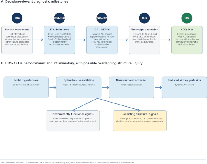
Figure 1: Historical evolution of the diagnostic criteria and pathophysiology of HRS. (A) A timeline of decision-relevant diagnostic milestones, from the 1978 Sassari consensus and the 1994–1996 ICA definitions, through the 2012–2015 integration of dynamic KDIGO criteria and the 2019 phenotype expansion, to the 2024 ADQI-ICA framework. (B) Portal hypertension and systemic inflammation lead to splanchnic vasodilation, neurohumoral activation, and reduced kidney perfusion; predominantly functional and coexisting structural signals may overlap in the same patient, without implying inevitable progression1,6,9. Abbreviations: HRS = hepatorenal syndrome; ICA = International Club of Ascites; ADQI = Acute Disease Quality Initiative; KDIGO = Kidney Disease: Improving Global Outcomes; AKI = acute kidney injury; AKD = acute kidney disease; CKD = chronic kidney disease; sCr = serum creatinine. Original schematic created by the authors. Please click here to view a larger version of this figure.
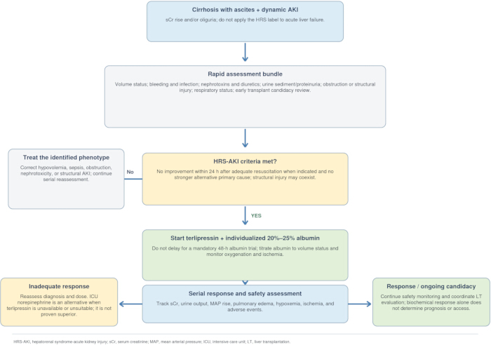
Figure 2: Evidence-based HRS-AKI assessment, treatment, and transplant pathway. A decision algorithm beginning with dynamic AKI confirmation, rapid assessment of volume status and precipitants, and exclusion of a stronger alternative primary cause. The No branch directs treatment of the identified phenotype with serial reassessment1. After HRS-AKI is established, the workflow proceeds to terlipressin plus individualized 20%–25% albumin, serial response and safety assessment, dose adjustment, and early LT evaluation. Norepinephrine is an ICU alternative when terlipressin is unavailable or unsuitable, not a proven superior routine switch1,59. Abbreviations: HRS-AKI = hepatorenal syndrome-acute kidney injury; sCr = serum creatinine; MAP = mean arterial pressure; ICU = intensive care unit; LT = liver transplantation. Original schematic created by the authors. Please click here to view a larger version of this figure.
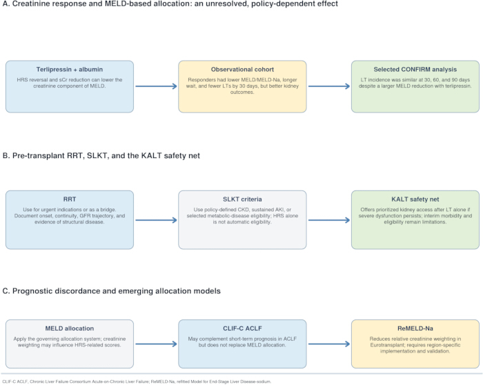
Figure 3: Transplant allocation and kidney-strategy considerations in HRS-AKI. (A) Terlipressin-associated sCr reduction can lower MELD, but observational and selected CONFIRM data do not show a uniform effect on LT access; this remains policy dependent14,15. (B) Kidney-injury type, duration, and severity inform recovery assessment and SLKT eligibility; no validated per-day estimate of irreversible injury is available, and OPTN criteria and the KALT safety net are summarized18,19,20,21. (C) CLIF-C ACLF may complement MELD in ACLF, while ReMELD-Na illustrates an emerging model with less creatinine weighting; neither independently determines transplantation22,76. Abbreviations: CLIF-C ACLF = Chronic Liver Failure Consortium Acute-on-Chronic Liver Failure; ReMELD-Na = refitted Model for End-Stage Liver Disease-sodium. Original schematic created by the authors. Please click here to view a larger version of this figure.
Table 1: Evidence-based prevention and early management of kidney dysfunction in cirrhosis with ascites. An evidence-based framework organized by recognized clinical setting, precipitating event, recommended action, supporting source, and evidentiary limitation. The table distinguishes guideline-supported prevention and early management from uncertain or unsupported proposals and summarizes the limited benefit of routine albumin in non-SBP infections, the controversy surrounding primary antibiotic prophylaxis for SBP, and the integration of artificial intelligence (AI) models for imminent HRS-AKI prediction. Abbreviations: ACLF = acute-on-chronic liver failure; SBP = spontaneous bacterial peritonitis; MAP = mean arterial pressure; MELD = Model for End-Stage Liver Disease; NGAL = neutrophil gelatinase-associated lipocalin; L-FABP = liver-type fatty acid-binding protein; AI = artificial intelligence; AUROC = area under the receiver operating characteristic curve. Please click here to download this Table.
Disclosures
The authors have no conflicts of interest to disclose.
Acknowledgements
This work was supported by the Jiuquan Science and Technology Plan Project (Grant No. 2024MA 1039).
References
- Nadim MK, et al. Acute kidney injury in patients with cirrhosis: Acute Disease Quality Initiative (ADQI) and International Club of Ascites (ICA) joint multidisciplinary consensus meeting. J Hepatol. 2024;81(1):163-83.
- Pose E, et al. Hepatorenal syndrome in cirrhosis. Gastroenterology. 2024;166(4):588-604.e1.
- Van Slambrouck C, et al. Bile cast nephropathy is a common pathologic finding for kidney injury associated with severe liver dysfunction. Kidney Int. 2013;84(1):192-7.
- Belcher JM, et al. Kidney biomarkers and differential diagnosis of patients with cirrhosis and acute kidney injury. Hepatology. 2014;60(2):622-32.
- Simonetto DA, Ginès P, Kamath PS. Hepatorenal syndrome: pathophysiology, diagnosis, and management. BMJ. 2020;370:m2687.
- Arroyo V, et al. Definition and diagnostic criteria of refractory ascites and hepatorenal syndrome in cirrhosis. International Ascites Club. Hepatology. 1996;23(1):164-76.
- Angeli P, Garcia-Tsao G, Nadim MK, Parikh CR. News in pathophysiology, definition and classification of hepatorenal syndrome: a step beyond the International Club of Ascites (ICA) consensus document. J Hepatol. 2019;71(4):811-22.
- Eknoyan G, Epstein M. Hepatorenal syndrome: a historical appraisal of its origins and conceptual evolution. Kidney Int. 2021;99(6):1321-30.
- Angeli P, et al. Diagnosis and management of acute kidney injury in patients with cirrhosis: revised consensus recommendations of the International Club of Ascites. J Hepatol. 2015;62(4):968-74.
- Biggins SW, et al. Diagnosis, evaluation, and management of ascites, spontaneous bacterial peritonitis and hepatorenal syndrome: 2021 practice guidance by the American Association for the Study of Liver Diseases. Hepatology. 2021;74(2):1014-48.
- Wong F, et al. Terlipressin plus albumin for the treatment of type 1 hepatorenal syndrome. N Engl J Med. 2021;384(9):818-28.
- Wong F, et al. Terlipressin improves renal function and reverses hepatorenal syndrome in patients with systemic inflammatory response syndrome. Clin Gastroenterol Hepatol. 2017;15(2):266-72.e1.
- Sanyal AJ, et al. A randomized, prospective, double-blind, placebo-controlled trial of terlipressin for type 1 hepatorenal syndrome. Gastroenterology. 2008;134(5):1360-8.
- Piano S, et al. Response to terlipressin and albumin is associated with improved liver transplant outcomes in patients with hepatorenal syndrome. Hepatology. 2021;73:1909-19.
- Rockey DC, et al. Terlipressin for hepatorenal syndrome in patients with early-stage acute-on-chronic liver failure. Liver Int. 2025;45:e70399.
- Jaques DA, et al. Biomarkers for acute kidney injury in decompensated cirrhosis: a prospective study. Nephrology (Carlton). 2019;24:170-80.
- Maiwall R, et al. Cystatin C predicts acute kidney injury and mortality in cirrhotics: a prospective cohort study. Liver Int. 2018;38:654-64.
- Organ Procurement and Transplantation Network. OPTN policies: Policy 9.9 and Table 9-17, medical eligibility criteria for liver-kidney allocation [Internet]. Rockville (MD): Health Resources and Services Administration; 2025 [cited 2026 Aug 25]. Available from: https://www.hrsa.gov/sites/default/files/hrsa/optn/optn_policies.pdf
- Wilk AR, et al. Developing simultaneous liver-kidney transplant medical eligibility criteria while providing a safety net: a 2-year review of the OPTN's allocation policy. Am J Transplant. 2021;21:3593-607.
- Eerhart MJ, et al. Kidney after liver transplantation matched-pair analysis: are kidneys allocated to appropriate patients to maximize their survival? Transplantation. 2020;104:804-12.
- Dixon W, et al. The type, duration, and severity of pretransplant kidney injury predict prolonged kidney dysfunction after liver transplantation. Liver Transpl. 2024;30:1159-68.
- Jalan R, et al. Development and validation of a prognostic score to predict mortality in patients with acute-on-chronic liver failure. J Hepatol. 2014;61:1038-47.
- Khemichian S, Francoz C, Nadim MK. Advances in management of hepatorenal syndrome. Curr Opin Nephrol Hypertens. 2021;30(5):501-6.
- Epstein M, et al. Renal failure in the patient with cirrhosis. The role of active vasoconstriction. Am J Med. 1970;49(2):175-85.
- Bradley SE. The pathogenesis of renal insufficiency. N Engl J Med. 1945;233:498-504.
- Hecker R, Sherlock S. Electrolyte and circulatory changes in terminal liver failure. Lancet. 1956;271:1121-5.
- Schrier R, et al. Peripheral arterial vasodilation hypothesis: a proposal for the initiation of renal sodium and water retention in cirrhosis. Hepatology. 1988;8(5):1151-7.
- Ginès A, et al. Incidence, predictive factors, and prognosis of the hepatorenal syndrome in cirrhosis with ascites. Gastroenterology. 1993;105(1):229-36.
- Roberts L, Kamath P. Ascites and hepatorenal syndrome: pathophysiology and management. Mayo Clin Proc. 1996;71(9):874-81.
- Ruiz-del-Arbol L, et al. Circulatory function and hepatorenal syndrome in cirrhosis. Hepatology. 2005;42(2):439-47.
- Arroyo V, Terra C, Ginès P. Advances in the pathogenesis and treatment of type-1 and type-2 hepatorenal syndrome. J Hepatol. 2007;46(5):935-46.
- Bernardi M, et al. Mechanisms of decompensation and organ failure in cirrhosis: from peripheral arterial vasodilation to systemic inflammation hypothesis. J Hepatol. 2015;63(5):1272-84.
- Kronsten V, Shawcross D. Clinical implications of inflammation in patients with cirrhosis. Am J Gastroenterol. 2025;120(1):65-74.
- Ginès P, et al. Hepatorenal syndrome. Nat Rev Dis Primers. 2018;4(1):23.
- Khemichian S, Nadim M, Terrault N. Update on hepatorenal syndrome: from pathophysiology to treatment. Annu Rev Med. 2025;76(1):373-87.
- Bairaktari E, et al. Partially reversible renal tubular damage in patients with obstructive jaundice. Hepatology. 2001;33(6):1365-9.
- Flint A. Clinical report on hydro-peritoneum, based on an analysis of forty-six cases. Am J Med Sci. 1863;45(90):306-39.
- Eknoyan G. On the etymology of nephritis: a historical appraisal of its origins. J Am Soc Nephrol. 2020;31(6):1170-3.
- Cardi E. The hepatorenal syndrome; a historical review. AMA Arch Surg. 1956;73(2):224-7.
- Orr T, Helwig F. Liver trauma and the hepatorenal syndrome. Ann Surg. 1939;110(4):682-92.
- Levy R, Simons D, Aronson S. Fatal hepatorenal syndrome associated with phenurone therapy. N Engl J Med. 1950;242(24):933-6.
- Wong F, Blendis L. Historical aspects of ascites and the hepatorenal syndrome. Clin Liver Dis (Hoboken). 2021;18(Suppl 1):14-27.
- James M, et al. Canadian Society of Nephrology commentary on the 2012 KDIGO clinical practice guideline for acute kidney injury. Am J Kidney Dis. 2013;61(5):673-85.
- Gambino C, et al. Diagnostic and prognostic performance of urinary neutrophil gelatinase-associated lipocalin in patients with cirrhosis and acute kidney injury. Hepatology. 2023;77(5):1630-8.
- Girish V, Maiwall R. Tracking the trajectory of kidney dysfunction in cirrhosis: the acute kidney injury-chronic kidney disease spectrum. Clin Mol Hepatol. 2025;31(3):730-52.
- Yang H, et al. Advances in the diagnosis of early biomarkers for acute kidney injury: a literature review. BMC Nephrol. 2025;26(1):115.
- Ostermann M, et al. Acute kidney injury. Lancet. 2025;405(10474):241-56.
- Patidar K, et al. Acute kidney disease is common and associated with poor outcomes in patients with cirrhosis and acute kidney injury. J Hepatol. 2022;77(1):108-15.
- Xu X, et al. Chinese guidelines on the management of ascites in cirrhosis: Chinese Society of Hepatology, Chinese Medical Association. Hepatol Int. 2024;18(4):1071-89.
- Thévenot T, et al. Effect of albumin in cirrhotic patients with infection other than spontaneous bacterial peritonitis. A randomized trial. J Hepatol. 2015;62(4):822-30.
- Guevara M, et al. Albumin for bacterial infections other than spontaneous bacterial peritonitis in cirrhosis. A randomized trial. J Hepatol. 2012;57(4):759-65.
- China L, et al. A randomized trial of albumin infusions in hospitalized patients with cirrhosis. N Engl J Med. 2021;384(9):808-17.
- Trebicka J, Garcia-Tsao G. Controversies regarding albumin therapy in cirrhosis. Hepatology. 2025;81(1):288-303.
- Schleicher EM, et al. Effect of albumin treatment duration on response rates and outcomes in patients with cirrhosis and acute kidney injury. J Hepatol. 2025;83(3):682-91.
- Karvellas CJ, et al. AASLD Practice Guidance on acute-on-chronic liver failure and the management of critically ill patients with cirrhosis. Hepatology. 2024;79(6):1463-502.
- Silvey S, et al. Spontaneous bacterial peritonitis is associated with high mortality, which interacts with antibiotic prophylaxis in a national cirrhosis cohort. Aliment Pharmacol Ther. 2026;63(8):1109-18.
- Thursz MR, et al. Prednisolone or pentoxifylline for alcoholic hepatitis. N Engl J Med. 2015;372(17):1619-28.
- Tian J, Cui R, Song H, Zhao Y, Zhou T. Prediction of acute kidney injury in patients with liver cirrhosis using machine learning models: evidence from the MIMIC-III and MIMIC-IV. Int Urol Nephrol. 2024;56(1):237-47.
- Olson JC, Subramanian RM. Comparative efficacy of terlipressin and norepinephrine for treatment of hepatorenal syndrome-acute kidney injury: a systematic review and meta-analysis. PLoS One. 2024;19:e0296690.
- Maiwall R, et al. A randomized controlled trial (TARGET-C) of high vs. low target mean arterial pressure in patients with cirrhosis and septic shock. J Hepatol. 2023;79(2):349-61.
- Cullaro G, et al. The relationship between mean arterial pressure and terlipressin in hepatorenal syndrome-acute kidney injury reversal: a post hoc analysis. Hepatology. 2025 Mar 7.
- Danielsen K, et al. Mapping the hemodynamic effects of terlipressin in patients with hepatorenal syndrome using advanced magnetic resonance imaging. JHEP Rep. 2025;7(8):101452.
- Bajaj JS, et al. Safety and efficacy of continuous infusion terlipressin (BIV201): a phase 2 trial in patients with decompensated cirrhosis and refractory ascites. Liver Transpl. 2025.
- Wong F, et al. Terlipressin use and respiratory failure in patients with hepatorenal syndrome type 1 and severe acute-on-chronic liver failure. Aliment Pharmacol Ther. 2022;56(8):1284-93.
- Allegretti AS, et al. From past to present to future: terlipressin and hepatorenal syndrome-acute kidney injury. Hepatology. 2025;81(6):1878-97.
- Utako P, et al. Clinical outcomes after liver transplantation for hepatorenal syndrome: a systematic review and meta-analysis. Biomed Res Int. 2018;2018:5362810.
- Park SS, et al. Kidney after liver transplantation (KALT) in the post-safety net era. Am J Transplant. 2025;25(8 Suppl 1):S470-1.
- Boyer TD, et al. Impact of liver transplantation on the survival of patients treated for hepatorenal syndrome type 1. Liver Transpl. 2011;17:1328-32.
- Weinberg EM, et al. Decreased need for renal replacement therapy in liver transplant recipients after pretransplant treatment of hepatorenal syndrome type 1 with terlipressin. Liver Transpl. 2024;30:347-55.
- Rössle M, Gerbes A. TIPS for the treatment of refractory ascites, hepatorenal syndrome and hepatic hydrothorax: a critical update. Gut. 2010;59(7):988-1000.
- Gonzalez-Garay A, et al. Transjugular intrahepatic portosystemic shunts for adults with hepatorenal syndrome. Cochrane Database Syst Rev. 2024;1(1):CD011039.
- Jung C, Chang J. Hepatorenal syndrome: current concepts and future perspectives. Clin Mol Hepatol. 2023;29(4):891-908.
- European Association for the Study of the Liver. EASL Clinical Practice Guidelines for the management of patients with decompensated cirrhosis. J Hepatol. 2018;69(2):406-60.
- Ramzan M, et al. Comparison of CLIF-C ACLF score and MELD score in predicting ICU mortality in patients with acute-on-chronic liver failure. Cureus. 2020;12(2):e7087.
- Kwon HM, et al. Benefits of liver transplant in critically ill patients with acute-on-chronic liver failure: implementation of an urgent living-donor program. Am J Transplant. 2025;25(1):150-63.
- Goudsmit BFJ, et al. Refitting the Model for End-Stage Liver Disease for the Eurotransplant region. Hepatology. 2021;74:351-63.
- Francoz C, Nadim MK, Durand F. Kidney biomarkers in cirrhosis: determining the threshold for simultaneous liver and kidney transplantation. J Hepatol. 2016;65(4):809-24.