This protocol describes the construction and molecular evaluation of a marker-free, multigene recombinant Escherichia coli strain designed for the chromosomal integration of genes associated with heavy-metal resistance and sequestration.
Method Article
Marker-Free Chromosomal Integration of a Multigene Cassette in Escherichia coli for Heavy-Metal Bioremediation Development
In This Article
Summary
Abstract
Heavy metal contamination remains a significant environmental challenge due to the persistence, toxicity, and bioaccumulation of metal pollutants. Genetically engineered bacteria offer a potential approach to heavy metal remediation; however, plasmid-based expression systems can present limitations associated with plasmid maintenance and antibiotic selection. This study describes a protocol for constructing a marker-free, multigene recombinant E. coli strain designed for the chromosomal integration of genes associated with heavy-metal resistance and sequestration. A 4,741 bp multigene cassette comprising cadD, cadA, and metallothionein (mt), together with a lipoprotein outer membrane protein A (Lpp-OmpA) surface display system and a green fluorescent protein (gfp) reporter gene under the control of the constitutive pyruvate dehydrogenase (Ppdh) promoter, was designed and synthesized. The construct was cloned into the pKIKOArsBKm integration vector and introduced into E. coli MG1655 using lambda (λ) Red-mediated recombination. The kanamycin resistance cassette was subsequently excised using the flippase–flippase recognition target (FLP-FRT) recombination system. Construct assembly and chromosomal integration were evaluated by restriction enzyme analysis, plasmid isolation, and polymerase chain reaction (PCR) amplification, while loss of antibiotic resistance was assessed by antibiotic sensitivity testing. Amplification of the expected cadD, cadA, and mt fragments from genomic DNA, together with the absence of detectable plasmid DNA, provided evidence consistent with chromosomal integration of the multigene cassette. Although this protocol establishes a framework for constructing and molecularly evaluating the recombinant strain, definitive confirmation of the integration locus, functional gene expression, long-term genetic stability, and heavy metal biosorption performance requires further experimental validation.
Introduction
Heavy metal contamination has become a major environmental concern due to the persistence, non-biodegradability, and toxicity of metal pollutants. Unlike organic contaminants, heavy metals accumulate in ecosystems and enter the food chain through bioaccumulation and biomagnification, posing serious risks to environmental and human health. Conventional remediation technologies, including chemical precipitation, ion exchange, and membrane filtration, are often associated with high operational costs, limited efficiency at low metal concentrations, and the generation of secondary pollutants. Consequently, biological remediation strategies have gained increasing attention as environmentally sustainable and cost-effective alternatives for heavy metal removal1.
Among these biological approaches, bacterial biosorption has emerged as a promising technology for heavy metal remediation. Biosorption involves the passive uptake of metal ions by biological materials through physicochemical interactions between metal ions and functional groups such as carboxyl, hydroxyl, amino, and phosphate groups present on the microbial cell surface2. These interactions facilitate metal removal through ion exchange, complexation, coordination, and precipitation mechanisms. Owing to their high surface-area-to-volume ratio, rapid growth, and diverse cell surface chemistries, bacteria are particularly effective biosorbents. The efficiency of bacterial biosorption depends on multiple factors, including bacterial species, metal ion characteristics, pH, temperature, and the presence of competing ions3. In addition to passive adsorption, bacteria can actively remove heavy metals through intracellular sequestration, bioaccumulation, biomineralization, and other detoxification mechanisms, thereby enhancing their metal removal capacity4.
Bacterial adaptation to heavy metal-contaminated environments is mediated by diverse resistance mechanisms, including membrane-associated efflux pumps, metal-binding proteins, enzymatic detoxification systems, and oxidation-reduction pathways that collectively maintain intracellular metal homeostasis. Several heavy metal resistance operons, including the mer, chr, ars, and cad systems, have been extensively characterized and represent valuable genetic resources for engineering bacterial biosorbents. The mer operon mediates mercury detoxification by reducing toxic Hg2⁺ to volatile elemental mercury (Hg°)5, whereas the chr operon confers chromate resistance by reducing Cr6⁺ to the less toxic Cr3⁺ 6. Similarly, the ars operon detoxifies arsenate through reduction to arsenite followed by active efflux7. The cadA gene, identified in Staphylococcus aureusRN4220 and Bacillus megaterium TWSL_4, encodes a P-type ATPase that actively exports cadmium ions from the cytoplasm, thereby reducing intracellular toxicity and contributing to bacterial metal resistance8,9. Metal binding proteins such as metallothioneins and phytochelatins further enhance heavy metal tolerance by sequestering metal ions and preventing their interaction with essential cellular components10.
Recent advances in synthetic biology have enabled the rational engineering of bacterial biosorbents with enhanced metal binding capabilities. Previous studies have demonstrated improved heavy metal removal through overexpression of metal-binding proteins in Bacillus subtilis and Pseudomonas putida11, immobilization of bacterial cells within alginate matrices or biofilms to improve stability and reusability12, and the introduction of synthetic metal-binding peptides into E. coli to increase mercury and arsenic biosorption13. Despite these advances, most engineered biosorbents rely on plasmid-based expression systems that require continuous antibiotic selection and are susceptible to plasmid instability, segregational loss, variable gene copy number, and increased metabolic burden. These limitations reduce their suitability for environmental and industrial applications where long-term genetic stability and biosafety are critical.
The strategic development of bacterial biosorbents, therefore, requires integrating microbiology, molecular biology, synthetic biology, and environmental engineering. Advances in genomics, proteomics, and metabolomics have further improved our understanding of bacterial responses to heavy metal stress, facilitating the identification of novel genes and regulatory pathways for engineering improved biosorbents.
Unlike conventional plasmid-based approaches, this study establishes an engineering platform to construct a chromosomally integrated, marker-free, multigene recombinant E. coli strain harboring genes associated with heavy metal resistance and sequestration. The engineering strategy combines λ Red recombineering, FLP-FRT-mediated marker excision, a constitutive multigene cassette, and an Lpp-OmpA-based surface-display design. The multigene cassette incorporated cadD (619 bp), cadA (2,564 bp), and metallothionein (mt) (154 bp), selected based on their previously reported roles in heavy metal resistance and metal sequestration. The genes were organized under the constitutive Ppdh promoter, with individual ribosome-binding sites, and mt was fused to Lpp-OmpA for intended surface localization. A downstream gfp reporter gene was incorporated into the construct.
The cassette was designed for chromosomal integration into E. coli MG1655 rather than continued maintenance on a multicopy plasmid. This strategy was selected to establish an engineering framework that may reduce dependence on plasmid maintenance and continuous antibiotic selection. FLP-FRT-mediated excision of the selectable antibiotic resistance marker was incorporated into the workflow, and antibiotic sensitivity testing was used to assess the resulting phenotype. The present study focuses on the assembly of constructs and the molecular evaluation of the recombinant strain. Long-term genetic stability, functional expression of the integrated genes, protein localization, heavy metal removal, and environmental performance were not evaluated and require further experimental validation.
This protocol is intended for researchers in molecular biology, microbial biotechnology, and synthetic biology who aim to construct marker-free, chromosomally integrated multigene bacterial strains. The method is particularly suitable for laboratory-scale genetic engineering and molecular evaluation of recombinant E. coli strains designed for future heavy-metal bioremediation research, rather than for direct environmental application.
Protocol
All experiments were conducted in accordance with institutional laboratory biosafety guidelines and applicable regulations for handling genetically modified microorganisms. All materials and software used in the current study are listed in the Table of Materials.
1. Design and in silico analysis of the multigene construct.
- Select the heavy metal resistance genes.
- Select the heavy metal resistance genes cadD (619 bp), cadA (2,564 bp), and metallothionein (mt; 154 bp), previously identified from Staphylococcus epidermidis TWSL_17, Bacillus megaterium TWSL_4, and Staphylococcus warneri TWSL_6, respectively.
- Retrieve the nucleotide sequences of cadD, cadA, and mt from the NCBI GenBank database using accession numbers MK900473.1, MK327282.1, and MK900456.1, respectively.
NOTE: These genes were selected based on previous characterization of the source bacterial strains for heavy metal tolerance and removal, and the sources of nucleotide sequences of each genetic element incorporated into the biosorbent construct are listed in Table 1.
- Perform in silico analysis of the selected genes and encoded proteins.
- Import the nucleotide sequences of cadD, cadA, and mt into the software (e.g., SnapGene) and analyze the coding sequences.
- Translate the nucleotide sequences into their corresponding amino acid sequences using the ExPASy translation server.
- Submit the predicted amino acid sequences of CadD, CadA, and Mt to I-TASSER and predict the three-dimensional protein structures and potential ligand-binding sites.
- Analyze the CadD, CadA, and Mt protein sequences using TMHMM Server to predict transmembrane domains.
- Analyze the protein sequences using the ExPASy TMPRED server to predict hydrophobicity profiles and membrane-associated regions.
NOTE: Record the predicted ligand-binding residues, structural parameters, transmembrane domains, and hydrophobicity profiles for subsequent interpretation of the predicted functions and localization of the encoded proteins.
- Design the multigene cassette.
- Design a 4,741-bp multigene cassette containing the constitutive pyruvate dehydrogenase operon promoter (Ppdh), cadD, cadA, Lpp-OmpA, mt, and gfp reporter gene.
- Place the selected genes downstream of the constitutive Ppdh promoter and incorporate a ribosome-binding site (RBS; 5′-AGGAGG-3′) upstream of each coding sequence.
- Fuse the metallothionein sequence to the Lpp-OmpA sequence to generate the Lpp-OmpA-Mt component designed for localization of metallothionein at the bacterial outer membrane.
- Incorporate gfp downstream of the heavy metal-associated genes as a reporter for subsequent evaluation of construct expression.
- Verify the orientation, nucleotide positions, and overall organization of the genetic elements in the final 4,741-bp construct using SnapGene.
- Synthesize and clone the multigene construct.
- Submit the finalized 4,741-bp multigene construct for commercial synthesis and cloning into the pET-21a vector between the BamHI and XhoI restriction sites.
- Upon receipt of the recombinant pET-21a construct, reconstitute the plasmid according to the supplier's instructions and proceed with transformation into E. coli BL21(DE3).
NOTE: E. coli BL21(DE3) was used for the initial propagation of the commercially synthesized multigene construct supplied in the pET-21a expression vector. Although BL21(DE3) is primarily used for recombinant protein expression, it is genotypically compatible with pET-21a and was used successfully in this study. Alternatively, dedicated cloning strains such as E. coli DH5α or TOP10 may be used for initial plasmid propagation, as T7 RNA polymerase expression is not required at this stage.
2. Construct the multigene biosorbent cassette.
- Transform the synthesized construct into E. coli BL21(DE3).
- Thaw E. coli BL21(DE3) competent cells on ice.
- Add 10–100 ng of the synthesized recombinant pET-21a plasmid containing the biosorbent construct to 50 µL of E. coli BL21(DE3) competent cells. Gently mix the contents without repeated pipetting.
- Incubate the cell suspension on ice for 30 min.
- Heat shock the cells at 42 °C for 45 s.
- Immediately transfer the tubes to ice and incubate for 2 min.
- Add 500 µL of prewarmed (37 °C), sterile SOC medium (Super optimal medium with catabolic repressor) without antibiotics. Incubate the culture at 37 °C with shaking (180 rpm) for 1 h to allow recovery.
- Spread 100 µL of the transformed culture onto LB (Luria-Bertani) agar plates containing ampicillin (100 µg/mL). Incubate the plates 16–18 h at 37 °C.
- Verify positive transformants harboring the recombinant plasmid.
- Select a well-isolated single colony from the selective agar plate and inoculate it into 5 mL of LB broth containing ampicillin (100 µg/mL).
- Incubate the culture 16-18 h at 37 °C with shaking (180 rpm).
- Isolate plasmid DNA from the 16–18 h-grown bacterial culture.
- Transfer 5 mL of a 16–18 h-grown bacterial culture into a centrifuge tube and harvest the cells by centrifugation at 11,000 × g for 1 min. Discard the supernatant completely.
- Resuspend the cell pellet thoroughly in 250 µL of ice-cold ET buffer.
- Add 250 µL of freshly prepared lysis buffer and mix gently by inverting (do not vortex) the tube several times. Incubate the mixture on ice for 5 min.
- Add 300 µL of neutralization solution and mix gently by inverting the tube several times (do not vortex).
- Centrifuge the mixture at 11,000 × g for 10 min, then carefully transfer the supernatant to a new microcentrifuge tube without disturbing the pellet.
- Add TE-equilibrated phenol to the collected supernatant at a 1:1 volume ratio and mix thoroughly. Centrifuge at 11,000 × g for 10 min, then carefully transfer the upper aqueous layer to a fresh microcentrifuge tube without disturbing the interface.
CAUTION: Phenol is highly corrosive and toxic and can cause severe chemical burns. Handle all phenol-containing solutions inside a certified chemical fume hood while wearing appropriate personal protective equipment. - Add absolute ethanol at twice the volume of the recovered aqueous phase, followed by 0.1 volume of 0.1 N sodium acetate. Mix thoroughly and incubate at −20 °C for 15 min to allow DNA precipitation.
- Centrifuge the sample at 11,000 × g for 20 min. Remove the supernatant carefully without dislodging the DNA pellet.
- Rinse the DNA pellet with 70% (v/v) ethanol and centrifuge to re-pellet the DNA. Remove the ethanol carefully and leave the pellet to air dry.
- Dissolve the dried DNA pellet in 20 µL of TE buffer or nuclease-free water.
PAUSE POINT: Store the isolated plasmid DNA at −20 °C until required for restriction enzyme digestion and verification of the recombinant plasmid.
- Confirm the isolated plasmid by restriction enzyme digestion.
- Prepare a 50 µL restriction digestion reaction in a sterile microcentrifuge tube containing 500 ng of plasmid DNA, 5 µL of 10× restriction enzyme buffer, 10 U of each of BamHI and XhoI, and nuclease-free water to a final volume of 50 µL.
NOTE: BamHI and XhoI were selected because these restriction sites were used to clone the synthesized biosorbent gene cassette into the pET-21a vector.
- Prepare a 50 µL restriction digestion reaction in a sterile microcentrifuge tube containing 500 ng of plasmid DNA, 5 µL of 10× restriction enzyme buffer, 10 U of each of BamHI and XhoI, and nuclease-free water to a final volume of 50 µL.
- Incubate the reaction mixture at 37 °C for 1 h.
- Separate and assess the digested DNA fragments by agarose gel electrophoresis to verify the presence and expected size of the biosorbent gene cassette.
- Prepare a 1% (w/v) agarose solution using 100 mL of 1× TAE buffer.
- Heat the agarose suspension in a microwave until the agarose is fully dissolved and the solution appears clear. Cool the molten agarose to approximately 60 °C.
- Add 10× DNA Safe Stain to the cooled molten agarose to achieve a final concentration of 1×. Mix gently to avoid introducing air bubbles.
- Pour the agarose mixture into a gel casting tray containing an appropriate comb. Remove visible air bubbles and allow the gel to set completely at room temperature.
- Transfer the solidified gel into the electrophoresis chamber and add sufficient 1× TAE running buffer to cover the gel. Carefully remove the comb.
- Mix 5 µL of each DNA sample with 1 µL of 6× DNA loading buffer and load the mixture into individual wells.
- Perform electrophoresis at 100 V for 40 min or until adequate separation of the DNA fragments is achieved.
- Visualize the DNA bands using a UV transilluminator and record the gel image using an appropriate gel documentation system.
CAUTION: Ultraviolet radiation can cause eye and skin injury. Use the protective shield and appropriate UV-protective equipment when operating the UV transilluminator.
NOTE: Include an undigested plasmid sample as a control to verify complete restriction enzyme digestion on the agarose gel.
- Preserve the confirmed recombinant strain.
- Inoculate a confirmed recombinant colony into 5 mL of LB broth containing ampicillin (100 µg/mL) and incubate 16–18 h at 37 °C with shaking (180 rpm).
- Mix the 16–18 h-grown culture with sterile glycerol to obtain a final glycerol concentration of 15% (v/v).
- Aliquot the glycerol stock into sterile cryovials and store at −80 °C.
PAUSE POINT: Store glycerol stocks at −80 °C for long-term preservation before proceeding with chromosomal integration.
3. Cloning of the biosorbent cassette into the chromosomal integration vector (pKIKOArsBKm) and integration into the host genome (E. coli MG1655)
- Clone the biosorbent gene cassette into the pKIKOArsBKm integration vector.
- Digest the recombinant pET-21a plasmid harboring the biosorbent gene cassette and the pKIKOArsBKm integration vector separately with SacI and BamHI.
- Prepare a 50 µL restriction digestion reaction containing 500 ng of plasmid DNA, 1× restriction enzyme buffer, 1 U each of SacI and BamHI, and nuclease-free water to a final volume of 50 µL. Incubate the reaction mixture at 37 °C for 1 h.
- Separate the restriction enzyme-digested DNA fragments by agarose gel electrophoresis, as described in Step 2.2.6.
- Excise the bands corresponding to the 4,741 bp biosorbent gene cassette and the linearized pKIKOArsBKm vector. Transfer each excised gel slice into a pre-weighed microcentrifuge tube.
NOTE: Minimize the exposure of the target DNA fragments to UV light during visualization and excision. - Purify the excised biosorbent gene cassette and linearized pKIKOArsBKm vector from the agarose gel.
- Weigh the tube containing the gel slice and calculate the mass of the excised agarose by subtracting the weight of the empty tube.
- Add 1× TE buffer at a volume equivalent to five times the mass of the gel slice, using a ratio of 5 µL of TE buffer per 1 mg of agarose (e.g., add 500 µL of TE buffer to a 100 mg gel slice).
- Incubate the tube in a water bath at 65 °C until the agarose is completely melted. Allow the sample to cool to room temperature before proceeding with phenol extraction.
- Follow Steps 2.2.3.6–2.2.3.10 to perform phenol extraction and DNA precipitation of the target DNA fragments.
PAUSE POINT: Store the purified DNA at −20 °C until required for subsequent cloning or ligation.
- Ligate the purified biosorbent gene cassette into the linearized pKIKOArsBKm vector using T4 DNA ligase at an insert-to-vector mass ratio of approximately 3:1.
- Prepare a 10 µL ligation reaction containing 150 ng of the purified biosorbent gene cassette, 49.2 ng of the linearized pKIKOArsBKm vector, 1 µL of 10X T4 DNA ligase buffer, and 3 U of T4 DNA ligase. Add nuclease-free water to a final volume of 10 µL, maintaining an insert-to-vector mass ratio of approximately 3:1.
- Mix the reaction gently by pipetting and incubate 16–18 h at 4 °C.
- Transform 5 µL of the ligation reaction mixture into E. coli Pir1 cells.
- Thaw E. coli Pir1 competent cells on ice.
- Add 5 µL of the ligation reaction mixture to 50 µL of E. coli Pir1 competent cells. Gently mix the contents without repeated pipetting.
- Keep the cell suspension on ice for 30 min.
- Subject the cells to heat shock at 42 °C for 45 s.
- Return the tubes immediately to ice and maintain them there for 2 min.
- Add 500 µL of sterile, prewarmed (37 °C) SOC medium without antibiotics. Allow the cells to recover for 1 h at 37 °C with shaking at 180 rpm.
- Plate 100 µL of the transformation mixture onto LB agar containing kanamycin (50 µg/mL) using a sterile spreader. Incubate the plates at 37 °C for 16–18 h.
- Select well-isolated kanamycin-resistant recombinant colonies from the LB agar plates.
- Confirm the presence of the recombinant integration plasmid by plasmid isolation as described in Step 2.2.3, followed by agarose gel electrophoresis as described in Step 2.2.6.
PAUSE POINT: Store the completed ligation reaction at −20 °C until required for subsequent transformation and verification of the recombinant pKIKOArsBKm construct.
- Digest the recombinant pET-21a plasmid harboring the biosorbent gene cassette and the pKIKOArsBKm integration vector separately with SacI and BamHI.
- Transform the ligation product containing the recombinant pKIKOArsBKm construct into E. coli MG1655.
- Prepare λ Red recombination host cells.
- Prepare chemically competent E. coli MG1655 cells.
- Select a well-isolated single colony of E. coli MG1655 from an LB agar plate and inoculate it into 5 mL of LB broth. Incubate the culture at 37 °C for 16–18 h with shaking.
- Transfer 0.5 mL of the overnight culture into 50 mL of sterile Super Optimal Broth (SOB) medium. Incubate the culture at 37 °C with shaking at 180 rpm until the OD₆₀₀ reaches 0.35–0.45.
- Transfer the culture immediately to an ice bath and chill thoroughly.
- Transfer the chilled culture into a sterile, prechilled 50 mL centrifuge tube and centrifuge at 10,000 × g for 10 min at 4 °C.
- Remove the supernatant carefully and gently resuspend the cell pellet in 20 mL of ice-cold deionized water.
- Centrifuge the cell suspension at 10,000 × g for 10 min at 4 °C. Carefully discard the supernatant.
- Gently resuspend the cell pellet in 2 mL of ice-cold 0.1 M calcium chloride and keep the suspension on ice for 30 min.
- Centrifuge the suspension at 10,000 × g for 5 min at 4 °C, then carefully remove the supernatant.
- Gently resuspend the final cell pellet in 1 mL of ice-cold 0.1 M CaCl₂. Dispense 80 µL aliquots of the chemically competent E. coli MG1655 cells into sterile, pre-chilled microcentrifuge tubes. Use the chemically competent E. coli MG1655 cells immediately for transformation or store the aliquots at −80 °C until use.
- Transform chemically competent E. coli MG1655 cells with the λ Red recombination plasmid pKD46 using the heat-shock transformation procedure.
- Add 100 ng of pKD46 plasmid to 80 µL of a chilled microcentrifuge tube containing chemically competent E. coli MG1655 cells. Gently mix the contents without repeated pipetting.
- Incubate the cell suspension on ice for 30 min.
- Expose the cells to a heat shock at 42 °C for 45 s.
- Transfer the tubes immediately to ice and keep them there for 2 min.
- Add 500 µL of sterile SOC media without antibiotics. Incubate the culture at 30 °C with shaking (180 rpm) for 1 h to allow recovery.
- Spread 100 µL of the transformed culture onto LB agar plates containing ampicillin (100 µg/mL). Incubate the plates 16–18 h at 30 °C.
NOTE: Maintain E. coli MG1655(pKD46) at 30 °C until use in step 3.2.2 to retain the temperature-sensitive pKD46 plasmid.
- Prepare chemically competent E. coli MG1655 cells.
- Prepare electrocompetent E. coli MG1655(pKD46) cells.
- Inoculate 1 mL of a 16–18 h-grown E. coli MG1655(pKD46) culture into an Erlenmeyer flask containing 250 mL of LB broth supplemented with ampicillin (100 µg/mL) and incubate the culture at 30 °C with shaking at 180 rpm until the OD₆₀₀ reaches 0.35–0.40.
- Add filter-sterilized 10% (w/v) L-arabinose to a final concentration of 0.1% (w/v) to induce expression of the λ Red recombination proteins.
NOTE: For a 250 mL culture, add 2.5 mL of 10% (w/v) L-arabinose to obtain a final concentration of approximately 0.1% (w/v). - Incubate the induced culture for an additional 15 min at 30 °C with shaking at 180 rpm.
- Transfer the Erlenmeyer flask containing the culture to an ice bath and incubate for 40 min.
- Transfer the chilled culture into prechilled centrifuge tubes and collect the cells by centrifuging at 1,800 × g for 15 min at 4 °C. Carefully remove the supernatant.
- Gently resuspend the cell pellet in 30 mL of ice-cold 10% (v/v) glycerol, then centrifuge at 3,000 × g for 5 min at 4 °C.
- Remove the supernatant and repeat the wash described in Step 3.2.2.6 twice more, giving a total of three washes with ice-cold 10% (v/v) glycerol. After the final wash, carefully discard the supernatant and resuspend the pellet in 200 µL of ice-cold 10% (v/v) glycerol.
- Dispense the electrocompetent E. coli MG1655(pKD46) cells into 50 µL aliquots and keep the cells on ice until electroporation.
NOTE: Use the freshly prepared electrocompetent cells immediately for electroporation. Alternatively, aliquot the cells into sterile microcentrifuge tubes, freeze at −80 °C, and thaw on ice before subsequent use.
- Linearize the recombinant integration construct obtained from Step 3.1.5.
- Digest the recombinant pKIKOArsBKm plasmid harboring the biosorbent gene cassette with NotI restriction enzyme to linearize the integration construct.
- Confirm complete digestion of the recombinant plasmid by agarose gel electrophoresis as described in Step 2.2.6.
- Purify the linearized DNA fragment from the digestion reaction mixture by phenol extraction and DNA precipitation as described in Steps 2.2.3.6–2.2.3.10.
- Electroporate the linearized integration construct into electrocompetent E. coli MG1655(pKD46).
- Add approximately 250 ng of the purified linearized DNA from Step 3.2.3.3 to 50 µL of freshly prepared electrocompetent E. coli MG1655(pKD46) cells. Mix gently and transfer the cell-DNA mixture into a pre-chilled electroporation cuvette.
- Electroporate the cells at 1.8 kV, 600 Ω, and 25 µF using an electroporator.
- Immediately add 500 µL of SOC medium to the electroporation cuvette and gently resuspend the cells.
- Transfer the recovered cell suspension into a sterile microcentrifuge tube and allow the cells to recover for 1 h at 37 °C with shaking at 180 rpm.
- Spread 100 µL of the recovered cell suspension onto LB agar plates supplemented with kanamycin (50 µg/mL) and incubate 16-18 h at 37 °C.
- Screen the recombinant colonies for chromosomal integration.
- Pick individual kanamycin-resistant colonies and inoculate each into a separate tube containing 10 mL of LB broth with kanamycin at a final concentration of 50 µg/mL.
- Grow the cultures for 16–18 h at 37 °C with shaking at 180 rpm.
- Transfer 5 mL of each culture into a separate sterile tube and isolate the plasmid DNA as described in Step 2.2.3.
- Analyze the isolated DNA by agarose gel electrophoresis as described in Step 2.2.6.
- Identify samples in which no plasmid DNA is detectable by agarose gel electrophoresis and retain these samples for subsequent evaluation of chromosomal integration.
- Isolate genomic DNA from the remaining 5 mL of each culture retained in Step 3.2.5.3, and use the isolated genomic DNA as the template for PCR amplification of the cadD, cadA, and mt genes as described in Step 3.2.7.
- Transfer 5 mL of the 16–18 h-grown bacterial culture to a sterile centrifuge tube and pellet the cells by centrifuging at 11,000 × g for 10 min at 4 °C. Discard the supernatant.
- Gently resuspend the cell pellet in 5 mL of sterile phosphate-buffered saline (PBS). Centrifuge at 11,000 × g for 10 min at 4 °C, then remove the supernatant completely.
- Resuspend the washed cell pellet in 100 µL of lysozyme solution (50 mg/mL) and incubate the suspension at 37 °C for 1 h.
- Add 500 µL of 5 M Guanidine thiocyanate solution to the lysozyme-treated cells. Mix gently and incubate at room temperature for 10 min.
CAUTION: Guanidine thiocyanate is harmful and may release toxic gases when mixed with acids or bleach. Wear suitable personal protective equipment and handle guanidine-containing solutions according to institutional chemical safety procedures. - Place the sample on ice for 10 min, then add 250 µL of ice-cold 7.5 M ammonium acetate while mixing gently. Keep the mixture on ice for a further 10 min.
- Add TE-equilibrated phenol to the lysate at a 1:1 (v/v) ratio. Vortex thoroughly and centrifuge at 10,000 × g for 15 min.
CAUTION: Phenol and chloroform are toxic and corrosive. Handle phenol inside a certified chemical fume hood while wearing appropriate personal protective equipment. - Transfer the upper aqueous layer carefully to a new sterile microcentrifuge tube without disturbing the interphase.
- Add 0.1 volume of sodium acetate solution and 2 volumes of absolute ethanol to the recovered aqueous phase. Mix gently by inversion, then incubate at −20 °C for 20 min.
- Centrifuge the sample at 10,000 × g for 15 min. Carefully remove the supernatant without dislodging the DNA pellet.
- Rinse the DNA pellet with 100 µL of 70% (v/v) ethanol.
- Centrifuge at 10,000 × g for 10 min, remove the remaining ethanol, and air-dry the DNA pellet at room temperature for 10 min.
- Resuspend the dried DNA pellet in 20–50 µL of TE buffer or nuclease-free water.
PAUSE POINT: Store the isolated genomic DNA at −20 °C until required for downstream molecular analysis.
- Evaluate chromosomal integration by gene-specific PCR and plasmid DNA analysis.
- Prepare separate 25 µL PCR reaction mixtures for amplification of the cadD, cadA, and mt genes.
- For each reaction, add the corresponding forward and reverse primers to a final concentration of 0.4 µM each, together with 1× PCR buffer, 200 µM of each dNTP, 1.5 mM MgCl₂, 100 ng of genomic DNA isolated in Step 3.2.6, and Taq DNA polymerase at 0.2 U/µL. Add nuclease-free water to a final reaction volume of 25 µL.
- Amplify the target genes by PCR using the following cycling program: denature initially at 95 °C for 5 min; carry out 35 cycles of denaturation at 94 °C for 30 s, annealing for 30 s at the gene-specific temperature listed in Table 2, and extension at 72 °C for 1 min; then perform a final extension at 72 °C for 5 min.
- Analyze the PCR products by agarose gel electrophoresis as described in Step 2.2.6, along with 1 kB DNA ladder as the molecular size marker.
- Evaluate the PCR products by comparing their migration with the 1 kb DNA ladder and identify bands corresponding to the expected amplicon sizes of 619 bp for cadD, 2,564 bp for cadA, and 154 bp for mt.
NOTE: The absence of detectable plasmid DNA combined with amplification of the expected cadD, cadA, and mt fragments from genomic DNA provides evidence supporting chromosomal integration of the biosorbent gene cassette.
- Prepare separate 25 µL PCR reaction mixtures for amplification of the cadD, cadA, and mt genes.
- Prepare λ Red recombination host cells.
4. Excise the kanamycin resistance marker.
- Prepare electrocompetent recombinant E. coli MG1655 cells with Biosorbent gene cassette.
- Inoculate a recombinant E. coli MG1655 colony carrying the chromosomally integrated biosorbent cassette confirmed in Step 3.2.7, into 5 mL of LB broth supplemented with kanamycin (50 µg/mL).
- Incubate the culture at 37 °C with shaking at 180 rpm until the OD₆₀₀ reaches 0.4–0.6.
- Transfer the culture to ice immediately after the OD₆₀₀ reaches 0.4–0.6 and incubate on ice for 15 min.
- Harvest the cells by centrifugation at 4,000 × g for 10 min at 4 °C and discard the supernatant.
- Wash the cell pellet twice with ice-cold deionized water, followed by one wash with ice-cold 10% (v/v) glycerol.
- Resuspend the final cell pellet in 50 µL of ice-cold 10% (v/v) glycerol.
NOTE: Keep the cells and all solutions ice-cold throughout the washing procedure to maintain electrocompetency.
- Electroporation of the helper plasmid pCP20
- Add 50 ng of the pCP20 helper plasmid to 50 µL of electrocompetent cells. Mix gently and transfer the mixture to a pre-chilled electroporation cuvette.
- Electroporate the cells using the following settings: 2.5 kV, 600 Ω, and 25 µF.
- Immediately add 1 mL of SOC medium to the cuvette and gently resuspend the cells.
- Transfer the recovered cells to a sterile culture tube and incubate at 30 °C with shaking at 180 rpm for 1 h.
- Spread 100 µL of the recovered culture onto LB agar plates supplemented with ampicillin (100 µg/mL). Incubate the plates 16–18 h at 30 °C.
- Select well-isolated single colonies to confirm the positive recombinants.
- Confirm the presence of the pCP20 plasmid by plasmid isolation as described in Step 2.2.3, followed by agarose gel electrophoresis as described in Step 2.2.6.
NOTE: Maintain the transformed cells at 30 °C to preserve the temperature-sensitive pCP20 plasmid.
- Excise the kanamycin resistance cassette.
- Inoculate a confirmed ampicillin-resistant colony from Step 4.2 into antibiotic-free LB broth and incubate 16–18 h at 42 °C with shaking at 180 rpm to induce FLP recombinase expression and cure the temperature-sensitive pCP20 plasmid.
- Streak the 16–18 h-grown culture separately onto LB agar plates containing no antibiotic, kanamycin (50 µg/mL), and ampicillin (100 µg/mL). Incubate each plate for 16–18 h at 37 °C.
- Identify colonies that grow only on antibiotic-free LB agar but fail to grow on kanamycin or ampicillin-containing plates. Select these colonies as putative marker-free recombinants.
- Purify the marker-free recombinants by re-streaking a single colony onto fresh antibiotic-free LB agar plates.
PAUSE POINT: Store purified marker-free recombinant strains as glycerol stocks at −80 °C until required for downstream molecular confirmation or functional characterization.
5. Analyze GFP expression of the constructed whole cell biosorbent by fluorescence microscopy.
- Prepare bacterial cultures for heavy metal exposure.
- Inoculate 5 µL of a 16–18 h-grown culture of recombinant marker-free E. coli MG1655 harboring the chromosomally integrated biosorbent construct from step 4.3.4 into LB broth containing Cd2⁺, Zn2⁺, or Pb2⁺ at final concentrations of 1, 5, 10, 50, 100, 500, or 1000 ppm.
- To prepare heavy metal solutions, first prepare sterile 2,000 ppm stock solutions of Cd2⁺, Zn2⁺, and Pb2⁺ by dissolving analytical grade cadmium chloride (CdCl₂), zinc chloride (ZnCl₂), and lead nitrate [Pb (NO₃)₂], respectively, in sterile LB broth.
- Sterilize each stock solution by filtration through a 0.22 µm membrane filter. Store the sterile stock solutions at 4 °C until use.
- Prepare fresh working solutions by diluting each stock solution with sterile LB broth to final metal ion concentrations of 1, 5, 10, 50, 100, 500, and 1,000 ppm.
NOTE: Prepare each metal solution separately to avoid precipitation or interactions between different metal ions.
- Prepare four control conditions: wild-type E. coli MG1655 cultured under the same conditions as the recombinant strain, marker-free recombinant E. coli MG1655 harboring the chromosomally integrated multigene cassette cultured in LB broth without added heavy metals as the recombinant no-metal control, a GFP-expressing E. coli JM109 strain containing pQBIT-GFP-T7 plasmid as the GFP-positive biological control, and uninoculated LB broth containing the corresponding heavy metal concentrations as the abiotic control.
- Incubate all cultures 16–18 h at 37 °C with shaking at 180 rpm.
- Inoculate 5 µL of a 16–18 h-grown culture of recombinant marker-free E. coli MG1655 harboring the chromosomally integrated biosorbent construct from step 4.3.4 into LB broth containing Cd2⁺, Zn2⁺, or Pb2⁺ at final concentrations of 1, 5, 10, 50, 100, 500, or 1000 ppm.
- Prepare microscope slides.
- Pipette 1 µL of each bacterial culture onto a separate clean glass microscope slide. Spread the culture to form a thin, even cell layer, and allow the smear to air-dry completely.
- Acquire fluorescence images.
- Observe the prepared samples using a fluorescence microscope at 600× magnification equipped with an FITC filter set (excitation wavelength, 460–490 nm).
- Capture fluorescence and corresponding bright field images using identical microscope settings for all recombinant and control samples.
NOTE: Maintain identical exposure time, gain, illumination intensity, and magnification for all samples to enable direct comparison of GFP fluorescence intensity between treatments.
CAUTION: Avoid prolonged exposure of the samples to excitation light to minimize GFP photobleaching.
Results
In-silico design of the multigene biosorbent construct
The designed multigene biosorbent cassette had a total length of 4,741 bp and consisted of the constitutive Ppdh promoter followed by the heavy metal resistance genes cadD, cadA, and mt, arranged as a polycistronic operon. Individual ribosome-binding sites (RBS) were incorporated upstream of each coding sequence to facilitate efficient translation, while a gfp reporter gene was included downstream of the biosorbent genes to enable monitoring of construct expression (Figure 1).

Figure 1: Design and organization of the multigene construct. The 4,741-bp multigene cassette was designed with the constitutive pyruvate dehydrogenase (Ppdh) promoter followed by an operator region to drive expression of genes associated with heavy metal resistance and sequestration. The construct contains the heavy metal resistance genes cadD and cadA, the metallothionein (mt) gene, a lipoprotein outer membrane protein A (Lpp-OmpA) surface-display component fused to mt for intended cell-surface localization, and the gfp reporter gene. Individual ribosome-binding sites (RBS; 5′-AGGAGG-3′) were incorporated upstream of the respective coding sequences to support translation within the multigene cassette. A terminator sequence is located downstream of gfp. The complete cassette was designed as a 4,741-bp construct for subsequent cloning and chromosomal integration in E. coli. Abbreviations: Ppdh = pyruvate dehydrogenase promoter; RBS = ribosome-binding site; Lpp-OmpA = lipoprotein–outer membrane protein A; mt = metallothionein; gfp = green fluorescent protein gene; bp = base pairs; E. coli = Escherichia coli. Please click here to view a larger version of this figure.
To facilitate extracellular metal sequestration, the mt gene was fused to the Lpp-OmpA outer membrane anchor to enable surface localization of the metallothionein protein. Based on the construct design, CadD and CadA were targeted to the inner membrane as transmembrane metal transport proteins, whereas the Lpp-OmpA-Mt fusion protein was targeted to the outer membrane. The GFP reporter protein was designed for cytoplasmic expression to facilitate monitoring of biosorbent gene expression (Figure 2).
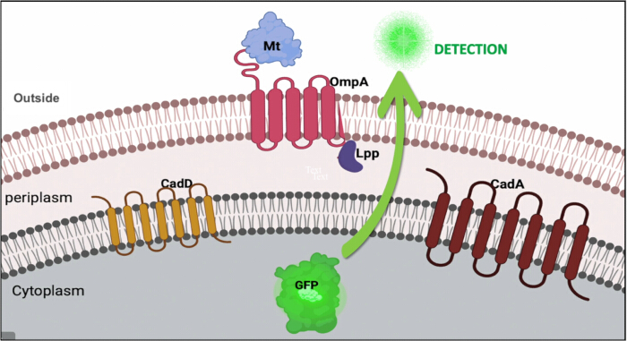
Figure 2: Predicted localization of proteins expressed from the multigene biosorbent construct. CadD and CadA were designed for localization within the inner membrane, the lipoprotein outer membrane protein A (Lpp-OmpA)-metallothionein fusion was targeted to the outer membrane, and green fluorescent protein was included as a cytoplasmic reporter protein. Abbreviations: Lpp-OmpA = lipoprotein–outer membrane protein A; Mt = metallothionein; GFP = green fluorescent protein. Please click here to view a larger version of this figure.
The sources of nucleotide sequences of each genetic element incorporated into the biosorbent construct are listed in Table 1.
| Genetic element in the biosorbent construct | Location in the construct (bp) | Source/Reference |
| Ppdh | 7-74 | Gao et al. 2002.14 |
| Operator | 75-92 | Operator region of cad D of Staphylococcus epidermidis strain TWSL_22 : NCBI Gene Bank accession no: MK900473.1 |
| cadD | 97-716 | NCBI Gene Bank accession no: MK900473.1 |
| cadA | 770-3334 | NCBI Gene Bank accession no: MK327282.1 |
| Lpp-OmpA | 3385-3754 | Stathopoulo et al. 1995. 15 |
| mt | 3755-3909 | NCBI Gene Bank accession no: MK900456.1 |
| gfp | 3960-4679 | TPMV_P-EGFP_H-His6 vector, NCBI Gene Bank accession no:MF379627.1 |
| Terminator | 4684-4741 | Terminator region of cadAC operon of Bacillus megaterium strain TWSL-4 : NCBI Gene Bank accession no: MK327282.1 |
Table 1: Sources of nucleotide sequences for the genetic elements in the biosorbent construct. The table summarizes the genetic elements incorporated into the 4,741-bp multigene construct, including their sequence sources, accession numbers or references, and nucleotide positions within the gene construct. Abbreviations: Ppdh = pyruvate dehydrogenase promoter; Lpp-OmpA = lipoprotein–outer membrane protein A; mt = metallothionein; gfp = green fluorescent protein gene; NCBI = National Center for Biotechnology Information; bp = base pairs.
Transformation and verification of the synthesized construct into E. coli BL21(DE3)
The recombinant pET-21a plasmid harboring the biosorbent gene cassette was successfully verified by restriction enzyme digestion (Figure 3A). Double digestion with BamHI and XhoI generated two DNA fragments corresponding to the pET-21a vector backbone (5,331 bp) and the biosorbent gene cassette (4,741 bp), consistent with the expected construct size. Although the two DNA fragments exhibited limited separation on the 1% agarose gel due to their similar sizes, both fragments were clearly distinguishable, confirming the integrity of the recombinant construct.
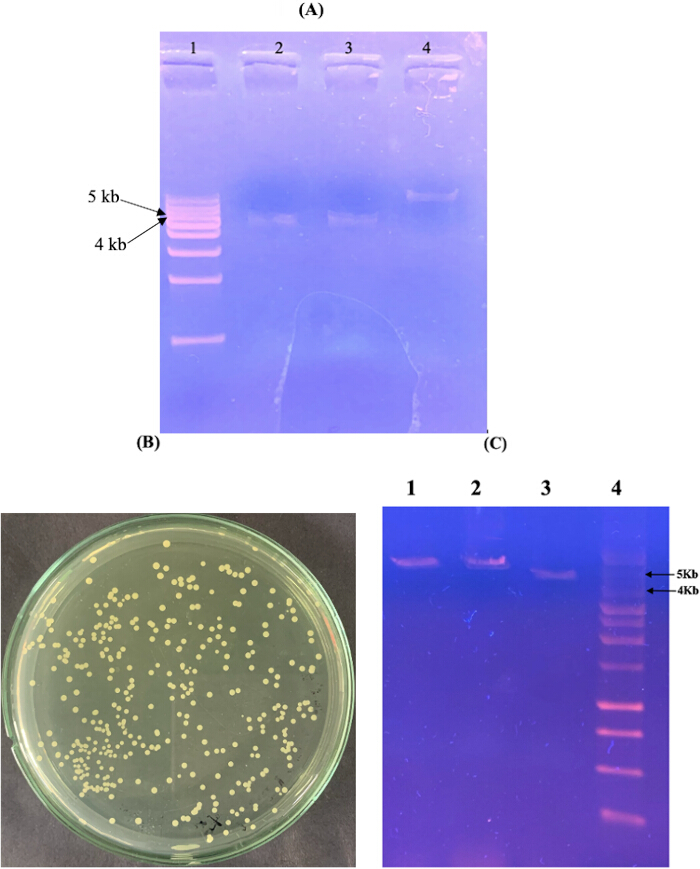
Figure 3: Construction and verification of the recombinant biosorbent plasmid. (A) Restriction enzyme analysis of the synthesized biosorbent construct cloned into the pET-21a expression vector. Lane 1, 1 kb DNA ladder; lanes 2–3, double-digested recombinant plasmid showing the pET-21a vector backbone (5,331 bp) and the biosorbent gene cassette (4,741 bp); lane 4, undigested recombinant plasmid. (B) Ampicillin-resistant E. coli BL21(DE3) transformants carrying the recombinant biosorbent plasmid. (C) Restriction enzyme analysis of plasmids isolated from representative transformants. Lane 1, BamHI-digested plasmid; lane 2, XhoI-digested plasmid; lane 3, BamHI/XhoI double-digested plasmid; lane 4, 1 kb DNA ladder. Abbreviations: DNA = deoxyribonucleic acid; bp = base pairs; kb = kilobases; E. coli = Escherichia coli. Please click here to view a larger version of this figure.
Following the transformation of the recombinant plasmid into E. coli BL21(DE3), ampicillin-resistant colonies were successfully recovered (Figure 3B). Plasmid DNA isolated from representative transformants was subsequently analyzed by restriction enzyme digestion. Digestion with BamHI, XhoI, and the combined double digestion generated the expected restriction profiles, confirming the successful propagation of the recombinant biosorbent plasmid in E. coli BL21(DE3) (Figure 3C).
Evaluation of chromosomal integration of the biosorbent gene cassette into E. coli MG1655
The biosorbent gene cassette was successfully cloned into the chromosomal integration vector pKIKOArsBKm. Recombinant clones were initially selected on LB agar supplemented with kanamycin (50 µg/mL) (Figure 4A). Restriction enzyme analysis confirmed successful cloning of the biosorbent cassette into the integration vector (Figure 4B). Linearization of the recombinant plasmid with NotI produced the expected DNA fragment of approximately 8.9 kb, while double digestion with BamHI and SacI released the 4.7-kb biosorbent cassette from the 4.2-kb vector backbone, confirming correct insertion of the construct into pKIKOArsBKm. The undigested plasmid exhibited the expected nicked and supercoiled conformations.
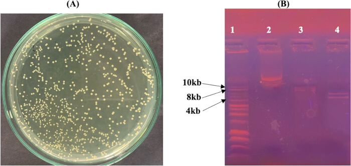
Figure 4: Construction of the recombinant chromosomal integration vector carrying the biosorbent gene cassette. (A) Kanamycin-resistant E. coli Pir1 colonies carrying the recombinant pKIKOArsBKm plasmid. (B) Restriction enzyme analysis of recombinant pKIKOArsBKm. Lane 1, 1 kb DNA ladder; lane 2, undigested recombinant plasmid; lane 3, NotI-linearized plasmid (~8.9 kb); lane 4, BamHI/SacI double-digested plasmid showing the 4.2-kb vector backbone and the 4.7-kb biosorbent cassette. Abbreviations: DNA = deoxyribonucleic acid; kb = kilobases; E. coli = Escherichia coli. Please click here to view a larger version of this figure.
The linearized recombinant pKIKOArsBKm construct was subsequently introduced into electrocompetent E. coli MG1655(pKD46) cells to facilitate λ Red-mediated chromosomal integration. Following selection on kanamycin-containing medium, genomic DNA and plasmid DNA were isolated from representative recombinant colonies to verify successful integration.
As shown in Figure 5, genomic DNA was isolated from representative recombinant colonies (Lane 1), whereas no detectable plasmid DNA was observed following plasmid isolation (Lane 2). PCR amplification using the gene-specific primers described in Table 2 generated amplicons of the expected sizes for cadD (619 bp), cadA (2,564 bp), and mt (154 bp) from the genomic DNA preparation (Lanes 4–6). Together, the absence of detectable plasmid DNA and detection of the three target genes in the genomic DNA preparation provided evidence consistent with chromosomal integration of the multigene biosorbent cassette in E. coli MG1655.
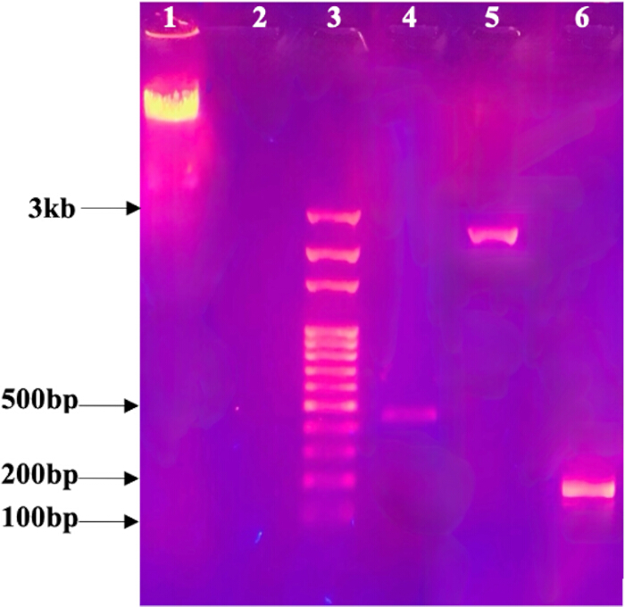
Figure 5: Molecular evaluation of the recombinant E. coli strain harboring biosorbent gene cassette. Lane 1, genomic DNA isolated from recombinant E. coli MG1655; lane 2, plasmid isolation from the corresponding recombinant colony showing the absence of detectable plasmid DNA; lane 3, 100 bp DNA ladder; lane 4, PCR amplification of cadD (619 bp); lane 5, PCR amplification of cadA (2,564 bp); lane 6, PCR amplification of mt (154 bp). Abbreviations: DNA = deoxyribonucleic acid; PCR = polymerase chain reaction; bp = base pairs; mt = metallothionein; E. coli = Escherichia coli. Please click here to view a larger version of this figure.
| Target DNA sequence | Primer name | Oligonucleotide sequence (5’→3’) | *Tan (℃) | Expected size of the PCR product (bp) |
| cadD | SD-EF | GAATTCATGATTCA GACTATTGTAACTG | 50 | 619 |
| SE-XHR | CTCGAGAAATATAA ATCCTAAAATTGTTTG | |||
| cadA | PD-BF | GGATCCGACACAAT GAAACACGAAGA | 56 | 2564 |
| PF-XHR | CCGCTCGAGCGGCAT GTAATTTTTCATTTTGA GTAAGCGAAG | |||
| mt | SEmtF | GGAATTCATGGAAAAAT GTGCGAGACCGAAC | 48 | 154 |
| SEmtR | CCGCTCGAGTGCAGTT GCACAGCTACAATC |
Table 2: Primer details for amplification of cadD, cadA, and mt. The table lists the forward and reverse primer sequences used for PCR amplification of cadD, cadA, and mt, together with the corresponding annealing temperatures and expected amplicon sizes. *Tan- Annealing temperature for each primer pair applied in the PCR.
Evaluation of kanamycin resistance marker excision from recombinant E. coli MG1655
Following transformation with the pCP20 helper plasmid, ampicillin-resistant colonies were recovered on LB agar supplemented with ampicillin (100 µg/mL). Plasmid DNA corresponding to pCP20 was detected following plasmid isolation from representative transformants (Figure 6).
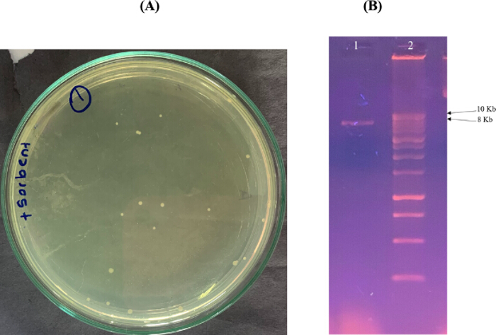
Figure 6: Confirmation of pCP20 transformation into recombinant E. coli MG1655 carrying the chromosomally integrated biosorbent cassette. (A) Ampicillin-resistant transformants following introduction of the pCP20 helper plasmid. (B) Agarose gel electrophoresis confirming the presence of the pCP20 plasmid. Lane 1, isolated pCP20 plasmid (~9.3 kb); lane 2, 10 kb DNA ladder. Abbreviations: DNA = deoxyribonucleic acid; kb = kilobases; E. coli = Escherichia coli. Please click here to view a larger version of this figure.
Following FLP recombinase-mediated marker excision and temperature treatment for pCP20 curing, recombinant colonies were evaluated by antibiotic sensitivity testing. As shown in Figure 7, the tested colonies grew on antibiotic-free LB agar but did not grow on LB agar supplemented with either kanamycin (50 µg/mL) or ampicillin (100 µg/mL). This antibiotic sensitivity phenotype was consistent with loss of kanamycin resistance and loss of ampicillin resistance following the marker-excision and plasmid curing procedure.
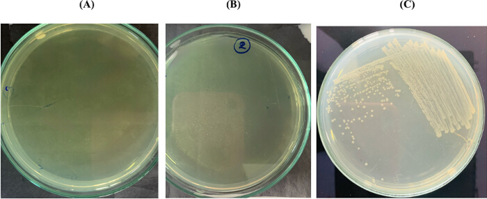
Figure 7: Confirmation of flippase-mediated excision of the kanamycin resistance marker and curing of the pCP20 helper plasmid. (A) No bacterial growth on Luria-Bertani agar supplemented with kanamycin (50 µg/mL). (B) No bacterial growth on LB agar supplemented with ampicillin (100 µg/mL). (C) Normal bacterial growth on antibiotic-free LB agar, confirming cell viability following marker excision. Abbreviations: LB = Luria-Bertani; E. coli = Escherichia coli. Please click here to view a larger version of this figure.
Fluorescence microscopy analysis of recombinant E. coli MG1655 carrying the biosorbent construct
Recombinant E. coli MG1655 generated through the chromosomal integration procedure was evaluated for detectable green fluorescence following exposure to Cd2⁺, Zn2⁺, and Pb2⁺ at concentrations ranging from 1 to 1,000 ppm. No detectable green fluorescence was observed in recombinant cells at any of the tested metal concentrations. No fluorescence signal was detected in the all four control samples.
Discussion
The protocol presented here describes a workflow for constructing a chromosomally integrated, marker-free, multigene recombinant E. coli strain containing genes associated with heavy metal resistance and sequestration. The method combines assembly of a multigene cassette, cloning into the pKIKOArsBKm integration vector, λ Red-mediated chromosomal integration, FLP-FRT-mediated selectable marker excision, molecular evaluation of recombinant colonies, and fluorescence microscopy. The approach was designed to avoid maintenance of the final construct on a multicopy plasmid and to generate a recombinant strain without the kanamycin resistance marker.
Several steps are critical for the successful implementation of this protocol. During λ Red-mediated integration, maintaining the temperature-sensitive pKD46 plasmid at 30 °C before recombination and inducing the λ Red recombination proteins with L-arabinose are important for efficient recombineering16,17. Preparation of electrocompetent E. coli MG1655(pKD46) cells also requires maintaining the cells and washing solutions at low temperature and minimizing residual salts before electroporation. The recombinant pKIKOArsBKm construct should be fully linearized and purified before electroporation, as linear DNA serves as the substrate for λ Red-mediated recombination. The pKIKO integration system has previously been reported to accommodate relatively large DNA inserts and to support efficient chromosomal integration18,19. Following selection, candidate colonies should be evaluated using both plasmid and genomic DNA preparations. In the present study, the absence of detectable plasmid DNA together with amplification of cadD, cadA, and mt from the genomic DNA preparation provided evidence consistent with chromosomal integration. However, these assays alone do not definitively establish the precise genomic integration event. Junction-specific PCR, sequencing across the integration boundaries, or whole-genome sequencing could provide more direct confirmation of the integration site in future applications of the protocol.
Marker excision is another critical component of the method. The FLP-FRT recombination system provides a means of removing selectable antibiotic resistance markers following genome engineering16,20. Following the introduction of the temperature-sensitive pCP20 helper plasmid, transformants are maintained at 30 °C, then incubated at 42 °C to promote FLP-mediated marker excision and loss of pCP20. Colonies should then be evaluated on antibiotic-free, kanamycin-containing, and ampicillin-containing media. Growth on antibiotic-free medium, accompanied by the absence of growth on kanamycin- and ampicillin-containing media, provides a practical screening approach for identifying candidate marker-free colonies following the excision and curing procedure. If antibiotic-resistant colonies remain, additional single-colony purification and temperature-dependent curing may be required before further characterization. The chromosomal integration strategy used here differs from conventional plasmid-based expression methods.
Plasmid systems can provide higher gene copy numbers and may therefore be advantageous when strong recombinant protein expression is required. However, plasmid-based systems can be affected by plasmid segregation, variable copy number, metabolic burden, and dependence on selective pressure for plasmid maintenance. Chromosomal integration eliminates the need to maintain the final biosorbent cassette as an extrachromosomal plasmid and was selected in this protocol to provide a more persistent genetic configuration. Previous studies have described chromosomal integration as an approach for improving genetic maintenance during cell division21,22. However, the long-term genetic stability of the recombinant strain generated in the present study was not directly evaluated. Therefore, improved genetic stability should not be considered an experimentally demonstrated outcome of this protocol. The multigene design is another feature of the method. Rather than introducing a single metal-associated gene, the cassette combines cadA, cadD, and metallothionein (mt) with an Lpp-OmpA surface-display design and a gfp reporter. Previous recombinant bacterial biosorbent strategies have commonly focused on individual metal-binding proteins or resistance determinants11,13,23,24. The modular configuration used here allows multiple genetic components to be incorporated into a single chromosomal integration workflow. The protocol could therefore be adapted for other multigene constructs by modifying the genes, promoter, reporter, or surface-display components according to the intended application. Nevertheless, increasing cassette size and the number of expressed genes may affect recombination efficiency, transcription, translation, and cellular metabolic burden, and should be considered when adapting the method.
An important limitation of the present study is that functional expression of the engineered cassette was not demonstrated under the experimental conditions tested. No detectable GFP fluorescence was observed following exposure of the recombinant cells to Cd2⁺, Zn2⁺, or Pb2⁺. Consequently, the present data do not establish functional expression of the complete multigene cassette or demonstrate heavy metal biosorption by the recombinant strain. Chromosomal integration generally yields lower gene copy numbers than multicopy plasmid systems, and recombinant expression can also be influenced by the genomic insertion site and promoter activity22,25. Cellular responses to heavy metal stress may additionally influence heterologous gene expression26,27. These factors represent possible considerations rather than experimentally established explanations for the absence of detectable fluorescence in the present study. When applying this protocol, the absence of detectable fluorescence should prompt evaluation of culture conditions, metal exposure conditions, promoter activity, and reporter expression. Independent measurements of transcription and protein production would help distinguish inadequate expression from limitations of fluorescence detection. Simultaneous expression of multiple recombinant proteins may also impose additional metabolic demands on the host, particularly under stressful culture conditions28.
These possibilities require direct experimental evaluation before the cause of the absence of detectable GFP fluorescence can be established. The protocol also has limitations in characterizing the final recombinant strain. Gene-specific PCR and the absence of detectable plasmid DNA provide evidence consistent with chromosomal integration but do not establish the precise integration-site sequence or long-term genetic stability. Antibiotic sensitivity provides a practical screen following marker excision but can be supplemented with molecular analysis of the FRT-containing region when more direct confirmation is required. In addition, fluorescence microscopy provides a qualitative assessment of detectable GFP fluorescence but does not substitute for quantitative measurements of transcription or protein abundance. Quantitative reverse-transcription PCR, protein analysis, quantitative fluorescence measurements, and sequencing-based confirmation could therefore strengthen characterization of strains generated using this protocol.
Further optimization and functional characterization are required before the engineered strain can be evaluated as a functional heavy metal biosorbent. Direct measurements of metal uptake or removal are necessary to establish biosorption performance. Protein expression and localization should also be experimentally verified, particularly for the Lpp-OmpA-metallothionein fusion. Long-term passaging experiments would be required to determine whether chromosomal integration provides the expected genetic stability under relevant culture conditions. Marker-free genome engineering may also facilitate subsequent rounds of chromosomal modification without accumulation of multiple selectable markers, providing opportunities for further development of multigene constructs15,27.
Overall, this protocol provides a methodological framework for constructing and evaluating a chromosomally integrated, marker-free, multigene recombinant bacterial strain. Its modular design may be adapted for other combinations of metal-associated genes, reporter systems, or synthetic biology applications. The current work establishes the engineering and screening workflow; functional expression, long-term genetic stability, protein localization, and heavy metal biosorption performance remain to be experimentally validated before conclusions can be drawn regarding environmental or bioremediation applications.
Disclosures
The authors have no conflicts of interest to declare.
Acknowledgements
This work was supported by a research grant, AP/3/2/2018/CG/33, University of Colombo.
Materials
| Name | Company | Catalog Number | Comments |
|---|---|---|---|
| 0.22 μm membrane filter | ThermoFisher Scientific | 596-3320 | None |
| 1 kb DNA marker | Promega Corporation | G5711 | None |
| 10 kb DNA ladder | N/A | N/A | Molecular size marker used for Figure 6 |
| 100 bp DNA ladder | N/A | N/A | Molecular size marker used for Figure 5 |
| 10X T4 DNA ligase buffer | Promega Corporation | C1263 | None |
| 6× DNA loading buffer | N/A | N/A | Used for agarose gel electrophoresis |
| Agarose | Molecular Designs | A113-3 | None |
| Ammonium acetate | N/A | N/A | Prepared as 7.5 M solution for genomic DNA extraction |
| Ampicillin | ThermoFisher Scientific | J60977.14 | None |
| BamHI restriction enzyme | New England BioLabs | R0136S | None |
| Cadmium chloride | ThermoFisher Scientific | 219141000 | None |
| Calcium chloride | ThermoFisher Scientific | 012316.A7 | None |
| Centrifuge | ThermoFisher Scientific | Heraeus Fresco 21 / 75002478 | None |
| Deionized water | ThermoFisher Scientific | 751-610 | None |
| DNA Safe Stain | Molecular Designs | C138 | None |
| dNTP Mix | Promega Corporation | U1511 | None |
| Electrophoresis equipment | BioRad | 1704405 | None |
| Electroporation cuvette | BioRad | 1652086 | None |
| Electroporator | BioRad | 1652100 | None |
| Escherichia coli BL21(DE3) | University of Colombo, Department of Chemistry | N/A | None |
| Escherichia coli MG1655 | Department of Life Science and Technology, Institute of Science Tokyo, Yokohama, Japan | N/A | None |
| Escherichia coli Pir1 competent cells | ThermoFisher Scientific | C101010 | None |
| Escherichia coli BL21(DE3) competent cells | ThermoFisher Scientific | C600003 | None |
| Escherichia coli JM109 strain containing pQBIT-GFP-T7 plasmid | Department of Life Science and Technology, Institute of Science Tokyo, Yokohama, Japan | N/A | None |
| ET Buffer | N/A | N/A | To prepare 1 L of ET buffer, dissolve 6.06 g of Tris base and 3.72 g of EDTA in approximately 800 mL of distilled water. Adjust the pH to the required value using HCl, mix thoroughly, and bring the final volume to 1 L with distilled water |
| Ethanol | ThermoFisher Scientific | T038181000CS | None |
| Ethylenediaminetetraacetic Acid (EDTA) | ThermoFisher Scientific | 17892 | None |
| Fluorescence microscope | Coastal Microscopes | Olympus BX53/ 11679 | None |
| Gel electrophoresis apparatus | Bio Rad | 1704405 | None |
| Glacial acetic acid | MilliporeSigma | PHR1748 | None |
| Glycerol | MilliporeSigma | G5516 | None |
| Guanidine thiocyanate | ThermoFisher Scientific | 433941000 | None |
| Hydrochloric acid (HCl) | MilliporeSigma | 1.00317 | None |
| Kanamycin | ThermoFisher Scientific | 15160054 | None |
| L-Arabinose | ThermoFisher Scientific | 365185000 | None |
| Lead nitrate | ThermoFisher Scientific | 211565000 | None |
| Luria–Bertani (LB) agar | Molecular Designs | C120 | None |
| Luria–Bertani (LB) broth | Molecular Designs | C121 | None |
| Lysis buffer | N/A | N/A | To prepare the lysis buffer, combine the appropriate volumes of 5 M NaOH and 20% (w/v) SDS stock solutions to obtain final concentrations of 0.2 N NaOH and 1% (w/v) SDS. Adjust to the required final volume with distilled water and mix thoroughly. |
| Lysozyme | ThermoFisher Scientific | 89833 | None |
| Magnesium chloride (MgCl2) | N/A | N/A | Used at 1.5 mM final concentration in PCR reactions |
| Neutralization solution | N/A | N/A | Prepare the neutralization buffer, using 3 M potassium acetate and glacial acetic acid. Adjust the solution to the required final volume with distilled water and mix thoroughly. |
| NotI restriction enzyme | New England BioLabs | R0189S | None |
| pCP20 | Department of Life Science and Technology, Institute of Science Tokyo, Yokohama, Japan | N/A | None |
| PCR machine | ThermoFisher Scientific | Applied Biosystems MiniAmp / A37834 | None |
| pET-21a plasmid in E. coli BL21(DE3) | University of Colombo, Department of Chemistry | N/A | None |
| Phosphate buffered saline | MilliporeSigma | P4474 | None |
| pKD46 | Creative Biogene | OVT2612 | None |
| pKIKOArsBKm | Addgene | 46766 | None |
| Potassium acetate | MilliporeSigma | 236497 | None |
| Restriction enzyme buffer | N/A | N/A | Used for restriction enzyme digestion reactions |
| SacI restriction enzyme | New England BioLabs | R0156 | None |
| Shaker incubator | Crystal technology and industries | IS-A6 | None |
| Sodium acetate | ThermoFisher Scientific | A13184.30 | None |
| Sodium dodecyl sulphate (SDS) | ThermoFisher Scientific | 15525017 | None |
| Sodium hydroxide (NaOH) | MilliporeSigma | 567530 | None |
| Super optimal broth (SOB) medium | MilliporeSigma | H8032 | None |
| Super optimal medium with catabolic repressor (SOC) Media | ThermoFisher Scientific | J60255.AK | None |
| T4 DNA ligase | Promega Corporation | M1801 | None |
| TAE Buffer | ThermoFisher Scientific | B49 | None |
| Taq DNA polymerase | Promega Corporation | M3001 | None |
| TE buffer | MilliporeSigma | 93283 | None |
| TE equilibrated phenol | ThermoFisher Scientific | 15513039 | None |
| Tris base | N/A | N/A | Used to prepare ET buffer |
| Universal PCR buffer | Promega Corporation | M7911 | None |
| UV trans-illuminator | Labexchange | TFX-35M, 021425 | None |
| UV/Visible spectrophotometer | Microbiotests | Jenway 6300, TE15 | None |
| Water bath | ThermoFisher Scientific | TSGP05 | None |
| XhoI restriction enzyme | New England BioLabs | R0146S | None |
| Zinc chloride | ThermoFisher Scientific | A16281.36 | None |
| Primers | |||
| cadD forward primer (SD-EF) | N/A | N/A | 5′-GAATTCATGATTC AGACTATTGTAACTG-3′ |
| cadD reverse primer (SE-XHR) | N/A | N/A | 5′-CTCGAGAAATATAA ATCCTAAAATTGTTTG-3′ |
| cadA forward primer (PD-BF) | N/A | N/A | 5′-GGATCCGACACAAT GAAACACGAAGA-3′ |
| cadA reverse primer (PF-XHR) | N/A | N/A | 5′-CCGCTCGAGCGGCA TGTAATTTTTCATTTTGA GTAAGCGAAG-3′ |
| mt forward primer (SEmtF) | N/A | N/A | 5′-GGAATTCATGGAAAAAT GTGCGAGACCGAAC-3′ |
| mt reverse primer (SEmtR) | N/A | N/A | 5′-CCGCTCGAGTGCAGTTG CACAGCTACAATC-3′ |
| Software/Tool Name | Version/Server | URL | Access date |
| ExPASy tools | TMPRED Server | https://embnet.vital-it.ch/cgi-bin/TMPRED_form_parser | 30-01-2023 |
| ExPASy Translate | ExPASy Translation Tool | https://web.expasy.org/translate/ | N/A |
| I-TASSER | N/A | https://zhanggroup.org/I-TASSER/ | 24-02-2023 |
| NCBI GenBank | GenBank database | https://www.ncbi.nlm.nih.gov/genbank/ | N/A |
| SnapGene | v.7.1.2 | N/A | N/A |
| TMHMM | v. 2.0 | https://services.healthtech.dtu.dk/services/TMHMM-2.0/ | 16-01-2023 |
References
- Volesky B. Detoxification of metal-bearing effluents: biosorption for the next century. Hydrometallurgy. 2001;59(2-3):203-16.
- Ayangbenro AS, Babalola OO. A new strategy for heavy metal polluted environments: a review of microbial biosorbents. Int J Environ Res Public Health. 2017;14(1):94.
- Das S, Dash HR, Chakraborty J. Genetic basis and importance of metal resistant genes in bacteria for bioremediation of contaminated environments with toxic metal pollutants. Appl Microbiol Biotechnol. 2016;100(7):2967-84.
- Volesky B. Biosorption of heavy metals. Biotechnol Prog. 1995;11(3):235-50.
- Barkay T, Miller SM, Summers AO. Bacterial mercury resistance from atoms to ecosystems. FEMS Microbiol Rev. 2003;27(2-3):355-84.
- Cervantes C, et al. Interactions of chromium with microorganisms and plants. FEMS Microbiol Rev. 2001;25(3):335-47.
- Cai L, Liu G, Rensing C, Wang G. Genes involved in arsenic transformation and resistance associated with different levels of arsenic-contaminated soils. BMC Microbiol. 2009;9:4.
- Silver S, Phung LT. Genes and enzymes involved in bacterial oxidation and reduction of inorganic arsenic. Appl Environ Microbiol. 2005;71(2):599-608.
- Kumari WMNH, et al. Characterization of a Bacillus megaterium strain with metal bioremediation potential and in silico discovery of novel cadmium binding motifs in the regulator, CadC. Appl Microbiol Biotechnol. 2021;105(6):2573-86.
- Hamer DH. Metallothionein. Annu Rev Biochem. 1986;55:913-51.
- Kazy SK, D’Souza SF, Sar P. Uranium and thorium sequestration by a Pseudomonas sp.: mechanism and chemical characterization. J Hazard Mater. 2009;163(1):65-72.
- Yan G, Viraraghavan T. Heavy-metal removal from aqueous solution by fungus Mucor rouxii. Water Res. 2003;37(18):4486-96.
- Pazirandeh M, Wells B, Ryan RL. Development of bacterium-based heavy metal biosorbents: enhanced uptake of cadmium and mercury by Escherichia coli expressing a metal binding motif. Appl Environ Microbiol. 1998;64(10):4068-72.
- Gao H, Jiang X, Pogliano K, Aronson AI. The E1β and E2 subunits of the Bacillus subtilis pyruvate dehydrogenase complex are involved in regulation of sporulation. J Bacteriol. 2002;184(10):2780-8.
- Stathopoulos C, Georgiou G, Earhart CF. Characterization of Escherichia coli expressing an Lpp′OmpA(46-159)-PhoA fusion protein localized in the outer membrane. Appl Microbiol Biotechnol. 1996;45(1-2):112-9.
- Datsenko KA, Wanner BL. One-step inactivation of chromosomal genes in Escherichia coli K-12 using PCR products. Proc Natl Acad Sci U S A. 2000;97(12):6640-5.
- Yu D, et al. An efficient recombination system for chromosome engineering in Escherichia coli. Proc Natl Acad Sci U S A. 2000;97(11):5978-83.
- Sabri S, et al. Knock-in/Knock-out (KIKO) vectors for rapid integration of large DNA sequences, including whole metabolic pathways, onto the Escherichia coli chromosome at well-characterised loci. Microb Cell Fact. 2013;12:60.
- Wang J, et al. Converting Escherichia coli MG1655 into a chemical overproducer through inactivating defense system against exogenous DNA. Synth Syst Biotechnol. 2020;5(4):333-42.
- Cherepanov PP, Wackernagel W. Gene disruption in Escherichia coli: TcR and KmR cassettes with the option of Flp-catalyzed excision of the antibiotic-resistance determinant. Gene. 1995;158(1):9-14.
- Liu M, et al. Metabolic engineering of Escherichia coli to improve recombinant protein production. Appl Microbiol Biotechnol. 2015;99(24):10367-77.
- Persad AK, Williams ML, LeJeune JT. Rapid loss of a green fluorescent plasmid in Escherichia coli O157. AIMS Microbiol. 2017;3(4):872-84.
- Jeiranikhameneh M, et al. Designing novel construction for cell surface display of protein E on Escherichia coli using non-classical pathway based on Lpp-OmpA. AMB Express. 2017;7(1):53.
- Dissanayake DMDC, Kumari WMNH, Chandrasekharan NV, Wijayarathna CD. Isolation of heavy metal-resistant Staphylococcus epidermidis strain TWSL_22 and evaluation of heavy metal bioremediation potential of recombinant E. coli cloned with isolated cadD. FEMS Microbiol Lett. 2023;370:fnad092.
- Bryant JA, Sellars LE, Busby SJW, Lee DJ. Chromosome position effects on gene expression in Escherichia coli K-12. Nucleic Acids Res. 2014;42(18):11383-92.
- Busuioc M, Ciofu O, Høiby N. Cellular responses to environmental stress in bacteria. FEMS Microbiol Rev. 2010;34(6):1042-64.
- Outten FW, Outten CE, Hale J, O'Halloran TV. Transcriptional activation of an Escherichia coli copper efflux regulon by the chromosomal MerR homologue, CueR. J Biol Chem. 2001;276(9):6346-54.
- Zaslaver A, et al. Just-in-time transcription program in metabolic pathways. Nat Genet. 2004;36(5):486-91.
- Thomason LC, Costantino N, Shaw DV, Court DL. Multicopy plasmid modification with phage lambda Red recombineering. Plasmid. 2007;58(2):148-58.