This protocol describes Stem Cell Educator therapy for ex vivo immune modulation of peripheral blood mononuclear cells and its application in patients with long COVID to evaluate safety, clinical outcomes, and immunological changes.
Method Article
Stem Cell Educator Therapy for Ex Vivo Immune Modulation of Peripheral Blood Mononuclear Cells in Patients with Long COVID
In This Article
Summary
Abstract
Long COVID, generally defined as clinical symptoms persisting for 3 months or longer following severe acute respiratory syndrome coronavirus 2 (SARS-CoV-2) infection, affects multiple organs and systems. At least 400 million people globally are living with long COVID, with symptoms including persistent fatigue and brain fog. Currently, long COVID care mainly consists of symptomatic management and supportive approaches, with no validated effective treatments available that address the underlying inflammation. Stem Cell Educator (SCE) therapy is a treatment developed to modulate immune responses. This protocol describes SCE therapy and its application in patients with long COVID to evaluate safety, clinical outcomes, and immunological changes. During SCE therapy, a patient’s peripheral blood mononuclear cells (PBMC) are collected by leukapheresis and co-cultured with adherent cord-blood-derived multipotent stem cells (CB-SC) in vitro, after which the “educated” PBMC are returned to the patient’s circulation via infusion. SCE therapy has previously been evaluated in clinical studies for type 1 diabetes and other autoimmune diseases. Twenty-five patients with long COVID (15 males and 10 females) received SCE therapy. Five patients experienced mild adverse effects during apheresis, and one patient reported itching during infusion. Brain fog and fatigue disappeared in 21 of 25 patients following treatment, and most participants reported feeling refreshed, energized, and mentally clear. Flow cytometry demonstrated reductions in inflammatory markers, including interleukin-1 beta (IL-1β)-positive cells and C-X-C motif chemokine ligand 16 (CXCL16)-positive cells. These findings demonstrate the application of SCE therapy in patients with long COVID and provide preliminary clinical and immunological observations supporting further evaluation of this approach.
Introduction
The global outbreak of coronavirus disease 2019 (COVID-19), caused by severe acute respiratory syndrome coronavirus 2 (SARS-CoV-2), has devastated populations worldwide, with over 651 million confirmed cases. Long COVID, generally defined as clinical symptoms persisting for 3 months or longer following SARS-CoV-2 infection, affects multiple organs and systems1,2,3. Based on a conservative estimate, at least 400 million people globally are living with long COVID4,5. Many patients experience severe, multisystem symptoms, including persistent fatigue, brain fog, high sensitivity to light and sound, loss of smell and taste, and various autonomic dysfunctions, that significantly impair their quality of life. One prospective study of low-risk individuals, assessing the heart, lungs, liver, kidneys, pancreas, and spleen, noted that 70% of 201 patients exhibited damage to at least one organ and 29% had multiorgan involvement6,7. Currently, long COVID care mainly consists of symptomatic management and supportive approaches, with no validated effective treatments available1,2,8,9.
Clinical studies have demonstrated that patients with long COVID exhibit significant increases in the number of non-conventional monocytes (CD14lowCD16high)10, activated B cells, and exhausted T cells10, along with elevated levels of inflammatory cytokines, such as interleukin-1β (IL-1β) and IL-611. Further investigations have identified high levels of autoantibodies (autoAbs) in subjects with long COVID, including autoAbs targeting angiotensin-converting enzyme 2 (ACE2)12, five antinuclear autoAbs commonly associated with systemic lupus erythematosus (SLE)13, and a broad range of autoAbs against 2,770 extracellular and secreted proteins. A particularly high prevalence of autoAbs against immunomodulatory proteins (e.g., cytokines, chemokines, complement components, and cell surface proteins) was also detected14. Collectively, these findings indicate that subjects with long COVID are characterized by multiple immune dysfunctions that share key features with autoimmune diseases15,16.
Angiotensin-converting enzyme 2 (ACE2), a negative regulator of the renin-angiotensin system with diverse physiological functions, has been recognized as an entry receptor for SARS-CoV-2 into host cells17, together with other host factors and potential pathways involved in viral attachment or entry, including transmembrane protease, serine 2 (TMPRSS2) and cathepsin B/L18, CD14719, O-acetylated sialic acid20, the antibody-dependent enhancement (ADE) pathway21, and CD2622. ACE2 may act as a limiting factor for viral infection at the initial stage of infection23. Notably, our previous studies24 demonstrated that ACE2 expression was markedly upregulated on activated type 1 macrophages (M1) and on human tissue macrophages, such as alveolar macrophages, Kupffer cells within the liver, and microglial cells in the brain at steady state. These findings suggest that macrophages, as frontline immune defenders, may be directly targeted by SARS-CoV-2 infection, resulting in activation of the macrophage system24. Activated macrophages, in turn, can trigger broad immune responses by releasing the inflammatory cytokine IL-1β and other cytokines and chemokines, leading to chronic, severe clinical symptoms such as brain fog, fatigue, and multiorgan dysfunction. Therefore, for the treatment of long COVID, it is critical to protect and restore the functions of the monocyte/macrophage system through immune modulation. This could potentially reduce virus-induced inflammation, improve antiviral immunity, reset the immune system, improve clinical outcomes, and expedite patient recovery.
To treat patients with autoimmune diseases, we have developed a unique and innovative technology designated Stem Cell Educator (SCE) therapy, which is based on immune cell education by cord-blood-derived multipotent stem cells (CB-SC)25,26,27. Over the last 15 years, this technology has been evaluated through international multicenter clinical studies conducted in the United States, China, and Spain. These studies have demonstrated the clinical safety and efficacy of SCE therapy in treating type 1 diabetes (T1D), alopecia areata (AA)28, and other immune dysfunction-associated diseases, such as type 2 diabetes (T2D)29, and Crohn’s disease30. Mechanistic studies demonstrated that CB-SC could release exosomes31. Functional analysis revealed that CB-SC-derived exosomes inhibited the proliferation of activated peripheral blood mononuclear cells (PBMC), reduced the production of inflammatory cytokines, decreased the proportions of activated CD4+ T and CD8+ T cells, and increased the proportions of naïve CD4+ T and CD8+ T cells31. Using the fluorescent dye DiO to label exosomes, flow cytometry revealed that exosomes preferentially bound to monocytes in PBMC, leading to the differentiation of purified monocytes into spindle-like type 2 macrophages (M2) displaying anti-inflammatory markers31,32. Based on these findings, we hypothesized that SCE therapy may effectively address the multiple immune cell dysfunctions underlying the pathogenesis of patients with long COVID. In this pilot open-label clinical study, we aimed to evaluate safety, clinical efficacy, and immunological changes in patients with long COVID after treatment with SCE therapy. The overall goal of this method is to apply SCE therapy for ex vivo immune modulation of patient PBMC and to evaluate its use in patients with long COVID.
Protocol
Conduct this open-label, single-site clinical study in accordance with the applicable institutional and regulatory requirements. Enroll and treat patients with long COVID (N = 25) from November 9, 2023, to June 11, 2026, at Throne’s outpatient facility through the Right-to-Try Act using the clinical protocol for type 1 diabetes under Investigational New Drug application IND 19247. Use the consent form approved by the central Institutional Review Board (IRB) at Advarra (Columbia, MD, USA; approval code CR00425169), and obtain signed informed consent from all participants before treatment.
Obtain human cord blood units collected from healthy donors through Life Line Stem Cell Tissue, Cord Blood Bank (New Haven, IN). Use cord blood units collected and distributed under the bank’s required accreditations, IRB approval, and signed donor consent. Treat enrolled subjects according to IND 19247, as summarized in Figure 1.
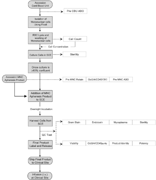
Figure 1. Workflow of SCE therapy and quality control testing. Please click here to view a larger version of this figure.
Cord blood mononuclear cells are isolated, washed, and cultured in the SCE device. After the culture reaches ≥80% confluence, the patient mononuclear cell (MNC) apheresis product is added to the SCE device for immune education. Following overnight incubation, the SCE-treated MNC apheresis product is collected and subjected to quality control (QC) testing before final-product labeling, release, and intravenous infusion. RBCs, red blood cells; MNC, mononuclear cell; SCE, Stem Cell Educator; QC, quality control.
Enroll patients aged ≥18 years with a confirmed diagnosis of long COVID and clinical symptoms persisting for ≥3 months. Exclude patients with clinically significant liver, kidney, or heart disease, or pregnancy. Schedule follow-up testing at months 1, 3, 6, 9, and 12 after SCE therapy to assess the durability of immune markers.
1. Preparation of SCE cultures
- Receive the cord blood unit and release it for good manufacturing practice (GMP) production.
- Transfer 30 mL of cord blood over 20 mL of density gradient medium (γ = 1.077) in a 50 mL conical tube.
- Centrifuge at 1,690 × g for 15 min at 20°C in a swinging-bucket rotor without braking.
- Carefully transfer the mononuclear cell layer to a new 50 mL conical tube containing 10 mL of 0.9% sodium chloride. Centrifuge at 751 × g for 10 min at 20°C to pellet the cells.
- Discard the supernatant and add 10 mL of ACK lysis buffer to the cell pellet. Resuspend the cells by pipetting and incubate for 2–4 min at room temperature to remove red blood cells.
- Fill the conical tube with 35 mL of 0.9% sodium chloride. Centrifuge at 751 × g for 5 min and discard the supernatant to obtain pelleted mononuclear cells.
- Wash the cells once with 40 mL of 0.9% sodium chloride to remove the remaining lysis buffer.
- Centrifuge at 751 × g for 5 min to pellet the cells.
- Discard the supernatant and resuspend the cord blood mononuclear cells in 10 mL of chemically defined serum-free medium per tube.
- Combine the cord blood mononuclear cell suspensions into a single tube.
- Transfer 1 mL of the cell suspension into a new 50 mL conical tube and add 29 mL of chemically defined serum-free medium to prepare a 1:30 dilution.
- Load 20 µL of the diluted cell suspension into the counting chamber. Quantify the cell number using an automated cell counter.
- Set up TC20 Automated Cell Counter, select cell size gating from 4–12 µm, and count cell concentration.
NOTE: Cord blood units are freshly collected with high viability (≥95%). There is no need to test cell viability. - Seed the mononuclear cells into each layer of the SCE device (12-layer immune-education culture device) at 3–4 mL per layer. Add 25 mL of chemically defined serum-free cell culture medium to each layer. Use ≥ 240 × 106 total nucleated cells (TNC) for preparation of one 12-layer device.
- Target 20–40 × 106 cord blood mononuclear cells per layer.
- Incubate the 12-layer immune-education culture device at 37°C under 8% CO₂ for 10–14 days until the CB-SC reach ≥ 80% confluence.
- Do not change the medium during the 10–14-day incubation. Add 20 mL medium/layer to keep CB-SC at good viability if the SCE cannot be used for clinical treatment within 20 days after the start of incubation.
- Observe the CB-SC cultures under the microscope, randomly count the cell confluence from five fields (center, left, top, right and bottom of top layer), and then calculate the average of cell confluence. SCE will be ready for clinical treatment once the CB-SC reach ≥ 80% confluence.
- Using a 10 mL syringe, inoculate 9 mL of the 1:30 diluted suspension into each BD BACTEC aerobic and anaerobic sterility-test bottle (commercial blood-culture bottles).
- Load the sterility-test bottles into the BD BACTEC FX40 automated blood culture system (automated sterility-testing system) and perform sterility testing for 14 days.
- The SCE product will be used for clinical treatment only if the sterility test results are negative after 14 days of incubation. If any bottle tests positive for microbial contamination, the entire SCE product will be discarded and will not be used for clinical treatment.
2. Peripheral blood vessel access and cannulation
NOTE: Perform peripheral blood vessel access and cannulation before apheresis. Perform this as a clean procedure using sterile draping to minimize field contamination; the degree of sterile preparation required for central venous catheter insertion is not required.
- Prepare the tray
- Cover a standard Mayo stand with a disposable absorbent underpad.
- Cover the underpad with a sterile drape. Maintain the sterility of the upper surface while unfolding the drape.
- Open the sterile gloves, folded in sterile paper, in the center of the sterile drape. Keep the upper surface of the glove paper sterile.
- Place 5–6 separate spots of sterile ultrasound gel onto the side of the sterile glove paper without contaminating the gel.
- NOTE: Maintain the sterility of the ultrasound gel because it contacts the patient’s skin within the sterile field and is used near the IV needle puncture site.
- Place two Tegaderm transparent film dressings onto the sterile field while maintaining sterility.
- Place two pieces of sterile gauze onto the sterile field.
- Place two sterile insulin syringes for administration of local anesthetic onto the sterile field.
- Prepare the arm and ultrasound probe for cannulation
- Apply a single-use tourniquet to the patient’s arm.
- Don sterile gloves.
- Ask an assistant to hold the single-use lidocaine vial upside down. Draw 1 mL of local anesthetic into each sterile disposable insulin syringe.
- The concentration of lidocaine is 2%.
- Disinfect the anticipated cannulation sites using chlorhexidine sterile swabs and cover the area with a sterile fenestrated drape, positioning the opening over the cannulation sites. Allow the chlorhexidine to dry.
- Ask an assistant to sanitize the ultrasound probe using CaviWipes surface disinfectant and hand the sanitized probe to the operator.
- Hold the ultrasound probe in the left hand and dip the probe footprint into one of the sterile ultrasound gel spots. Do not reuse the gel spot.
- Apply a transparent film dressing to the probe footprint using the right hand to create a sterile ultrasound contact surface.
- Dip the covered ultrasound probe footprint into a new spot of sterile ultrasound gel.
- Cannulate the vein
- Ask an assistant to hold the monitor displaying the ultrasound image. Use the ultrasound probe to identify an antecubital vein suitable for cannulation while ensuring that only the sterile ultrasound probe footprint contacts the prepared cannulation site.
- Use the predrawn syringe containing local anesthetic to anesthetize the skin at the cannulation site.
- Cannulate the vein using a 16 G IV catheter.
- Ask the assistant to remove the tourniquet.
- Secure the IV cannula while placing sterile gauze beneath the cannula opening.
- Clamp the catheter, remove the needle, and immediately connect a flushed needleless connector/extension set.
- Unclamp the catheter and flush it with 3–5 mL of normal saline using the push-pause method to confirm successful venous access and maintain catheter patency.
- Cover and secure the cannulation site with a transparent film dressing.
- Remove the sharps from the sterile field and tray, and dispose of them appropriately. Remove the draping from the patient’s arm.
- Repeat the procedure on the opposite arm
- Remove the gloves and perform hand hygiene.
- Apply a new single-use tourniquet to the opposite arm.
- Don a new pair of sterile gloves.
- Reuse the prepared sterile tray only if it has remained uncontaminated. Retain the prepared anesthetic syringe, transparent film dressing, and gauze on the sterile tray.
- Ask the assistant to sanitize the ultrasound probe again before returning it to the operator. Prepare a new sterile ultrasound contact surface using a new transparent film dressing as described in steps 2.2.6–2.2.9.
- Repeat steps 2.3.1–2.3.9 on the opposite arm.
3. Mononuclear cell (MNC) apheresis
- Set up the Spectra Optia blood cell separator and select the Continuous CMNC apheresis protocol in the system software version 12.1.
- Set up the initial whole blood-to-ACD-A ratio at 12:1 and target the collected volume between 250 and 300 mL.
- Prepare the patient for apheresis. Record the patient’s body weight and height, and review the complete blood count (CBC) laboratory report for the hematocrit value.
- Enter the patient’s weight, height, and hematocrit into the blood cell separator.
- Confirm reliable venous access by flushing the catheter with 3–5 mL of normal saline.
- Connect the catheter to the blood cell separator and initiate the MNC apheresis procedure.
- Prepare the calcium gluconate solution by adding 20 mL of 10% calcium gluconate (2 g) to 250 mL of physiological saline. Connect the infusion line to the Return line of MNC apheresis at the distal portion of the circuit before the fluid re-enters the patient's venous access device and infuse the solution into the patient at 50 mL/h.
NOTE: Administer calcium gluconate to reduce adverse effects associated with acid citrate dextrose solution A (ACD-A) anticoagulation during collection. - Manually adjust the colorgram and flow rate during collection to optimize product purity.
- Inspect the color of the collection line and visually compare the color of the blood component flowing through the collection line against the Terumo BCT Spectra Optia Collection Preference Tool Colorgram to target a dark-red color with Collection Preference between 25 and 35.
- Maintain the whole blood-to-ACD-A ratio at 12:1.
NOTE: Adjust the whole blood-to-ACD-A ratio when the patient’s CBC indicates a high hematocrit level or when clotting occurs. - Adjust the whole blood-to-ACD-A ratio to 10:1 if platelet aggregation, clumping, or fiber/clot formation is observed inside the tubing set or near the collection port.
- Monitor the patient’s blood pressure and heart rate every 30 min during apheresis.
- Continue apheresis until the target blood volume has been processed.
- Normally process the target blood volume at 2.5 total blood volume (TBV).
- Seal the collection bag and label the collected MNC product.
- Return the residual blood to the patient.
- Transfer the apheresis product to the GMP laboratory at the outpatient treatment facility.
- After collection, monitor the patient’s vital signs twice at 15 min intervals. Evaluate the venous access site and discharge the patient after confirming clinical stability.
NOTE: Document and manage any adverse events occurring during or after leukapheresis according to the study protocol.
4. Immune education of patient mononuclear cells collected by apheresis
- Purify mononuclear cells from the apheresis product
NOTE: Remove contaminating red blood cells from the apheresis product before immune education by CB-SC.- Layer 30–35 mL of the apheresis product over 15 mL of density gradient medium (γ = 1.077) in a 50 mL conical tube.
- Centrifuge at 1,690 × g for 15 min at 20°C in a swinging-bucket rotor without braking.
- Carefully transfer the mononuclear cell layer to a new 50 mL conical tube containing 10 mL of 0.9% sodium chloride. Centrifuge at 751 × g for 10 min at 20°C to pellet the cells.
- Discard the supernatant.
- Wash the cells once with 40 mL of 0.9% sodium chloride. Centrifuge at 751 × g for 5 min and discard the supernatant.
- Resuspend the cell pellet in 40 mL of 0.9% sodium chloride.
- Determine the cell concentration using a hemocytometer after preparing the 1:100 dilution.
- Prepare a 1:100 dilution by adding 100 µL of the cell suspension to 9,900 µL of 0.9% sodium chloride in a new 15 mL conical tube.
- Calculate the total cell number and the volume of the mononuclear cell suspension required for addition to the 12-layer immune-education culture device.
- Adjust cell concentration to 25–85 × 107 cells /mL.
- Prepare the immune-education culture device
- Wipe the 12-layer immune-education culture device with an alcohol swab and place it in a biosafety cabinet.
- Remove all caps from the device sequentially from top to bottom. Avoid wetting the filters.
- Gently mix the device two to three times using a circular motion and pour the culture medium into a sterile tray.
- Wash each layer of the immune-education culture device
- Add 20 mL of 0.9% sodium chloride to each layer using a 25 mL plastic pipette.
- Tilt the device forward to visually confirm that 0.9% sodium chloride has been added to each layer.
- Gently mix the device three times using a circular motion and pour the 0.9% sodium chloride into the sterile tray.
- Repeat steps 4.3.1–4.3.3 to perform a total of two complete washes.
- Wash the bottom chamber of the immune-education culture device
- Clean the bottom port with an alcohol preparation pad.
- Remove the cap from the bottom port using an alcohol preparation pad. Position the device over the sterile tray or on a sterile towel.
- Allow residual 0.9% sodium chloride to drain into the sterile tray.
- Transfer 45 mL of 0.9% sodium chloride into a 50 mL conical tube using a 25 mL plastic pipette.
- Attach an 18 G needle or Monoject blunt cannula to a 50 mL syringe.
- Draw 20 mL of 0.9% sodium chloride from the 50 mL conical tube into the syringe.
- Insert the needle or blunt cannula into the bottom port and dispense the 0.9% sodium chloride into the device.
- Gently mix the device three times using a circular motion and pour the 0.9% sodium chloride into the sterile tray.
- Repeat steps 4.4.6–4.4.8 to perform a total of two complete washes.
- Replace the cap on the bottom port.
- Prepare the cell culture suspension
- Obtain a new 500 mL centrifuge bottle.
- Add 247.6 mL of 0.9% sodium chloride to the bottle.
- Add 66.4 mL of the patient’s plasma.
- Before transferring the mononuclear cell layer (see Step 4.1.3), collect 10–15 mL of plasma from each centrifuged 50 mL conical tube and transfer the plasma into the 500 mL centrifuge bottle (see Step 4.5.1), resulting in a total patient plasma volume of 66.4 mL.
- Add 10 mL of the patient’s mononuclear cell suspension to the prepared cell culture medium and mix.
- Load 27 mL of the cell suspension into each layer of the 12-layer CB-SC-coated immune-education culture device using a 25 mL plastic pipette.
- Incubate the device at 37°C under 8% CO₂ for 17 ± 1 h.
- Perform sterility testing
- Using a 10 mL syringe, inoculate 9 mL of the cell suspension into each aerobic and anaerobic sterility-test bottle.
- Load the sterility-test bottles into the automated blood culture system and initiate sterility testing.
NOTE: Use this sterility test to determine whether the apheresis product was contaminated during collection or processing. - After 17 h of sterility testing (see step 5.3.7), review the real-time sterility results and assess whether microbial contamination is present in the apheresis product.
5. Collect the final product and perform quality control (QC) testing
- Verify the sterility-test result
- Check the sterility-test result for the apheresis product before collecting the final product.
- Discard the product and do not collect or infuse it if the sterility-test result is positive for bacterial contamination.
- Prepare the immune-education culture device for final-product collection
- Wipe the 12-layer immune-education culture device with a 70% alcohol preparation pad and place it in the biosafety cabinet.
- Clean the top port with an alcohol preparation pad and remove the white cap.
- Attach the Plastisol Horseshoe Y connector from the loading set to the top port.
- Close all pinch clamps on the Y connector of the loading set.
- Using the spike with chamber, connect a 250 mL bag of 0.9% sodium chloride to the loading set.
- Hang the 0.9% sodium chloride bag from the hook inside the biosafety cabinet.
- Clean the bottom port with an alcohol preparation pad and remove the cap.
- Attach a Plastisol Horseshoe Y connector to the bottom port.
- Connect a 1,000 mL collection bag to the bottom Y connector.
- Place the 1,000 mL collection bag in a tray outside the biosafety cabinet and leave the corresponding clamp open. Position the collection bag below the level of the biosafety cabinet to permit continuous gravity flow.
- Unclamp the 0.9% sodium chloride bag and allow the solution to drain into the immune-education culture device.
- Swirl the device using a circular motion to allow the 0.9% sodium chloride to wash downward through the device and drain into the collection bag.
- Collect the final product
- Tilt the immune-education culture device to drain the SCE-treated MNC apheresis product into the collection bag.
- After the product has completely drained into the collection bag, disconnect the loading set from the bottom Y connector and connect a new 50 mL syringe.
- Gently mix the final product 10 times and withdraw 25 mL of the final product for QC testing.
- Allocate final-product samples for QC testing
- Transfer 7 mL of the final product into a new 15 mL conical tube for flow cytometry by using a Gallios flow cytometer.
- Transfer 0.5 mL of the final product for manual cell counting and cell viability testing by using the trypan blue exclusion test under a microscope. The acceptance criterion is a cell viability of ≥85%.
- Transfer 0.5 mL of the final product for endotoxin testing by using the Endosafe nexgen-PTS with a cartridge sensitivity of 0.005–0.5 EU/mL. The acceptance criterion is an endotoxin level of ≤0.5 EU/mL.
- Transfer 1 mL of the final product for mycoplasma testing by using the 7500 Fast Real-Time PCR System. The acceptance criterion is a negative mycoplasma test result.
- Transfer 8 mL of the final product into each of two sterility-test bottles and perform sterility testing using the BD BACTEC FX40. The acceptance criterion is a negative sterility test result.
- Seal, label, and release the final product
- Seal the collection bag using a tube sealer and disconnect the final product from the immune-education culture device.
- Prepare and label the final product.
- Confirm a cell viability of ≥85%, an endotoxin level of ≤0.5 EU/mL, and a negative overnight sterility-test result for the apheresis product before releasing the final product. These QC tests must be completed and meet acceptance criteria before releasing for infusion.
- Perform flow cytometry and mycoplasma testing on final product.
- Release the final product for infusion after all required pre-release QC criteria have been satisfied.
6. Infusion of SCE-treated MNC apheresis product
- Perform hand hygiene before handling the infusion product.
- Don a new pair of sterile gloves.
- Measure the patient’s temperature and blood pressure before initiating the infusion.
- Connect the patient’s IV catheter to the infusion bag containing the SCE-treated MNC apheresis product.
- Initiate the infusion at 100 mL/h and maintain this rate for 30 min.
- After 30 min, assess the patient for adverse reactions. If no adverse reaction occurs, increase the infusion rate to 150 mL/h and maintain this rate for 1 h.
- If the patient tolerates the infusion at 150 mL/h, increase the infusion rate to a maximum of 200 mL/h and continue until the infusion is complete.
- If the patient develops adverse events such as chills, itching, or hives, administer 25 mg diphenhydramine (Benadryl) intravenously and reduce the infusion rate to 150 mL/h.
NOTE: If the patient develops nausea and vomiting, stop the infusion and administer 25 mg diphenhydramine and 10 mg dexamethasone intravenously. Resume the infusion at a lower rate of 120 mL/h only when clinically appropriate and after the patient’s symptoms have resolved. - After completing the infusion, rinse the infusion bag with 50 mL of normal saline and infuse the saline into the patient.
- Monitor the patient for adverse reactions for 15 min after completion of the infusion.
- If the patient remains clinically stable, with normal body temperature and blood pressure, the patient may be discharged.
- Discharge the patient after confirming that the post-infusion discharge criteria have been met.
7. Flow cytometry analysis of immune markers
- Take the cell suspension (step 5.4.1) and collect cells at 751 × g for 5 min.
- Resuspend the cells in 200 µL of 0.9% sodium chloride and transfer cells into a 5 mL conical tube.
- Add 5 µL of Fc blocker per sample and incubate for 15 min at room temperature to block the non-specific binding.
- Aliquot 100 µL of cell suspension for intracellular staining and 100 µL of cell suspension for cell-surface staining.
- For cell surface staining: aliquot 30 μL of cell suspension into three 5 mL conical tubes and add 70 µL of 0.9% sodium chloride to each tube with final volume 100 µL/tube, and then add 5 µL/tube fluorescence-conjugated mouse anti-human monoclonal antibodies including human regulatory T cell cocktail (FITC Mouse anti-Human CD4, PE-Cy7 Mouse anti-Human CD25, and Alexa Fluor 647 Mouse anti-Human CD127); pacific blue (PB)-conjugated mouse anti-human CD3, Spark Violet 538 anti-human CD4, APC/Fire 750 anti-human CD8, PE/Cyanine7-conjugated anti-human CD45RO, PE-conjugated anti-human CCR7, and isotype-matched IgG controls to 100 μL volume of cells. Incubate for 30 min at room temperature with light protection.
- For intracellular staining: cells are fixed and permeabilized according to the PerFix-nc kit.
- Add 5 µL/tube fluorescence-conjugated mouse anti-human monoclonal antibodies including BV421-conjugated mouse anti-human IL-1β, PE-conjugated mouse anti-human CXCL16, and isotype-matched IgG controls mAbs. Incubate for 30 min at room temperature with light protection.
- After staining, wash cells with 4 mL of 0.9% sodium chloride and centrifuge at 751 × g for 5 min to pellet cells.
- Resuspend the cells with 200 µL of 0.9% sodium chloride.
- Collect and analyze cells using a Gallios Flow Cytometer, acquiring 100,000 events for surface staining and 500,000 events for intracellular staining. Gating strategies for flow cytometry analysis of different T-cell populations include CD3+CD4+ T cells, CD3+CD8+ T cells, CD4+CD25+CD127low/− Tregs, CD4+CD45RO+CCR7+ central memory T (TCM) cells, CD4+CD45RO+CCR7− effector memory T (TEM) cells, and CD4+CD45RO−CCR7+ naïve T cells. Use isotype-matched IgG controls to establish positive thresholds.
- Analyze the final data using the Kaluza Flow Cytometry Analysis Software.
8. Statistical analysis
- Assess data normality using the Shapiro–Wilk test. Use a paired t-test for normally distributed paired data and the Wilcoxon matched-pairs signed-rank test for nonparametric paired data. Define statistical significance as P < 0.05. The data are presented as mean ± standard deviation (SD).
- In the current clinical study, the primary efficacy endpoint was the change in the immune markers between baseline and follow-up month 1. Results from this long COVID study were analyzed using a significance level of 5% for paired comparisons between baseline and follow-up month 1 measurements. Multiple-comparison correction was not applicable to this study.
- Calculate 95% confidence intervals using GraphPad Prism (version 10.6.1).
Results
Twenty-five patients with long COVID (15 males and 10 females; age, 51.68 ± 14.14 years) received SCE therapy (Figure 1). Five subjects (5/25) experienced mild tingling and numbness during apheresis, which were attributed to reduced calcium levels associated with citrate anticoagulation. These symptoms resolved promptly following administration of oral calcium tablets or intravenous calcium gluconate. No significant adverse events were reported during the Gleukocell infusion. One subject (HU2291; 1/25, 4.0%) experienced mild itching during the infusion.
Flow cytometry demonstrated that the percentage of Tregs increased from 5.76% ± 1.21% at baseline to 7.53% ± 3.17% at the 1-month follow-up (Figure 2, P = 0.003). In the current clinical study, 17/25 subjects with long COVID displayed elevated percentages of IL-1β-positive cells, with a baseline value of 8.43% ± 10.12%. After treatment with SCE therapy, the percentage of IL-1β-positive cells decreased to 3.01% ± 6.07% (Figure 3, P = 0.0017).
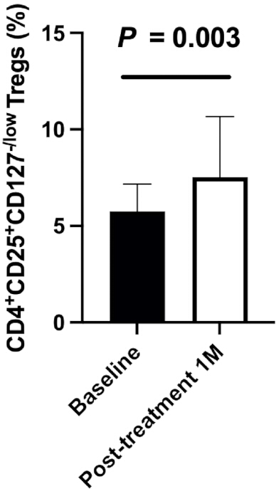
Figure 2. Increase in regulatory T cells in patients with long COVID one month after SCE therapy. Please click here to view a larger version of this figure.
All subjects received one treatment with SCE therapy. Peripheral blood mononuclear cells (PBMC) were analyzed at baseline and one month after treatment. Data normality was assessed using the Shapiro–Wilk test. Because the data were nonparametrically distributed, the Wilcoxon matched-pairs signed-rank test was used for statistical analysis in GraphPad Prism version 10.6.1. Statistical significance was defined as P < 0.05. The percentage of regulatory T cells (Tregs) increased significantly one month after SCE therapy (P = 0.003). Error bars represent SD. PBMC, peripheral blood mononuclear cells; SCE, Stem Cell Educator; Tregs, regulatory T cells.
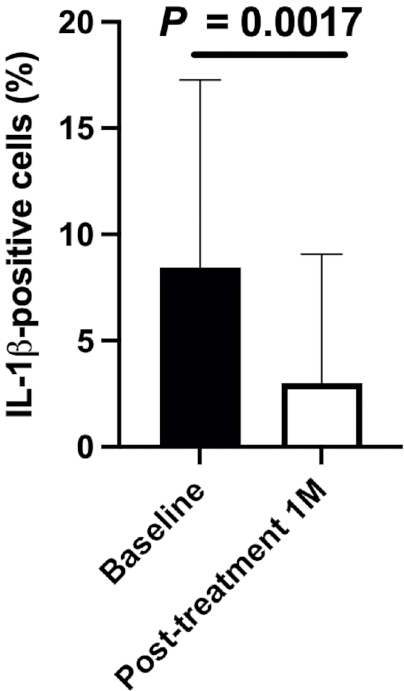
Figure 3. Reduction in IL-1β-positive cells in peripheral blood mononuclear cells of patients with long COVID after SCE therapy. Please click here to view a larger version of this figure.
All subjects received one treatment with SCE therapy. Peripheral blood mononuclear cells (PBMC) were isolated at baseline and one month after treatment and analyzed by intracellular flow cytometry using the PerFix-nc cellular staining kit and a Gallios flow cytometer equipped with 488 nm, 638 nm, and 405 nm lasers. Data were analyzed using Kaluza Flow Cytometry Analysis Software version 2.1. Isotype-matched immunoglobulin G (IgG) controls were used as controls. The Wilcoxon matched-pairs signed-rank test was used for statistical analysis in GraphPad Prism version 10.6.1. Statistical significance was defined as P < 0.05. The percentage of IL-1β-positive cells decreased significantly one month after SCE therapy (P = 0.0017). Error bars represent SD. IL-1β, interleukin-1 beta; PBMC, peripheral blood mononuclear cells; SCE, Stem Cell Educator.
CXCL16 is expressed by a variety of immune cells, including monocytes/macrophages, dendritic cells, and B cells. Recent studies suggest that CXCL16 contributes to the pathogenesis of autoimmune diseases such as Crohn’s disease, multiple sclerosis, rheumatoid arthritis, and lupus33,34,35, as well as long COVID36. Flow cytometry showed that PBMC from patients with long COVID displayed a high percentage of CXCL16-positive cells at baseline (63.98% ± 19.11%), which decreased to 44.1% ± 24.52% one month after SCE therapy (Figure 4, P = 0.03).
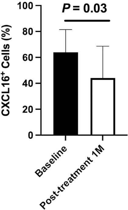
Figure 4. Reduction in CXCL16-positive cells in peripheral blood mononuclear cells of patients with long COVID one month after SCE therapy. Please click here to view a larger version of this figure.
All subjects received one treatment with SCE therapy. Peripheral blood mononuclear cells (PBMC) were isolated at baseline and one month after treatment and analyzed by flow cytometry using the PerFix-nc cellular staining kit and a Gallios flow cytometer. Isotype-matched immunoglobulin G (IgG) controls were used as controls. Data normality was assessed using the Shapiro–Wilk test, and a paired t-test was used for statistical analysis in GraphPad Prism version 10.6.1. Statistical significance was defined as P < 0.05. The percentage of CXCL16-positive cells decreased significantly one month after SCE therapy (P = 0.03). Error bars represent SD. CXCL16, C-X-C motif chemokine ligand 16; PBMC, peripheral blood mononuclear cells; SCE, Stem Cell Educator.
The percentage of CD4+CD45RO+CCR7− TEM cells decreased in subjects with long COVID one month after treatment with SCE therapy (Figure 5A, P = 0.0003). Conversely, the percentage of naïve CD4+CD45RO−CCR7+ T cells increased one month after SCE therapy (Figure 5B, P = 0.0026). These findings demonstrate changes in CD4+ effector memory and naïve T-cell populations following SCE therapy. The baseline and 1-month values, 95% confidence intervals, and corresponding P values for these immune-marker comparisons are summarized in Table 1.
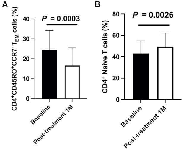
Figure 5. Changes in CD4+ effector memory and naïve T-cell populations in patients with long COVID after SCE therapy. Please click here to view a larger version of this figure.
All subjects (N = 25) received one treatment with SCE therapy. Peripheral blood mononuclear cells (PBMC) were isolated at baseline and one month after treatment and analyzed by flow cytometry using a Gallios flow cytometer. Data were analyzed using Kaluza Flow Cytometry Analysis Software version 2.1. CD3+ T cells were gated from total PBMC for analysis of T-cell subsets. Isotype-matched immunoglobulin G (IgG) controls were used as controls. (A) Percentage of CD4+CD45RO+CCR7− effector memory T (TEM) cells at baseline and one month after SCE therapy. TEM cells decreased significantly one month after treatment (P = 0.0003). (B) Percentage of CD4+CD45RO−CCR7+ naïve T cells at baseline and one month after SCE therapy. Naïve T cells increased significantly one month after treatment (P = 0.0026). A paired t-test was used for statistical analysis in GraphPad Prism version 10.6.1. Statistical significance was defined as P < 0.05. Error bars represent SD. PBMC, peripheral blood mononuclear cells; SCE, Stem Cell Educator; TEM, effector memory T cells.
| Immune markers in PBMC | Baseline (%) | 1 month post-treatment (%) | 95% confidence interval | P value | |
| Regulatory T cells (Tregs) | 5.76 ± 1.21 | 7.53 ± 3.17 | 0.11 to 3.81 | 0.003 | |
| IL-1β⁺ cells | 8.43 ± 10.12 | 3.01 ± 6.07 | −9.73 to −0.04 | 0.0017 | |
| CXCL16⁺ cells | 63.98 ± 19.11 | 44.1 ± 24.52 | −35.50 to −1.557 | 0.03 | |
| CD4⁺CD45RO⁺CCR7⁻ TEM cells | 24.48 ± 10.13 | 16.65 ± 8.83 | −15.27 to −5.686 | 0.0003 | |
| Naïve CD4⁺CD45RO⁻CCR7⁺ T cells | 42.95 ± 12.63 | 49.42 ± 12.6 | 4.016 to 15.39 | 0.0026 | |
Table 1: Comparison of immune markers in peripheral blood mononuclear cells (PBMCs) at baseline and 1 month after treatment.
Data are presented as mean ± SD. The 95% confidence intervals correspond to the baseline-to-1-month comparisons. Statistical analyses were performed as described in the Statistical Analysis section.
Twenty-one of 25 patients reported resolution of the common long COVID symptoms of brain fog and fatigue following treatment with SCE therapy. Most participants also reported feeling refreshed, energized, and mentally clear after therapy. Notably, subject HU2171, a medical resident who cared for patients with COVID-19 in the intensive care unit during the pandemic, had contracted COVID-19 in 2021. Over the subsequent four years, he underwent various treatments, including JAK inhibitors and seven infusions of autologous MSCs derived from abdominal adipose tissue, without reported improvement. His condition progressively worsened, eventually rendering him bedridden for two years. He developed severe sensitivity to light and sound and kept his room darkened with black tape sealing the window gaps. After receiving SCE therapy, he demonstrated clinical improvement over a 3-month period. His light and sound sensitivities resolved, without recurrence of headaches. Flow cytometry also showed immunological changes: the percentage of IL-1β-positive cells decreased from 8.84% at baseline to 1.7% at 3 months after treatment, and the percentage of CXCL16-positive cells decreased from 78.42% at baseline to 48.16%. These clinical observations were accompanied by reductions in the measured inflammatory immune-cell markers following SCE therapy.
Discussion
Given the millions of individuals affected by long COVID and the current lack of disease-modifying therapies, with care largely consisting of symptomatic and supportive approaches, we explored the therapeutic potential of SCE therapy in patients with long COVID using the previously described open-loop system25 and clinical protocols previously applied to T1D and AA. In this pilot study, 25 subjects with long COVID received SCE therapy, with a safety profile consistent with previously published clinical studies25,28. Clinical symptoms, including brain fog and fatigue, improved in most subjects with long COVID. Critical steps of the protocol include the isolation and culture of CB-SC, collection of the patient's mononuclear cells by apheresis, removal of contaminating red blood cells, co-culture of patient mononuclear cells with CB-SC in the SCE device for 17 ± 1 h, collection and QC testing of the final product, and reinfusion of the SCE-treated MNC apheresis product. Maintaining the specified culture conditions, cell-processing procedures, sterility testing, and final-product release criteria is important for consistent implementation of the method. Before treatment, patients with long COVID exhibited increased non-conventional monocytes (CD14lowCD16high)10. SARS-CoV-2 spike protein (S-protein) can prime inflammasome formation and the release of mature IL-1β in macrophages derived from patients with COVID-1937. IL-1β, produced by monocytes/macrophages, is a major regulator of inflammation that orchestrates various innate immune responses38. The current study demonstrated that most subjects with long COVID displayed increased percentages of IL-1β-positive cells, which significantly decreased by one month after SCE therapy, consistent with downregulation of the activated monocyte/macrophage system. Inflammatory microglia, the resident macrophages of the central nervous system, may also be involved in neurological manifestations; however, microglial changes were not directly measured in the current study. Therefore, the observed clinical improvements in brain fog, fatigue, and cognitive impairment cannot be directly attributed to changes in microglial activity. The detailed mechanisms underlying cognitive impairment remain unclear and may involve multiple pathological processes, including increased reactive oxygen species (ROS), iron accumulation39, and pyroptosis of neural stem cells40. Additionally, the percentages of CXCL16-positive cells were significantly reduced in subjects with long COVID one month after SCE therapy. CXCL16 is predominantly expressed by monocytes/macrophages, B cells, and dendritic cells and plays an important role in immune cell trafficking. The observed reduction in CXCL16-positive cells may therefore be associated with changes in immune-cell trafficking; however, migration across the blood–brain barrier was not directly evaluated in this study. These findings suggest that the percentages of IL-1β-positive and CXCL16-positive cells in PBMC may represent potential candidate biomarkers that warrant further investigation. Future prospective studies with larger cohorts and predefined clinical response criteria should evaluate whether baseline IL-1β and/or CXCL16 levels are associated with, or can predict, treatment response.
Adaptive immune responses are crucial for protection against viral reinfection through the establishment of immune memory. Clinical studies have demonstrated that robust mucosal SARS-CoV-2-specific T-cell responses can contribute to protection against COVID-19 and the establishment of polyfunctional resident memory T cells within the lungs41. Adamo et al. reported long-lived effector memory CD8+ T cells in the blood of patients with COVID-19 from the acute infection phase through 1 year after recovery42. The current clinical data demonstrated significant decreases in the percentages of CD4+ TEM cells, accompanied by significant increases in the percentages of CD4+ naïve T cells and CD4+CD25+CD127low/− Tregs. Although similar trends were observed for CD8+ TEM cells and CD8+ naïve T cells, these changes did not reach statistical significance, potentially because of the small sample size in the current clinical study. We acknowledge that the relatively small sample size and lack of a prospective power calculation are limitations of the current clinical study. Additional limitation of the present study is that these observations were obtained from a small, open-label, single-site cohort without a concurrent control group. The reported clinical outcomes were primarily symptom-based, and the possibility of placebo effects contributing to some of the observed symptomatic improvements cannot be completely excluded. However, most patients in the current study had previously failed to achieve satisfactory improvement with multiple conventional therapies. Notably, subject HU2171 had undergone several treatments, including JAK inhibitor therapy and autologous mesenchymal stem cell (MSC) therapy, without adequate clinical benefit. Therefore, while a placebo effect cannot be ruled out, the magnitude of the observed clinical improvements and restoration of immune markers to the normal levels may be less likely to be explained solely by placebo effects.
The present study does not establish whether the observed changes are specific to SCE therapy. Larger, well-controlled clinical studies are therefore warranted to validate these findings and further characterize the immunomodulatory effects of SCE therapy in patients with long COVID. Future studies should incorporate an appropriate control group to more rigorously assess treatment safety and clinical efficacy. Longer-term follow-up will also be important for determining the durability of the observed clinical and immunological changes. Future studies should incorporate randomized controlled trial designs with appropriate control groups, including sham apheresis and/or placebo controls, where feasible, to rigorously evaluate the efficacy of SCE therapy. Such controlled studies will help distinguish treatment-specific effects from potential nonspecific effects associated with the apheresis procedure, cell infusion, or placebo responses. We will consider these approaches in future clinical trials to more definitively establish the efficacy, safety, and clinical benefits of SCE therapy in patients with long COVID.
Subjects with long COVID exhibit multiple forms of immune dysfunction and clinical features resembling autoimmune disorders, involving several immune-cell compartments, including monocytes/macrophages, T cells, B cells, NKT cells, and Tregs. Targeting these diverse immune-cell populations simultaneously with conventional immunotherapies and pharmacological strategies is challenging in clinical management. Mesenchymal stem cells also have limitations in addressing multiple immune-cell dysfunctions43,44. SCE therapy may provide a broader approach to immune modulation, as suggested by previous clinical studies25,45. During the procedure, immune cells are collected from the patient's peripheral blood by leukapheresis and co-cultured with CB-SC for 17 ± 1 h within the SCE device. The CB-SC-educated immune cells are subsequently collected and reinfused into the patient's circulation. Previous studies have described multiple molecular mechanisms associated with CB-SC-mediated immune modulation, including AIRE transcription factor-mediated induction of immune tolerance in T cells, galectin-9-mediated modulation of B-cell function, and exosome-mediated regulation of monocytes/macrophages. Thus, SCE therapy may provide a multifaceted strategy for addressing the broad immune dysfunction associated with long COVID. Future mechanistic studies need to distinguish exosome-mediated from cell-mediated effects underlying SCE therapy in patients with long COVID. From a methodological perspective, the ex vivo approach allows patient immune cells to interact with CB-SC before the treated cells are returned to the patient, rather than administering CB-SC directly. Potential applications of this method therefore include further investigation of immune modulation in long COVID and other conditions characterized by broad immune dysfunction. Interestingly, patients with long COVID share clinical and biological features with other post-viral syndromes, including myalgic encephalomyelitis/chronic fatigue syndrome (ME/CFS). Although the present study focused exclusively on long COVID and does not provide evidence for the efficacy of SCE therapy in ME/CFS or other post-viral conditions, the immunomodulatory effects observed in this study, particularly the reduction of inflammatory monocyte/macrophage-associated CXCL16 and IL-1β responses, provide a rationale for further investigation of SCE therapy in patients with ME/CFS and potentially other post-viral syndromes in the future.
In the present study, we cannot definitively determine which subset of patients with long COVID is most likely to benefit from Stem Cell Educator therapy because of the limited sample size, lack of a randomized control group, and relatively short follow-up period. However, our findings suggest that patients with persistent long COVID symptoms accompanied by measurable immune dysregulation, particularly increased activated T-cell populations and elevated inflammatory markers such as IL-1β and CXCL16, may represent a population of interest for future studies. The observed reductions in CD4⁺ and CD8⁺ effector-memory T cells and inflammatory markers after therapy, together with increases in naïve T-cell populations, provide a rationale for investigating whether patients with a more pronounced inflammatory or immune-dysregulated phenotype are more likely to respond. Additionally, SCE therapy is more procedurally complex than conventional pharmacological therapies because it requires leukapheresis, ex vivo interaction of the patient’s mononuclear cells with cord-blood-derived stem cells in the SCE device, and subsequent reinfusion. Thus, widespread implementation would require enough supplies of cord blood units, appropriately equipped clinical centers, trained personnel, standardized operating procedures, and coordination of cell-processing GMP facilities and infusion activities.
The current proof-of-concept pilot study provides preliminary safety, clinical, and immunological observations following SCE therapy in patients with long COVID. Similar to our ongoing clinical studies in patients with recent-onset T1D, most patients experienced sustained clinical benefits and changes in immune markers after a single treatment without repeated treatment during the reported observation period. The potential durability of this response, as well as the optimal dosing regimen and retreatment intervals, warrants further investigation. SCE therapy differs methodologically from several conventional immunotherapies and other cell- or plasma-based approaches, including IVIG infusion, TPE, lymphoplasmapheresis46, MSC therapy, and EV-based therapies, because the patient's mononuclear cells undergo ex vivo immune education with CB-SC before reinfusion. However, direct comparative efficacy was not evaluated in the present study. Modifications and troubleshooting of the method should focus on maintaining reproducible cell preparation, culture conditions, apheresis procedures, sterility and QC testing, and infusion procedures described in the Protocol; deviations from these parameters may affect consistency of the final product. Taken together with our previous clinical studies of multiple autoimmune conditions, the present findings support further evaluation of SCE therapy as an ex vivo CB-SC-mediated immune-modulation approach for patients with long COVID. Its clinical efficacy, durability, and comparative advantages over alternative approaches require confirmation in larger controlled studies.
Disclosures
Yong Zhao is the inventor of the SCE technology and has a fiduciary role at Throne Biotechnologies (Throne). Boris Veysman, Richard Fox, Zhi Hua Huang, Chandler Judd, Jennifer Anti-Ampratwum, Rebecca Lackrajh, Ana Villagran, Honglan Wang, Yelu Zhao, Terri Tibbot, and Misha Votruba are employees of Throne. All other authors declare no financial interests that may be relevant to the submitted work.
Acknowledgements
Y.Z. supervised the experiments and contributed to the study concept, clinical and experimental design, data analysis and interpretation, manuscript preparation, and final approval of the manuscript. B.V. and R.F. contributed to patient care and final approval of the manuscript. N.M., K.A., and D.P. performed apheresis and contributed to technical support and patient care. Z.H.H., C.J., R.L., A.V., H.W., and Yelu Z. prepared the Stem Cell Educators, performed GMP production and QC testing, isolated PBMC, and performed flow cytometry analysis. J.A. and M.V. contributed to patient care and recruitment. T.T. provided materials. All authors read and approved the final version of the manuscript.
Materials
| Name | Company | Catalog Number | Comments |
|---|---|---|---|
| 0.9% sodium chloride, 250 mL IV bag | B. Braun Medical Inc. | L8002 | Product collection, device washing, and infusion |
| 0.9% sodium chloride, prefilled 10 mL syringe | Aquabiliti | 2T0806 | IV cannulation and flushing |
| 10 mL plastic pipette | MISSCI | PR-SERO-10 | Cell processing and device washing |
| 10 mL syringe | B. Braun | 4617100V-02 | Sterility-test sample inoculation |
| 15 mL conical tube | MISSCI | C15R | Cell dilution and QC sampling |
| 18 G needle | Henry Schein | 900-4469 | Washing the immune-education culture device |
| 25 mL plastic pipette | MISSCI | PR-SERO-25 | Cell processing and device washing |
| 50 mL conical tube | MISSCI | C50R | Cell isolation and washing |
| 50 mL syringe | BD | 309653 | Device washing and final-product sampling |
| 500 mL centrifuge bottle | TPP | 87450 | Preparation of cell culture suspension |
| 7500 Fast Real-Time PCR System | Thermo Scientific | E-PCR-7500-1 | Mycoplasma test |
| 1,000 mL collection bag | ICU Medical | 7951-19 | Final-product collection |
| Acid citrate dextrose solution A (ACD-A) | Terumo BCT | 40817 | Anticoagulant for MNC apheresis |
| Alcohol prep pads | Henry Schein | 570-1307 | Device preparation and IV procedures |
| APC/Fire 750-conjugated mouse anti-human CD8 mAb | BioLegend | 344746 | Flow cytometry |
| APC/Fire 750-conjugated mouse IgG1κ | BioLegend | 400196 | Flow cytometry, isotype control |
| Automated cell counter | BioRad | 1450102 | Cell counting |
| BD BACTEC aerobic blood culture bottle | BD | 442023 | Sterility testing |
| BD BACTEC anaerobic blood culture bottle | BD | 442022 | Sterility testing |
| BD BACTEC FX40 Automated Blood Culture System | BD | B442299 | Sterility testing |
| Biological Safety Cabinet, Class II | Thermo Scientific | Model 1387 | Cell processing and final-product collection |
| Blunt cannula, Monoject | Cardinal Health | 8881202322 | Washing the immune-education culture device |
| BV421-conjugated mouse anti-human IL-1β mAb | BD Biosciences | 567791 | Flow cytometry |
| BV421-conjugated mouse IgG1κ | BD Biosciences | 562438 | Flow cytometry, isotype control |
| Calcium gluconate, 10% | Fresenius Kabi | 63323036019 | Prevention/management of citrate-related adverse effects during apheresis |
| CaviWipes Bleach | Metrex | 13-9100 | Ultrasound probe disinfection |
| Cell-counting chamber slide | BioRad | 1450011 | Automated cell counting |
| Cord blood unit | Life Line Stem Cell Tissue, Cord Blood Bank | N/A | Source material for CB-SC culture |
| Criterion Earloop Mask, Level 3 | Henry Schein | 570-2044 | Personal protective equipment |
| Criterion Nitrile Exam Gloves | Henry Schein | N200 | Personal protective equipment |
| Density gradient medium, Ficoll-Paque Plus | VWR | 17-1440-03 | Mononuclear cell isolation |
| Disposable insulin syringe, sterile, 31 G × 5/16 in | EXEL | 26012 | Local anesthetic administration |
| Endosafe nexgen-PTS | Charles River | PTS150K | Endotoxin testing |
| Endotoxin test cartridge/reagent | Charles River | PTS20005F | Final-product endotoxin QC using the endotoxin-testing system |
| Gallios Flow Cytometer | Beckman Coulter | A94303 | Flow cytometry |
| Gauze pads, 2 × 2 in, 12-ply | Medsource | MS-GZ2212S | IV cannulation |
| Gleukocell | Throne Biotechnologies | GC-2001 | Commercial name of SCE-treated MNC apheresis product |
| GraphPad Prism, version 10.6.1 | Dotmatics | N/A | Statistical analysis |
| Hemocytometer | Cellop | DCC-010 | Cell counting |
| Human Regulatory T Cell Cocktail | BD Pharmingen | 560249 | Flow cytometry |
| Human TruStain FcX (Fc Blocker) | BioLegend | 422302 | Flow cytometry, block non-specific binding |
| Incontinence underpad, 17 × 24 in, light absorbency | Henry Schein | 1125633 | IV cannulation and infusion setup |
| Introcan Safety IV Catheter, 16 G | B. Braun | 4252586-02 | Peripheral venous cannulation |
| Kaluza Flow Cytometry Analysis Software, version 2.1 | Beckman Coulter | B16408 | Flow cytometry data analysis |
| Lidocaine, 2%, 100 mg/5 mL, sterile single-dose vial | Eugia | NDC 55150-165-05 | Local anesthesia for IV cannulation |
| Lysis Buffer | Lonza | BP10-548E | Red blood cell lysis |
| Mayo stand | GoBioMed | DK-4365 | IV cannulation setup |
| Microbore extension set, 8 in | Henry Schein | 570-0884 | IV cannulation |
| Mycoplasma detection test/kit | ThermoFisher Scientific | 4460626 | Final-product QC |
| Pacific Blue-conjugated mouse anti-human CD3 mAb | BioLegend | 300431 | Flow cytometry |
| Pacific Blue-conjugated mouse IgG1κ | BioLegend | 400131 | Flow cytometry, isotype control |
| PE-conjugated mouse anti-human CCR7 mAb | BioLegend | 353204 | Flow cytometry |
| PE-conjugated mouse anti-human CXCL16 mAb | BioLegend | 360804 | Flow cytometry |
| PE-conjugated mouse IgG1κ | BioLegend | 400114 | Flow cytometry, isotype control |
| PE/Cyanine 7-conjugated mouse anti-human CD45RO mAb | BioLegend | 304230 | Flow cytometry |
| PE/Cyanine 7-conjugated mouse IgG1κ | BioLegend | 400126 | Flow cytometry, isotype control |
| PerFix-nc Kit, no-centrifuge assay kit | Beckman Coulter | B31167 | Intracellular staining for flow cytometry |
| Single Transfer Set | Charter Medical | 03-220-90 | Product collection |
| Sorvall ST 16R Centrifuge | Thermo Scientific | 43471362 | Cell isolation and washing |
| Spark Violet 538-conjugated mouse anti-human CD4 mAb | BioLegend | 344674 | Flow cytometry |
| Spark Violet 538-conjugated mouse IgG1κ | BioLegend | 402418 | Flow cytometry, isotype control |
| Spectra Optia | Terumo BCT | 61000 | MNC apheresis |
| Spectra Optia IDL Set | Terumo BCT | 12320 | MNC apheresis |
| Stem Cell Educator device, 12 layers | Throne Biotechnologies | TH-002 | CB-SC culture and immune education |
| Steri-Cycle 370 CO2 Incubator | Thermo Scientific | 370 | CB-SC culture and immune education |
| Sterile Aquasonic 100 Ultrasound Transmission Gel | Parker Laboratories | 01-Jan | Ultrasound-guided IV cannulation |
| Sterile gloves | Henry Schein | 570-4500 | Sterile IV and product-handling procedures |
| Sterile tray | Sampling System | 8081P-3000S | Device washing and product-processing procedures |
| Strap tourniquet, 1 × 18 in | Cardinal Health | CH6064 | IV cannulation |
| Tegaderm Film | 3M Deutschland GmbH | 1624W | IV cannulation and ultrasound probe covering |
| Towel drape with 3 in fenestration, 18 × 26 in | Dynarex | 4409 | IV cannulation |
| Trypan Blue Solution, 0.4% | ThermoFisher Scientific | 15250061 | Final-product viability QC |
| T-Seal Mobile | Terumo BCT | T5460 | Final-product collection |
| Ultros X9HD wireless ultrasound | Ultros Ultrasound | X9HD | Ultrasound-guided IV cannulation |
| X-VIVO 15 | Lonza | BP04-744Q | CB-SC culture |
References
- Greenhalgh T, Sivan M, Perlowski A, Nikolich JZ. Long COVID: a clinical update. Lancet. 2024;404(10453):707-24.
- Davis HE, McCorkell L, Vogel JM, Topol EJ. Long COVID: major findings, mechanisms and recommendations. Nat Rev Microbiol. 2023;21(3):133-46.
- Oba S, Hosoya T, Iwai H, Yasuda S. Long COVID: mechanisms of disease, multisystem sequelae, and prospects for treatment. Immunol Med. 2026;49(1):35-58.
- Soares L, et al. Recommended long COVID outcome measures and their implications for clinical trial design, with a focus on post-exertional malaise. EBioMedicine. 2026;123:106083.
- Al-Aly Z, et al. Long COVID science, research and policy. Nat Med. 2024;30(8):2148-64.
- Dennis A, et al. Multiorgan impairment in low-risk individuals with post-COVID-19 syndrome: a prospective, community-based study. BMJ Open. 2021;11(3):e048391.
- Dennis A, et al. Multi-organ impairment and long COVID: a 1-year prospective, longitudinal cohort study. J R Soc Med. 2023;116(3):97-112.
- Cao B, et al. Clinical practice guideline for long COVID prevention and treatment. Eur Respir J. 2026;68(1):2502611.
- Alwan NA, Al-Aly Z. Learning from long COVID: conceptualising action to tackle infection-associated non-communicable diseases. Lancet. 2025;405(10476):360-2.
- Klein J, et al. Distinguishing features of long COVID identified through immune profiling. Nature. 2023;623(7985):139-48.
- Peluso MJ, et al. Markers of immune activation and inflammation in individuals with postacute sequelae of severe acute respiratory syndrome coronavirus 2 infection. J Infect Dis. 2021;224(11):1839-48.
- Arthur JM, et al. Development of ACE2 autoantibodies after SARS-CoV-2 infection. PLoS One. 2021;16(9):e0257016.
- Su Y, et al. Multiple early factors anticipate post-acute COVID-19 sequelae. Cell. 2022;185(5):881-95.e20.
- Wang EY, et al. Diverse functional autoantibodies in patients with COVID-19. Nature. 2021;595(7866):283-8.
- Talwar S, Harker JA, Openshaw PJM, Thwaites RS. Autoimmunity in long COVID. J Allergy Clin Immunol. 2025;155(4):1082-94.
- Skevaki C, et al. Long COVID: pathophysiology, current concepts, and future directions. J Allergy Clin Immunol. 2025;155(4):1059-70.
- Wrapp D, et al. Cryo-EM structure of the 2019-nCoV spike in the prefusion conformation. Science. 2020;367(6483):1260-3.
- Hoffmann M, et al. SARS-CoV-2 cell entry depends on ACE2 and TMPRSS2 and is blocked by a clinically proven protease inhibitor. Cell. 2020;181(2):271-80.e8.
- Ulrich H, Pillat MM. CD147 as a target for COVID-19 treatment: suggested effects of azithromycin and stem cell engagement. Stem Cell Rev Rep. 2020;16(3):434-40.
- Kim CH. SARS-CoV-2 evolutionary adaptation toward host entry and recognition of receptor O-acetyl sialylation in virus-host interaction. Int J Mol Sci. 2020;21(12):4549.
- Ulrich H, Pillat MM, Tarnok A. Dengue fever, COVID-19 (SARS-CoV-2), and antibody-dependent enhancement (ADE): a perspective. Cytometry A. 2020;97(7):662-7.
- Vankadari N, Wilce JA. Emerging WuHan (COVID-19) coronavirus: glycan shield and structure prediction of spike glycoprotein and its interaction with human CD26. Emerg Microbes Infect. 2020;9(1):601-4.
- Sungnak W, et al. SARS-CoV-2 entry factors are highly expressed in nasal epithelial cells together with innate immune genes. Nat Med. 2020;26(5):681-7.
- Song X, et al. Little to no expression of angiotensin-converting enzyme-2 on most human peripheral blood immune cells but highly expressed on tissue macrophages. Cytometry A. 2020;103(2):136-45.
- Zhao Y, et al. SCE therapy in type 1 diabetes: from the bench to clinical trials. Autoimmun Rev. 2022;21(5):103058.
- Zhao Y, et al. Immune regulation of T lymphocyte by a newly characterized human umbilical cord blood stem cell. Immunol Lett. 2007;108(1):78-87.
- Zhao Y, et al. Human cord blood stem cell-modulated regulatory T lymphocytes reverse the autoimmune-caused type 1 diabetes in nonobese diabetic (NOD) mice. PLoS One. 2009;4(1):e4226.
- Li Y, et al. Hair regrowth in alopecia areata patients following SCE therapy. BMC Med. 2015;13(1):87.
- Zhao Y, et al. Targeting insulin resistance in type 2 diabetes via immune modulation of cord blood-derived multipotent stem cells (CB-SCs) in SCE therapy: phase I/II clinical trial. BMC Med. 2013;11:160.
- Fox R, et al. Clinical, immunological, radiographic, and pathologic improvements in a patient with long-standing Crohn's disease after receiving SCE therapy. Int J Mol Sci. 2025;26(15):7292.
- Hu W, et al. Released exosomes contribute to the immune modulation of cord blood-derived stem cells. Front Immunol. 2020;11:165.
- Hu W, et al. Differentiation of monocytes into phenotypically distinct macrophages after treatment with human cord blood stem cell (CB-SC)-derived exosomes. J Vis Exp. 2020;(165):e61562.
- Bao N, et al. Role of the CXCR6/CXCL16 axis in autoimmune diseases. Int Immunopharmacol. 2023;121:110530.
- Hassan AM, et al. Serum-soluble CXCL16 in juvenile systemic lupus erythematosus: a promising predictor of disease severity and lupus nephritis. Clin Rheumatol. 2018;37(11):3025-32.
- O'Connor RA, et al. Adjuvant immunotherapy of experimental autoimmune encephalomyelitis: immature myeloid cells expressing CXCL10 and CXCL16 attract CXCR3+CXCR6+ and myelin-specific T cells to the draining lymph nodes rather than the central nervous system. J Immunol. 2012;188(5):2093-101.
- Scott NA, et al. Monocyte migration profiles define disease severity in acute COVID-19 and unique features of long COVID. Eur Respir J. 2023;61(5):2202226.
- Theobald SJ, et al. Long-lived macrophage reprogramming drives spike protein-mediated inflammasome activation in COVID-19. EMBO Mol Med. 2021;13(8):e14150.
- Kaneko N, et al. The role of interleukin-1 in general pathology. Inflamm Regen. 2019;39:12.
- Zhang X, et al. Iron/ROS/Itga3 mediated accelerated depletion of hippocampal neural stem cell pool contributes to cognitive impairment after hemorrhagic stroke. Redox Biol. 2024;71:103086.
- Chen J, et al. Healthy young human plasma-derived exosomes enhance neural stem cell therapy by suppressing pyroptosis via TXNIP/NLRP3 after intracerebral hemorrhage. J Nanobiotechnology. 2026;24(1):131.
- Zhu A, et al. Robust mucosal SARS-CoV-2-specific T cells effectively combat COVID-19 and establish polyfunctional resident memory in patient lungs. Nat Immunol. 2025;26(3):459-72.
- Adamo S, et al. Signature of long-lived memory CD8(+) T cells in acute SARS-CoV-2 infection. Nature. 2022;602(7895):148-55.
- Zaripova LN, et al. Mesenchymal stem cells in the pathogenesis and therapy of autoimmune and autoinflammatory diseases. Int J Mol Sci. 2023;24(22):16040.
- Lee OJ, Keating A. Mesenchymal stromal cells: an update. Curr Opin Hematol. 2025;32(5):270-8.
- Hu W, et al. Suppression of B-cell activation by human cord blood-derived stem cells (CB-SCs) through the galectin-9-dependent mechanism. Int J Mol Sci. 2024;25(3):1830.
- Zeng Q, et al. Lymphoplasmapheresis for steroid-refractory neuromyelitis optica spectrum disorder: a real-world multicenter study in China. CNS Neurosci Ther. 2025;31(10):e70628.
Reprints and Permissions
This article has been published
Video Coming Soon