
Guest Editors
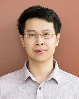
Kui Cui
Harvard Medical School/Boston Children’s Hospital
Dr. Kui Cui is a Research fellow in Dr. Hong Chen’s Lab at the Vascular Biology Program of Harvard Medical School and Boston Children’s Hospital, USA. He earned his master’s degree at Soochow University (mentor Dr. Zhenghong Qin) and PhD at East Tennessee State University (mentor Dr. Valentin Yakubenko). Dr. Cui is a vascular biologist interested in lipid metabolism and therapies for cardiovascular diseases and lymphatic dysfunction using nano-therapy, computational science, and cell molecular biology. His long-term goal is to develop new therapeutic strategies to improve human vascular health.
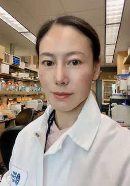
Beibei Wang
Harvard Medical School/Boston Children’s Hospital
Dr. Beibei Wang is a PhD and an MD in Dr. Hong Chen’s Lab at the Vascular Biology Program of Boston Children’s Hospital, Surgery Department of Harvard Medical School, USA. Dr. Wang is a vascular biologist, her study focuses on chronic inflammation, lipid metabolism, and lymphatic dysfunction in atherosclerosis and therapies for cardiovascular diseases caused by atherosclerosis.
Collection Overview
Atherosclerosis, a complex vascular disease characterized by plaque buildup within arteries, is a leading cause of cardiovascular diseases. Accurate collection and analysis of atherosclerotic lesions are crucial for advancing our understanding of disease progression and developing effective therapeutic strategies. In this JoVE collection, we detail a step-by-step protocol for the collection, preservation, and subsequent analysis of atherosclerotic specimens from animal models, providing valuable insights into lesion composition and molecular mechanisms.
This collection will encompass a wide range of methodological approaches, including but not limited to:
This collection encompasses a wide range of methodological approaches, including, but not limited to:
1. Animal Model Preparation:
Selecting appropriate animal models (e.g., ApoE-/- mice) with established susceptibility to atherosclerosis.
2. Tissue Harvesting:
Euthanizing animals using approved methods. Perfusing the cardiovascular system to remove blood contaminants and facilitate fixation. Excising target arteries (e.g., aorta, carotid) with minimal disruption.
3. Fixation and Preservation:
Optimizing fixation duration and the choice of fixative (e.g., paraformaldehyde) to maintain tissue morphology. Embedding specimens in paraffin or cryoprotectant for long-term preservation.
4. Histological Processing:
Sectioning tissues to obtain thin slices suitable for staining and analysis. Performing routine histological staining (e.g., Hematoxylin & Eosin) to visualize general morphology.
5. Advanced Staining Techniques:
Employing immunohistochemistry to detect specific cell types (macrophages, smooth muscle cells) and molecules (lipids, cytokines). Utilizing special stains like Oil Red O to highlight lipid-rich regions.
6. Microscopic Analysis:
Capturing high-resolution images of stained sections. Quantifying key parameters such as plaque size, composition, and inflammatory cell infiltration.
7. Data Interpretation:
Correlating histological findings with molecular data to gain insights into disease mechanisms.
Comparing results across experimental groups or treatment conditions.
This JoVE video article introduces a state-of-the-art method for collecting and analyzing atherosclerotic tissues, offering researchers a robust tool to investigate disease progression and potential therapeutic targets. By combining advanced histological techniques with molecular analyses, this protocol contributes to a deeper understanding of atherosclerosis and its underlying biology.
Articles

Abstracts
Influence of cell incubation parameters for molecular imaging of atherosclerosis by functionalized gold nanoshells
Emma Rodriguez*1
1Universidad Autonoma de Madrid