Guest Editor

Collection Overview
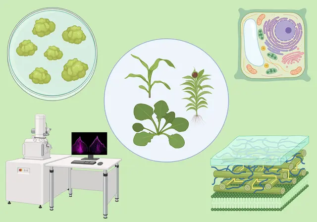
Plant cells serve as excellent model systems to study cellular events. Compared to animal systems, plant cells are more accessible for performing cellular imaging without moral obligation and ethical concerns. Plant live cell imaging provides the window to understand a cellular process in the in vivo condition over the entire life cycle. At the same time, some plant tissues can be challenging to image due to secondary metabolites, autofluorescence, and tissue thickness.
In this Methods Collection, we invite authors to submit their methods on plant cell imaging, including dynamic live cell imaging, immunostaining-based procedure, tissue sectioning, and electron microscopy. This collection will delve into cellular events, including cell division, protein trafficking, organelle movement, cell wall dynamics, and polarized growth. It will also explore diverse tissue types such as the primary root, pollen, stomata, embryo, lateral root, and root hair. The collection will cover various plant species, such as Arabidopsis thaliana, Physcomitrella patens, Marchantia polymorpha, and Zea mays. Through this collection, we aim to increase transparency and reproducibility and inspire more labs to adopt state-of-the-art plant cell imaging techniques in the lab.
