
Guest Editors
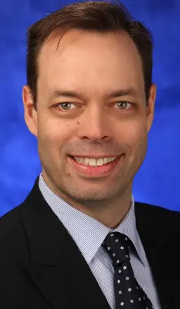
Salvatore L. Stella, Jr.
Penn State College of Medicine
Salvatore Stella is an Assistant Professor in the Department of Neural and Behavioral Sciences at Penn State College of Medicine in Hershey, PA. He received his BS in Biology from San Diego State University, followed by graduate training in the Departments of Pharmacology and Ophthalmology at the University of Nebraska Medical Center with Wallace B. Thoreson. He followed this with his post-doctoral training at the University of California, Los Angeles in neuroscience under Nicholas C. Brecha. He first joined the faculty at the University of Missouri-Kansas City, School of Medicine in 2010, and later moved to Penn State Hershey College of Medicine in 2015. His research combines various physiological and imaging methodologies to study retinal and central nervous system function in both health and disease. One of the main overarching themes in the lab focuses on purines and how they alter neurotransmission and visual processing in the retina and brain. Imaging methods in the lab encompass both in vitro through in vivo applications focused on the zebrafish visual system. Techniques used in the lab have centered on activity-dependent dyes for measuring exocytosis, fast calcium imaging and use of heritable genetic Ca2+ indicators as well as ion sensitive dyes for deciphering neuronal function. Other imaging approaches include super resolution techniques both in wide field and confocal microscopy in order to resolve the dynamics and changes of small synaptic structures in both health and disease states within the vertebrate visual system.

Wallace B. Thoreson
University of Nebraska Medical Center
Wallace B. Thoreson is the Gilmore Professor of Ophthalmology and Visual Sciences and Director of Research in the Truhlsen Eye Institute at the University of Nebraska Medical Center (UNMC), Omaha, NE. He received his BA from Carleton College, Minnesota followed by graduate training in the Departments of Physiology and Psychology at the University of Minnesota with Dwight Burkhardt and Richard Purple. He continued at the University of Minnesota with post-doctoral training in neuroscience and eye research under Robert F. Miller. He joined the faculty of the University of Nebraska in 1993. His research combines various imaging and electrophysiological methods to study mechanisms of neurotransmission and early visual processing in the vertebrate retina, particularly at rod and cone ribbon synapses. Electrophysiological techniques in the laboratory include whole cell patch clamp, single channel recordings, and membrane capacitance measurements. Imaging techniques in the laboratory include confocal Ca2+ imaging, total internal reflectance imaging of single vesicle fusion events, and imaging of individual calcium channels labeled with quantum dots.
Collection Overview
By exploring the structure and function of the brain, scientists can further elucidate the underlying mechanisms that contribute to its normal circuitry, development, and disease. Optical imaging has proven to be an indispensable tool for exploring the nervous system. Unlike traditional anatomical and physiological methods, optical imaging has the advantage of allowing scientists to observe neuronal function in real time without perturbing individual neurons or the neural network. From super resolution imaging to high speed optical approaches, we are currently sitting at the frontier of a dynamic and powerful series of optical techniques that will shape the future of neuroscience.
This Methods Collection covers fundamental approaches and cutting-edge technologies in the evolving field of optical techniques and is designed to be a go-to resource for junior and senior scientists. This collection can be utilized as a tool to for deciphering synaptic and neuronal changes that contribute to the physiological processes at the local level and that ultimately lead to disease development at the global level. Thus, further research in the optical imaging realm as applied to neurons in vitro, in toto, and in vivo will be of great help in answering fundamental questions about synapses and neuronal circuits with a particular emphasis on health and disease.
Articles

Abstracts
High-density functional near-infrared spectroscopy of subthalamic nucleus deep brain stimulation
Fabrice Wallois1,
Michel Lefranc1,
Mahdi Mahmoudzadeh*1
1INSERM U1105, GRAMFC, Univerity of Picardie Jules Verne, Amiens, France
Functional (optical-electrical) imaging of the human brain in early infancy A multimodal analysis using fNIRS, EEG and DCS
Fabrice Wallois1,
Mina Nourhashemi1,
Mahdi Mahmoudzadeh*1
1INSERM U1105, GRAMFC, Univerity of Picardie Jules Verne, Amiens, France
Multimodal (optical-electrical) analysis of interictal epileptic spikes using ECoG, MUA, FOS, fNIRS and DCS measurements in rats
Fabrice Wallois1,
Mana Manoochehri1,
Mina Nourhashemi1,
Mahdi Mahmoudzadeh*1,
Cristian Arnal-Real1
1INSERM U1105, GRAMFC, Univerity of Picardie Jules Verne, Amiens, France