A woman developed necrotizing fasciitis 20 days after abdominoplasty and liposuction. Successful management involved aggressive surgical debridement, antibiotic bone cement application, VSD, and skin grafting. Quinolone-susceptible pathogens were identified. Multidisciplinary care achieved infection control and recovery, highlighting cosmetic surgery risks and the critical need for prompt intervention and asepsis.
Case Report
Case Report on Necrotizing Fasciitis after Liposuction and Abdominoplasty
Chuze Xu1,2 , Weijie Zhou2 , Xiaodi Zou2 , Yanzhao Dong2 , Ahmad Alhaskawi2 , Haiying Zhou2 , Hui Lu2
huilu@zju.edu.cn
Corresponding Authors: Hui Lu <huilu@zju.edu.cn>
Opens in a new tab
In This Article
Summary
Loading...
$$\rightleftharpoonup{xx}$$
$$\longleftharp{xx}$$,
$$\longrightharp{xx}$$,
Abstract
Loading...
$$\rightleftharpoonup{xx}$$
$$\longleftharp{xx}$$,
$$\longrightharp{xx}$$,
Liposuction and abdominoplasty are common cosmetic procedures, but they can lead to severe complications such as necrotizing fasciitis (NF), which is a life-threatening infection with rapid progression and significant mortality rates. NF can develop after surgical procedures due to bacterial colonization or inadequate postoperative care. In the case shown here, a 33-year-old woman presented with skin flap fascial necrosis with abdominal wall infection 20 days after undergoing abdominoplasty with liposuction at another private clinic. Initial management included incision and drainage of an abdominal wall sinus tract, debridement of necrotic tissue, and application of antibiotic bone cement, followed by vacuum sealing drainage (VSD). Postoperative blood tests and bacterial culture identified multiple pathogens, all susceptible to quinolones. Subsequent surgeries involved re-debridement and skin grafting. The patient received a comprehensive rehabilitation program, resulting in effective infection control and favorable outcomes. The case underscores the importance of early diagnosis and aggressive surgical debridement in managing NF. The use of antibiotic bone cement provided local high-concentration antibiotics and supported granulation tissue formation, contributing to the treatment of the wound. This rare complication highlights the potential risks of cosmetic surgery and the necessity of strict adherence to aseptic techniques.
Introduction
Loading...
$$\rightleftharpoonup{xx}$$
$$\longleftharp{xx}$$,
$$\longrightharp{xx}$$,
In recent decades, the field of plastic surgery has undergone significant expansion. Liposuction has become a common procedure in the field of plastic surgery, employed to decrease locally excessive fatty tissue in specific areas of the body. It serves as a solution for patients who are dissatisfied with the fat accumulation in specific areas of their bodies1. The procedure can either be performed individually or in combination with other body contouring operations, such as abdominoplasty and mammoplasty, to achieve the ideal aesthetic form of the patient2. Liposuction surgery can lead to various types of complications, including asymmetry, infection, skin necrosis, hematoma, seroma, pigmentation, lymphedema, and venous thromboembolism2,3,4. Among these, infection is a relatively common complication, usually induced by Staphylococcus aureus, Streptococcus group A, and Streptococcus pyogenes5, with an incidence rate of 0.1% in single liposuction and 0.7% in combined surgeries6. Although the incidence rate is relatively low, for patients undergoing cosmetic surgery, any kind of complication is usually intolerable, and patients with severe infections may develop necrotizing fasciitis (NF), infectious shock, and even death5,7,8. Understanding the connection between liposuction and potential infectious complications is crucial for effective treatment and also serves as a warning to prevent the recurrence of similar infections. Introducing antibiotics into implant materials such as bone cement can achieve high-concentration aggregation of local antibiotics, which has a significant effect upon preventing and treating bacterial infections, and has been widely used in orthopedic surgeries9. In patients with abdominal infections, placing bone cement at the wound site not only helps prevent infection but can stimulate granulation tissue growth and create conditions for skin grafting10.
Here, we present a female patient who suffered a bacterial infection after liposuction and abdominoplasty, which eventually led to skin infection, abdominal wall abscess, abdominal wall infectious sinus tract, transplanted flap necrosis, lumbar fasciitis, and NF. Detailed admission history, physical examination findings (fever, hypotension), laboratory results (normal white blood cell count, hemoglobin level, and LRINEC score), and imaging studies (revealing multiple areas of subcutaneous exudation in the abdominal wall and lumbodorsal gluteal regions) provide a basis for the diagnosis of NF. On the day of admission, empirical carbapenem antibiotic treatment, such as ertapenem sodium 1.0 g intravenous driponce a day (ivgtt qd), was initiated on the premise that renal function permitted (creatinine clearance rate needed to be monitored). Three aggressive surgical debridements were performed on the 2nd, 9th, and 50th days after the first admission, and antibiotic bone cement was applied intraoperatively for localized sustained antibiotic release, thereby achieving the goals of anti-infection and stimulating granulation tissue growth. Postoperative pathogen identification was achieved through bacterial culture and metagenomic next-generation sequencing (mNGS). Active anti-infective therapy was pursued in the absence of relevant contraindications (e.g., a history of QT prolongation precluding the use of moxifloxacin Tablets (400 mg/tablet): 1 tablet once daily and Faropenem Sodium Granules (0.1 g/sachet): 0.1 g 3x daily. This comprehensive treatment regimen ultimately achieved effective infection control. Beyond its application in this specific case, this integrated strategy may also hold reference value for managing infections arising in general surgery or other cosmetic interventions.
Case Presentation:
A 33-year-old patient presented to the First Affiliated Hospital of Zhejiang University due to flap necrosis 20 days after abdominoplasty with liposuction. The patient underwent abdominoplasty with liposuction at another private clinic in China and subsequently developed partial flap necrosis. Without receiving any treatment, the patient came directly to our hospital for medical attention. The patient was previously healthy, without diseases such as hypertension, diabetes, viral hepatitis, heart disease, etc., and had no history of allergies or long-term medication.
On admission, the patient exhibited a body temperature of 39.2 °C and a blood pressure 89/54 mmHg. Physical examination revealed a conscious and alert patient with a flat, soft abdomen; the abdominal wall exhibited skin flap necrosis, which was covered with bandages, and there was no evidence of abdominal tenderness or rebound tenderness. No palpable liver or spleen was identified below the costal margins, and shifting dullness was negative. Abdominal wall reflexes were normal. Neurological examinations showed negative pathological signs. Initial blood tests exhibited a white blood cell (WBC) count of 6.64x 10⁹ cells/L, a red blood cell (RBC) count of 4.56 x 10¹² cells/L, neutrophils 58.8%, hemoglobin 122 g/L, sodium 142 mmol/L, glucose 5.21 mmol/L, creatinine 52 µmol/L, a platelet count of 339 x 109 platelets/L (normal: 101-320 x 109 platelets/L), eosinophils 0.4% (normal: 0.5%-5.0%), and high-density lipoprotein cholesterol (HDL-C) 0.77 mmol/L (normal: 0.88-2.04 mmol/L). Abdominal mass MRI plain scan combined with diffusion-weighted imaging (DWI) on admission demonstrated multiple areas of exudation within the subcutaneous soft tissues of the abdominal wall and lumbodorsal-gluteal regions, findings consistent with cellulitis. Correlation with clinical presentation is recommended (Figure 1).
In consideration of the patient's limited physiological tolerance and the need for effective infection control, a staged surgical approach was planned. Preoperatively, a standing order for ertapenem sodium injection 1.0 g ivgtt qd was initiated to mitigate disease progression. At 2 days after admission, surgical procedures were performed under general anesthesia, including incision and drainage of the abdominal wall sinus tract, repair of the chronic ulcer, and harvest and transposition of a pedicled fascial flap for soft tissue coverage. Intraoperative findings revealed an infected abdominal wall defect with swelling in the abdominal and lumbar regions, along with local purulent exudate. The necrotic margins were incised, exposing necrotic fascia and adipose tissue. Debridement was performed on the necrotic tissue. Considering the risk of reinfection in the wound and promoting the growth of granulation tissue, the area was covered and filled with antibiotic bone cement. A vacuum sealing drainage (VSD) device was applied, and two drainage tubes were placed (Figure 2).
Blood tests on the 1st day after the operation showed a WBC count of 5.01 x 10⁹ cells/L, RBC count of 3.85 x 10¹² cells/L, neutrophils 59.9%, eosinophils 0.8%, CRP 1.90 mg/L, HDL-C 0.65 mmol/L (normal: 0.88-2.04 mmol/L), hemoglobin 105 g/L (normal: 113-151 g/L), sodium 146 mmol/L, glucose 5.17 mmol/L and creatinine 54 µmol/L. Postoperative bacterial culture and metagenomic sequencing identified Corynebacterium glucuronolyticum, Fingoldia magna, Prevotella buccalis, Prevotella timonensis, Peptoniphilus rhinitidis, Anaerococcus prevotii, and Anaerococcus lactolyticus, confirming the diagnosis (Table 1). All identified bacterial species demonstrated susceptibility to quinolones; therefore, on the 6th day after admission, oral moxifloxacin at a dose of 400 mg 1x daily was initiated and continued for 3 consecutive days.
At 9 days after admission, the second surgery was performed, which was the same as the first one. During the surgery, the bone cement was removed. Fresh granulation tissue was observed in the local wound area, with a small amount of necrotic tissue on the surface. Upon exploration, the flap was found to be well-adhered to the subcutaneous tissue (Figure 3). On the 19th day after admission, the patient was temporarily discharged for conservative treatment and was prescribed two boxes of moxifloxacin tablets, each containing three 400 mg tablets, with instructions to take one tablet daily. At 50 days after the initial hospitalization, the patient was re-admitted to the hospital for the final debridement, and skin grafting was performed using the full-thickness skin from the right inguinal region as the donor site. Postoperative blood tests revealed a WBC count of 8.15 x 10⁹ cells /L, RBC count of 4.03 x 10¹² cells/L, neutrophils at 73.0% (normal: 50.0%-70.0%), lymphocytes at 22.6% (normal: 20.0%-40.0%), hemoglobin 107 g/L (normal: 113-151 g/L), sodium 138 mmol/L, glucose 5.51 mmol/L, creatinine 41 µmol/L and CRP level of 0.50 mg/L. After the operation, ertapenem sodium 1.0 g ivgtt qd was continued to be given as the routine anti-infection treatment, which proved effective. At 58 days after the initial hospitalization, the patient was discharged home. Upon discharge, the patient was administered faropenem sodium granules (0.1 g per sachet) 0.1 g per dose, 3x daily, to continue anti-infection therapy.
The patient participated in a comprehensive rehabilitation program incorporating physical therapy and therapeutic exercises, achieving progressive functional recovery. The infection was effectively controlled, resulting in a favorable outcome following the chronic ulcer repair surgery (Figure 4).
Diagnosis, Assessment, and Plan:
The patient was admitted to the hospital with a skin infection as the preliminary diagnosis. MRI+DWI revealed multiple areas of exudation in the subcutaneous soft tissues of the abdominal wall and lumbodorsal-gluteal regions, suggestive of cellulitis. However,a series of symptoms (fever 39.2 °C, hypotension 89/54 mmHg, and abdominal wall skin flap necrosis) indicated severe disease progression, suggesting the possibility of NF.Intraoperative exploration during the first debridementrevealed an infected abdominal wall defect with swelling in the abdominal and lumbar regions, along with local purulent exudate. The necrotic margins were incised, exposing necrotic fascia and adipose tissue. The exploration results changed the postoperative diagnosis to skin infection, abdominal wall abscess, abdominal wall infectious sinus tract, transplanted flap necrosis, lumbar fasciitis, and NF. The diagnosis was further improved through postoperative bacterial culture and metagenomic sequencing, identifying seven pathogens (Corynebacterium glucuronolyticum, Fingoldia magna, Prevotella buccalis, Prevotella timonensis, Peptoniphilus rhinitidis, Anaerococcus prevotii, and Anaerococcus lactolyticus) with universal quinolone susceptibility.
Management employed a multidisciplinary strategy: (1) Staged surgical intervention-radical debridement with vacuum-sealing drainage (VSD) and antibiotic bone cement for infection control (Nov 16), secondary exploration confirming viable granulation tissue (Nov 23), and definitive closure via skin grafting (Jan 4); (2) Culture-guided antimicrobial therapy-initial empirical coverage with ertapenem sodium injection 1.0 g ivgtt qd targeting NF, escalation to moxifloxacin tablets 400 mg, taken orally once a day (po qd) post-identification of quinolone-susceptible pathogens, and eventually switching to oral faropenem sodium granules 100 mg 3x a day (TID) after discharge; (3) Supportive care for symptoms such as fever and hypotension; and (4) Structured rehabilitation, achieving complete wound healing, functional recovery, and eradication of infection without recurrence.
Protocol
Loading...
$$\rightleftharpoonup{xx}$$
$$\longleftharp{xx}$$,
$$\longrightharp{xx}$$,
This study was approved by the Clinical Research Ethics Committee of The First Affiliated Hospital of Zhejiang University School of Medicine (Reference Number: 2024-0844). Written informed consent was obtained. The study complied with the Declaration of Helsinki.
1. Patient admission and clinical evaluation
- Initial presentation and history
- Skin flap necrosis was presented after the patient underwent prior cosmetic surgery and lasted for 20 days. Detailed medical history information was obtained.
- Clinical assessment
- Physical examination was conducted, focused on the affected skin area.
- The extent of skin necrosis was documented.
- Vital signs (fever, hypotension) and laboratory findings (normal WBC, Hb, LRINEC score) were recorded.
2. Imaging and empirical treatment
- Diagnostic imaging
- The extent of involvement of abdominal wall and lumbodorsal-gluteal subcutaneous soft tissues was evaluated using routine abdominal MRI plain scan and diffusion-weighted imaging (DWI, 3.0T).
- Multiple areas of exudation in the subcutaneous soft tissues of the abdominal wall and lumbodorsal-gluteal regions (hyperintensity on T2-weighted imaging (T2WI) and DWI), were revealed, suggestive of cellulitis. Combining clinical manifestations was recommended for evaluation and treatment.
- Empirical carbapenem therapy (e.g., ertapenem sodium 1.0 g ivgtt qd) was initiated.
3. Surgical debridement
- Preoperative preparation
- Patient was prepared for surgical exploration under combined intravenous-inhalational general anesthesia, with strict adherence to aseptic protocols.
- Operative procedure
- Incisions were made along necrotic margins using a scalpel. The necrotic tissue was then separated and debrided with surgical instruments, including a scalpel, tissue scissors, and forceps, until the absence of non-viable tissues that were identified by black/gray discoloration, leathery texture, and lack of bleeding.
- The wound was filled with vancomycin-loaded bone cement.
- VSD dressing (AD-1-15 x 5 x 1 cm) was trimmed and then tightly packed into the wound cavity, ensuring full contact with the entire wound bed. Embedded drainage tubes were exteriorized through a separate stab incision made in healthy skin and connected to a negative pressure suction device.
- The entire wound surface was subsequently sealed with a transparent occlusive dressing film without wrinkles, and a negative pressure of approximately 125 mmHg was maintained.
- Specimen collection
- More than three tissue specimens were collected from different wound sitesusing a scalpel for excision and scissors for dissection.
- The samples were placed in sterile containers for bacterial/fungal culture and histopathological analysis.
4. Pathogen identification and next-generation sequencing (NGS) testing
- Traditional microbiology
- Gram staining and bacterial/fungal culture were performed on tissue samples11.
- Molecular testing
- DNA/RNA was extracted from fresh tissue for NGS12.
- Pathogen confirmation
- A total of seven pathogens, including Fingoldia magna, were confirmed through bacterial culture and NGS.
5. Post-diagnosis management using a combination of antibacterial therapy and staged surgical debridement
- Treatment plan adjustment
- Moxifloxacin tablets (400 mg per tablet) were given at one tablet, 1x a day on the basis of the original ertapenem sodium administration following confirmed diagnosis.
- Subsequent debridements
- Second surgical debridement
- The implanted bone cement was separated from the surrounding soft tissues by using instruments such as tissue scissors and curved forceps along the surface of the bone cement. The bone cement was then removed by holding it with forceps like the Allis forceps, and the wound bed was assessed for fresh granulation tissue (wound bed coverage with beefy-red, granular, non-friable tissue; absence of fibrinopurulent exudate) with a few superficial necrotic tissues. The viability of the peripheral flap adherence to subcutaneous tissue was confirmed.
- Incisions were made along necrotic margins using a scalpel. The necrotic tissue was then separated and debrided with surgical instruments, including a scalpel, tissue scissors, and forceps, until all necrotic tissue was debrided, and the wound was covered with antibiotic bone cement.
- VSD dressing (AD-1-15 x 5 x 1 cm) was trimmed and then tightly packed into the wound cavity, ensuring full contact with the entire wound bed. And embedded drainage tubes were exteriorized through a separate stab incision made in healthy skin and connected to a negative pressure suction device. The entire wound surface is subsequently sealed with a transparent occlusive dressing film without wrinkles, and a negative pressure of approximately 125 mmHg is maintained.
- Third surgical debridement
- The implanted bone cement was removed, and the wound was assessed for mature scar tissue (tissue firm, non-fluctuant, epithelialized edges; no purulence/slough) with partial skin deficiency.
- Radical debridement of necrotic tissue was performed; incisions were made along necrotic margins using a scalpel. The necrotic tissue was then separated and debrided with surgical instruments, including a scalpel, tissue scissors, and forceps, until all necrotic tissue was debrided. Abdominal wall reconstruction was conducted, and the defect site was covered with a free skin graft.
- The VSD device was applied.
- Second surgical debridement
- Post-discharge therapy
- Faropenem sodium granules (0.1 g per sachet) 0.1 g each time was prescribed, 3x a day after discharge for continued anti-infective therapy.
6. Recovery and follow-up
- Definitive wound closure
- Skin grafting was performed using the full-thickness skin from the right inguinal region as the donor site after confirming complete necrotic tissue excision.
- Follow-up schedule
- Assessments were scheduled for 2 weeks, 1 month, and 3 months postoperatively. Wound healing, flap viability, recurrence, and infection signs were monitored.
- Outcomes documentation
- Clinical progression, imaging, lab results, and final outcomes were analyzed. Comprehensive case materials for reporting/publication were compiled.
Results
Loading...
$$\rightleftharpoonup{xx}$$
$$\longleftharp{xx}$$,
$$\longrightharp{xx}$$,
This case demonstrates the synergistic effect of stepwise surgical debridement combined with antimicrobial stepping therapy and also highlights the efficacy of novel diagnostic methods like pathogen mNGS.The patient successfully underwent surgical exploration. Preoperative broad-spectrum antibiotic coverage with ertapenem sodium (1.0 g ivgtt qd) provided a safe window for surgery. Three staged debridement procedures progressively and thoroughly removed necrotic tissue. Culturing and mNGS of samples obtained during the initial surgery identified seven pathogens: Corynebacterium glucuronolyticum, Fingoldia magna, Prevotella buccalis, Prevotella timonensis, Peptoniphilus rhinitidis, Anaerococcus prevotii, and Anaerococcus lactolyticus, all demonstrating universal susceptibility to quinolones. Based on etiological findings, moxifloxacin tablets at a dosage of one tablet daily were incorporated into the existing treatment regimen to provide targeted antimicrobial therapy.Definitive wound reconstruction was ultimately achieved using a skin graft harvested from the full-thickness skin of the right inguinal region, accomplishing both infection eradication and functional restoration. At follow-up evaluations conducted at 2 weeks, 1 month, and 3 months postoperatively, the surgical site showed satisfactory healing progress with no evidence of recurrence or postoperative complications.
Throughout the treatment protocol, timing of debridement and antimicrobial transitions were precisely guided by monitoring hemoglobin (Hb), the LRINEC score (based on WBC count, creatinine, glucose, hemoglobin, sodium, and CRP levels), and the progression of granulation tissue formation. Although the patient's preoperative laboratory parameters (WBC 6.64 x 10⁹ cells/L, Hb 122 g/L) did not meet the typical thresholds for NF, the diagnosis was confirmed based on the clinical presentation, imaging findings showing signs of infiltration, and intraoperative exploration. Granulation tissue formation commenced on the 9th day post-admission and had matured into stable scar tissue by the time of the third debridement, indicating effective infection control. This case offers a systematic solution for managing complex soft tissue infections.
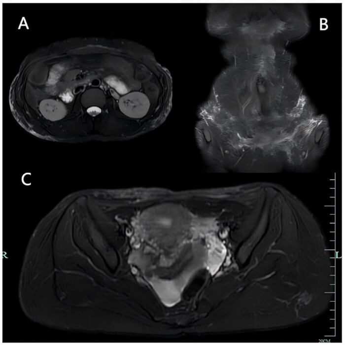
Figure 1: Admission MRI+DWI findings. (A-C) MRI+DWI revealed multiple areas of exudation in the subcutaneous soft tissues of the abdominal wall and lumbodorsal-gluteal regions. Please click here to view a larger version of this figure.
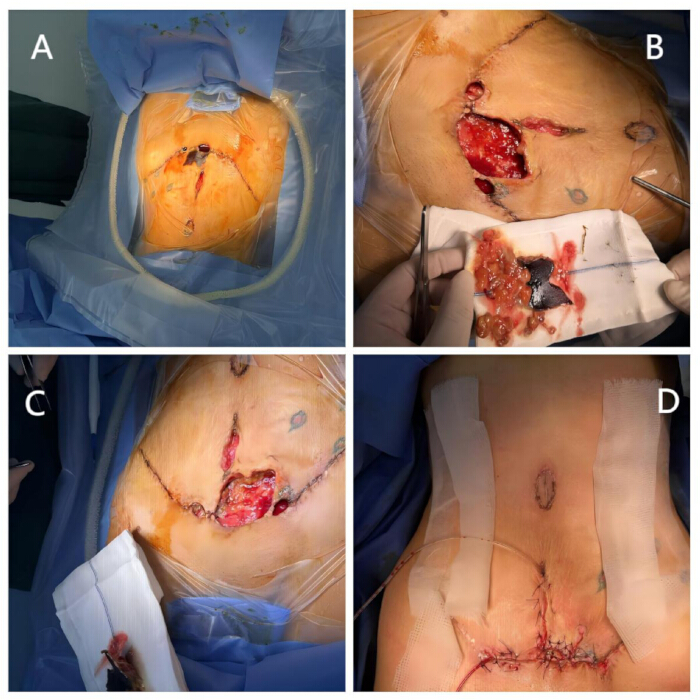
Figure 2: First debridement surgery. (A) Preoperative appearance of the affected abdominal wall. (B) Intraoperative view of the excised necrotic tissue. (C) Resultant defect following debridement of necrotic tissue. (D) Postoperative appearance. Please click here to view a larger version of this figure.
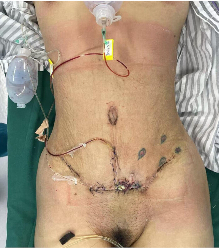
Figure 3: Panoramic view after the second debridement. The necrotic tissue was removed, bone cement was re-filled, and a VSD device was used for drainage. Please click here to view a larger version of this figure.
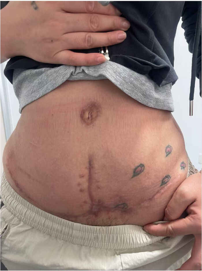
Figure 4: Frontal view of the abdominal wall 3 months after discharge. A favorable prognosis was achieved, characterized by effective control of the infection. Please click here to view a larger version of this figure.
| Detection process | Overall results | Pathogen detection | Sequence number |
| DNA process | Positive | Bacteria (6 types): | |
| 1. Fingoldia magna | 12,045 | ||
| 2. Prevotella buccalis | 690 | ||
| 3. Prevotella timonensis | 109 | ||
| 4. Peptoniphilus rhinitidis | 399 | ||
| 5. Anaerococcus prevotii | 92 | ||
| 6. Anaerococcus lactolyticus | 52 | ||
| Fungi/Viruses/Parasites/Mycobacteria/Mycoplasma and Chlamydia: Undetected | |||
| RNA process | Negative | All pathogens were not detected (including RNA viruses) | |
Table 1: Pathogen metagenomic sequencing detection report. Six types of pathogenic bacteria were detected through the DNA detection process.
Discussion
Loading...
$$\rightleftharpoonup{xx}$$
$$\longleftharp{xx}$$,
$$\longrightharp{xx}$$,
Liposuction and abdominoplasty are currently among the most common cosmetic surgical procedures. They achieve patients' desired aesthetic outcomes by reducing localized excess adipose tissue in specific body areas, making them highly popular1,2,13. The fact that patients receiving cosmetic surgery are typically in good health and have high expectations makes it challenging for them to accept complications. Therefore, despite the low incidence rate, potential complications of liposuction warrant significant attention3,4. The risk factors for postoperative complications are multifaceted. A prospective study indicated that age, BMI, and male gender are independent risk factors for complications. Combined surgical procedures can lead to an increased incidence of major complications14. While smoking has been well-documented to increase morbidity15, it was not found to be an independent risk factor in this study, which contrasts with previous findings.This discrepancy is attributed to the generally good health of patients undergoing cosmetic surgery and the cautious approach surgeons take when operating on smokers and diabetic patients.Furthermore, hospital medical equipment, the level of sterilization and hygiene, and the surgeon's technique also influence the complication rate.In recent years, medical tourism for cosmetic procedures has surged dramatically16. Many private clinics and companies attract patients with promises of short waiting times and low costs. This trend has contributed to a rise in associated complications. In addition to poor medical institution conditions, it may also be related to the risk of thromboembolism brought about by patients' long-distance travel, and even language barriers16.In this case, the patient underwent surgery at a private clinic prior to admission. As the patient's preoperative physical condition was relatively good, it is speculated that the cause of infection may be related to the substandard medical conditions at that clinic.
Complications resulting from liposuction and abdominoplasty are diverse, including common ones such as asymmetry, infection, hematoma, seroma, pigmentation, and lymphedema. However, beyond common complications, liposuction carries potentially life-threatening risks, including NF, toxic shock syndrome (TSS), toxicity or drug interactions, visceral organ perforation, and pulmonary embolism17,18,19,20. Among these, NF and TSS typically represent severe infections. In comparison with more prevalent complications, NF is exceedingly rare. In the preceding two decades, only a limited number of literature reports have documented NF associated with liposuction5,21,22,23,24.
NF is a severe and potentially fatal condition with a mortality rate as high as 25-35%25. Causative pathogens include various aerobic and anaerobic bacteria, Group A Streptococcus, and methicillin-resistant Staphylococcus aureus (MRSA), among others26,27,28. NF caused by fungal infections such as Candida species is exceedingly rare, with only a small number of cases reported to date29. Erythema, paresthesia, dusky discoloration, disproportionate pain, and fever present as the most prevalent early manifestations of NF. NF is characterized by infection of the superficial soft tissue with rapid dissemination along fascial planes. The infection progresses through the subcutaneous adipose tissue, reaching deeper fascial planes. This results in clinically occult tissue necrosis, initially not involving muscle, but eventually leading to systemic collapse and septic shock30,31. Any breach in the skin or mucous membranes, as well as various surgical procedures, increases patients' risk for NF. Patients suffering from comorbidities such as diabetes, alcoholism, obesity, malignancy, vascular disease, and immunosuppression exhibit an elevated risk of developing NF and progressing to severe sepsis and septic shock26.
No specific laboratory test has proven reliably diagnostic for NF32. The majority of reviewed articles employed a multifaceted approach to diagnosis, integrating clinical, radiological, microbiological, and laboratory findings. Surgical exploration remains the definitive method for confirming the diagnosis of necrotizing infection8,33,34. Differentiating NF from cellulitis is crucial, as the former requires surgical intervention while the latter can often be managed with antimicrobial therapy alone35. Similar to NF, a characteristic presentation of cellulitis is erythema, oedema, and warmth of the skin. However, patients with cellulitis generally exhibit stable hemodynamics, while those exhibiting NF might manifest significant tenderness or pain, ultimately progressing towards instability of the hemodynamics as well as tissue necrosis28. Unfortunately, the distinction is often challenging to identify, given that the initial NF clinical presentation is frequently vague. Factors such as nonsteroidal anti-inflammatory drug (NSAID) use can further complicate this differentiation35. Ultrasonography (US) may help distinguish between early NF and cellulitis, but in 64% of cases, clinical findings may be incorrect36. Wong et al. constructed the Laboratory Risk Indicator for Necrotizing Fasciitis (LRINEC) to assist clinicians in screening for NF by measuring serum levels of total WBC count, creatinine, glucose, hemoglobin, sodium, as well as CRP37. It is considered suggestive of NF if an LRINEC score is 6 or higher. Nevertheless, further reports have indicated that LRINEC score predictions were not accurate for NF38,39,40. This tool's sensitivity, specificity, and prognostic value remain subject to further evaluation. In this case, although surgical exploration confirmed the NF diagnosis, several LRINEC-related parameters in the patient's blood tests were within normal ranges, precluding its use for screening or assessment. Therefore, developing more accurate non-surgical diagnostic methods for NF may be a direction worthy of attention.
Initial drug treatment for NF ought to comprise empirical, broad-spectrum antibiotics until the availability of Gram staining, culture, and sensitivity results of the tissue, in conjunction with corresponding sensitive drugs28. In suspected or diagnosed cases of NF, surgical intervention is regarded as the gold standard treatment. An urgent surgical exploration and vigorous removal of necrotic tissue are critical to reducing morbidity and mortality41,42,43. During surgical exploration, specimens should be obtained for Gram staining and culture to facilitate the diagnosis of pathogenic bacteria. Based on traditional detection methods, this case adopted the pathogen mNGS technology, and six pathogenic bacteria were successfully detected, providing a basis for the precise selection of sensitive antibiotics. This technology has been proven to be an important tool for the diagnosis of infectious disease pathogens, especially suitable for difficult, rare, mixed infection, or culture-negative cases44,45. Based on its high efficiency and comprehensiveness, mNGS may also be used as an auxiliary diagnostic method for necrotizing fasciitis. However, mNGS results are susceptible to variables such as specimen collection (e.g., sampling depth, contamination), library preparation protocols, and bioinformatics analysis parameters. Different laboratories or platforms may report divergent pathogen profiles46. Patient-specific factors significantly impact detection reliability: immunocompromised individuals may yield false-positive results due to the identification of low-abundance commensal bacteria or environmental microorganisms, while early-stage infections or antibiotic pretreatment can reduce pathogen load, increasing false-negative risks47,48. Additionally, high costs and the need for specialized data analysis expertise limit its adoption in primary care facilities12. Thus, in resource-limited settings, traditional culture methods should remain the initial screening approach, with mNGS reserved for critically ill or diagnostically challenging cases. For NF patients, multiple debridements should be performed until no necrotic tissue is identified and only healthy tissue remains49. Once the patient is stable, secondary wound closure may be necessary using techniques like VAC, skin grafting, or local and free flaps. In most cases, the aesthetic outcome of NF involves disfigurement of the affected area.In cases requiring graft or flap reconstruction, significant scarring may also occur at the donor site level8. In this case, due to skin necrosis and increased tension in the defect following liposuction and abdominoplasty, direct suture was rendered impossible. Consequently, the full-thickness skin on the right inguinal region was chosen as the donor site for skin grafting.
Antibiotic bone cement is commonly used in orthopedic surgery for infection prevention and treatment9. In this case, after debridement of necrotic tissue, we opted to fill the defect with antibiotic bone cement. Compared with traditional treatment methods, this provided local sustained high concentrations of antibiotics, aiding infection control. Meanwhile, its space-occupying effect prevented wound collapse while simultaneously stimulating granulation tissue formation, creating favorable conditions for subsequent skin grafting. It may serve as an adjunctive treatment for necrotizing fasciitis. However, bone cement is primarily used for bone infections in areas with poor blood supply9. Whether its use in well-vascularized soft tissues like the abdominal wall offers superior outcomes compared to traditional debridement combined with VSD (vacuum sealing drainage) negative pressure therapy requires validation through studies with larger sample sizes.Furthermore, should infection persist, prompt debridement and cement replacement become necessary. However, bone cement embedded within soft tissues may become encased by granulation tissue, complicating complete removal and thereby impeding therapeutic efficacy.
To reduce complications from liposuction and abdominoplasty and enhance the management of severe infections such as NF, a multi-layered prevention and control system needs to be established, encompassing strict preoperative assessment to screen high-risk patients and control risk factors (such as smoking cessation and weight management),standardized medical facility credentials and surgical procedures to minimize complication risks, and enhanced postoperative monitoring for early identification of signs of serious complications. Once suspicious signs appear, prompt diagnosis and treatment should be achieved by integrating clinical evaluation, imaging, and laboratory testing, aided by rapid detection technologies like mNGS. Except for liposuction and abdominoplasty, this comprehensive treatment plan may also be of reference value for managing infections caused by general surgery or other cosmetic interventions. To address current limitations such as the high cost of mNGS and the difficulty of removing bone cement, future efforts should focus on optimizing mNGS by establishing regional testing centers and utilizing artificial intelligence-assisted analysis to reduce costs and improve accessibility, alongside developing point-of-care rapid sequencing devices. Exploring the replacement of bone cement with degradable carriers (such as thermosensitive hydrogels-antibiotic composites) to achieve injection filling and controlled sustained release, thus avoiding secondary removal surgery.
Conclusion
Although NF following routine cosmetic procedures such as liposuction is rare, this case, which progressed from skin flap necrosis to abdominal wall abscess, sinus tract formation, and ultimately NF, serves as a critical warning that surgical risks cannot be overlooked. Local tissue damage or inadequate postoperative care may trigger catastrophic outcomes. Given NF's high mortality, vigilance for early symptoms, such as erythema, disproportionate pain, diminished skin sensation, and rapidly progressive edema, is essential, emphasizing the urgency of early debridement and intervention. This case provides management insights for rare complications and further underscores that clinicians must fully understand postoperative complications and strictly adhere to intraoperative aseptic principles.
Disclosures
Loading...
$$\rightleftharpoonup{xx}$$
$$\longleftharp{xx}$$,
$$\longrightharp{xx}$$,
The authors have nothing to disclose.
Acknowledgements
Loading...
$$\rightleftharpoonup{xx}$$
$$\longleftharp{xx}$$,
$$\longrightharp{xx}$$,
The authors have no acknowledgments.
Materials
| Name | Company | Catalog Number | Comments |
|---|---|---|---|
| 3.0T MRI Scanner | General Electric Company | Not specified | |
| Allis tissue forceps | Tonglu Changsheng Medical Instrument Co., Ltd. | CS-AL012 | |
| Bandage 8*600 | Yongkang Weizhong Medical Instrument Co., LTD | WZ-BD822 | |
| Cotton balls | Henan Saizhong Medical Instrument Co., LTD | SZ-MQ100 | |
| Drain tube | Zhejiang Daweier Medical Technology Co., LTD | DW-YLF815 | |
| Dressing | Yongkang Weizhong Medical Instrument Co., LTD | WZ-FT326 | |
| Electrosurgical pencil | Xi 'an Surgical Medical Technology Co., LTD | XW-DDUNI25 | |
| Ertapenem sodium injection | Stone Pharmaceutical Group Ouyi Pharmaceutical Co., LTD | H20133325 | |
| Faropenem sodium granules | Hunan Warner Pharmaceutical Factory Co., LTD | H20080152 | |
| Fine gauze | Henan Saizhong Medical Instrument Co., LTD | SZ-SS422 | |
| Medium curved clamp | Tonglu Changsheng Medical Instrument Co., Ltd. | CS-ZW150 | |
| Mosquito forceps | Tonglu Changsheng Medical Instrument Co., Ltd. | CS-WS160 | |
| Moxifloxacin tablets | Bayer Pharma AG | H20140721 | |
| Nerve dissector | Shengxiang Medical Instrument Co., LTD | SX-BLZ280 | |
| Plastic surgery forceps, smooth | Shanghai Jinzhong Medical Instrument Co., LTD | JZ-YS344 | |
| Plastic surgery forceps, toothed | Shanghai Jinzhong Medical Instrument Co., LTD | JZ-WS368 | |
| Saline | CSPC Pharmaceutical Group Limited | CSPC-SS600 | |
| Scalpel handle #3 | Shanghai Jinzhong Medical Instrument Co., LTD | JZ-DB334 | |
| Scissors | Shanghai Jinzhong Medical Instrument Co., LTD | JZ-ZZ580 | |
| Small suction tip | Yangzhou Pengyan Biotechnology Co., LTD | PY-FSXT106 | |
| Two-prong retractor | Shanghai Jinzhong Medical Instrument Co., LTD | JZ-LG253 | |
| VSD(AD-1-15*5*1) | Shanghai XiuHui Technology Co., Ltd. | Not specified |
References
Loading...
$$\rightleftharpoonup{xx}$$
$$\longleftharp{xx}$$,
$$\longrightharp{xx}$$,
- Liposuction: Concepts, safety, and techniques in body-contouring surgery. Cleve Clin J Med. 87 (6), 367-375 (2020).">Wu, S., Coombs, D. M., Gurunian, R. Liposuction: Concepts, safety, and techniques in body-contouring surgery. Cleve Clin J Med. 87 (6), 367-375 (2020).
- Risks and complications rate in liposuction: A systematic review and meta-analysis. Aesthet Surg J. 44 (7), Np454-np463 (2024).">Comerci, A. J., et al. Risks and complications rate in liposuction: A systematic review and meta-analysis. Aesthet Surg J. 44 (7), Np454-np463 (2024).
- Analysis of postoperative complications for superficial liposuction: A review of 2398 cases. Plast Reconstr Surg. 127 (2), 863-871 (2011).">Kim, Y. H., Cha, S. M., Naidu, S., Hwang, W. J. Analysis of postoperative complications for superficial liposuction: A review of 2398 cases. Plast Reconstr Surg. 127 (2), 863-871 (2011).
- Updates and advances in liposuction. Aesthet Surg J. 30 (1), quiz 98-100 98-100 (2010).">Stephan, P. J., Kenkel, J. M. Updates and advances in liposuction. Aesthet Surg J. 30 (1), quiz 98-100 98-100 (2010).
- Infectious shock after liposuction. BMC Infect Dis. 22 (1), 617(2022).">Lu, J., et al. Infectious shock after liposuction. BMC Infect Dis. 22 (1), 617(2022).
- Cosmetic liposuction: Preoperative risk factors, major complication rates, and safety of combined procedures. Aesthet Surg J. 37 (6), 680-694 (2017).">Kaoutzanis, C., et al. Cosmetic liposuction: Preoperative risk factors, major complication rates, and safety of combined procedures. Aesthet Surg J. 37 (6), 680-694 (2017).
- Toxic shock syndrome complicated with symmetrical peripheral gangrene after liposuction and fat transfer: A case report and literature review. BMC Infect Dis. 21 (1), 1137(2021).">Liu, Z., et al. Toxic shock syndrome complicated with symmetrical peripheral gangrene after liposuction and fat transfer: A case report and literature review. BMC Infect Dis. 21 (1), 1137(2021).
- Necrotizing fasciitis in aesthetic surgery: A review of the literature. Aesthetic Plast Surg. 41 (2), 352-358 (2017).">Marchesi, A., et al. Necrotizing fasciitis in aesthetic surgery: A review of the literature. Aesthetic Plast Surg. 41 (2), 352-358 (2017).
- A review on the promising antibacterial agents in bone cement-from past to current insights. J Orthop Surg Res. 19 (1), 673(2024).">Lin, H., et al. A review on the promising antibacterial agents in bone cement-from past to current insights. J Orthop Surg Res. 19 (1), 673(2024).
- Application and clinical effectiveness of antibiotic-loaded bone cement to promote soft tissue granulation in the treatment of neuropathic diabetic foot ulcers complicated by osteomyelitis: A randomized controlled trial. J Diabetes Res. 2021, 9911072(2021).">Mendame Ehya, R. E., Zhang, H., Qi, B., Yu, A. Application and clinical effectiveness of antibiotic-loaded bone cement to promote soft tissue granulation in the treatment of neuropathic diabetic foot ulcers complicated by osteomyelitis: A randomized controlled trial. J Diabetes Res. 2021, 9911072(2021).
- Differential staining of bacteria: Gram stain. Curr Protoc Microbiol. Appendix 3 (Appendix 3C), (2009).">Moyes, R. B., Reynolds, J., Breakwell, D. P. Differential staining of bacteria: Gram stain. Curr Protoc Microbiol. Appendix 3 (Appendix 3C), (2009).
- Clinical metagenomic next-generation sequencing for pathogen detection. Annu Rev Pathol. 14, 319-338 (2019).">Gu, W., Miller, S., Chiu, C. Y. Clinical metagenomic next-generation sequencing for pathogen detection. Annu Rev Pathol. 14, 319-338 (2019).
- Liposuction. Obstet Gynecol Clin North Am. 37 (4), viii 507-519 (2010).">Pelosi, M. A. 3rd, Pelosi, M. A. 2nd Liposuction. Obstet Gynecol Clin North Am. 37 (4), viii 507-519 (2010).
- Abdominoplasty: Risk factors, complication rates, and safety of combined procedures. Plast Reconstr Surg. 136 (5), 597e-606e (2015).">Winocour, J., et al. Abdominoplasty: Risk factors, complication rates, and safety of combined procedures. Plast Reconstr Surg. 136 (5), 597e-606e (2015).
- Current evidence on the role of smoking in plastic surgery elective procedures: A systematic review and meta-analysis. J Plast Reconstr Aesthet Surg. 71 (5), 624-636 (2018).">Theocharidis, V., et al. Current evidence on the role of smoking in plastic surgery elective procedures: A systematic review and meta-analysis. J Plast Reconstr Aesthet Surg. 71 (5), 624-636 (2018).
- Infective complications of cosmetic tourism: A systematic literature review. J Plast Reconstr Aesthet Surg. 84, 9-29 (2023).">Gilardi, R., Galassi, L., Del Bene, M., Firmani, G., Parisi, P. Infective complications of cosmetic tourism: A systematic literature review. J Plast Reconstr Aesthet Surg. 84, 9-29 (2023).
- Life threatening complications post-liposuction. Aesthetic Plast Surg. 42 (2), 384-387 (2018).">Ezzeddine, H., et al. Life threatening complications post-liposuction. Aesthetic Plast Surg. 42 (2), 384-387 (2018).
- Fatal outcomes from liposuction: Census survey of cosmetic surgeons. Plast Reconstr Surg. 105 (1), discussion 447-438 436-446 (2000).">Grazer, F. M., De Jong, R. H. Fatal outcomes from liposuction: Census survey of cosmetic surgeons. Plast Reconstr Surg. 105 (1), discussion 447-438 436-446 (2000).
- and lethal complications of liposuction: A review of 72 cases in germany between 1998 and 2002. Plast Reconstr Surg. 121 (6), 396e-403e (2008).">Lehnhardt, M., et al. and lethal complications of liposuction: A review of 72 cases in germany between 1998 and 2002. Plast Reconstr Surg. 121 (6), 396e-403e (2008).
- Imaging findings of liposuction with an emphasis on postsurgical complications. Korean J Radiol. 16 (6), 1197-1206 (2015).">You, J. S., Chung, Y. E., Baek, S. E., Chung, S. P., Kim, M. J. Imaging findings of liposuction with an emphasis on postsurgical complications. Korean J Radiol. 16 (6), 1197-1206 (2015).
- Small intestinal perforation and necrotizing fasciitis after abdominal liposuction. Aesthetic Plast Surg. 30 (6), 712-716 (2006).">Sharma, D., Dalencourt, G., Bitterly, T., Benotti, P. N. Small intestinal perforation and necrotizing fasciitis after abdominal liposuction. Aesthetic Plast Surg. 30 (6), 712-716 (2006).
- Necrotizing fasciitis after liposuction. Acta Chir Plast. 49 (4), 99-102 (2007).">Gonzáles Alana, I., Marin De La Cruz, D., Palao Doménech, R., Barret Nerin, J. P. Necrotizing fasciitis after liposuction. Acta Chir Plast. 49 (4), 99-102 (2007).
- Blindness and necrotizing fasciitis after liposuction and fat transfer. Plast Reconstr Surg. 126 (4), 1358-1363 (2010).">Sherman, J. E., Fanzio, P. M., White, H., Leifer, D. Blindness and necrotizing fasciitis after liposuction and fat transfer. Plast Reconstr Surg. 126 (4), 1358-1363 (2010).
- Necrotizing fasciitis complicated with multiple organ dysfunction syndrome after breast augmentation with fat from the waist and lower extremities: A case report. J Int Med Res. 48 (7), 300060520937623(2020).">Zhang, M., Chen, L., Chi, K., Xu, L., Li, Y. Necrotizing fasciitis complicated with multiple organ dysfunction syndrome after breast augmentation with fat from the waist and lower extremities: A case report. J Int Med Res. 48 (7), 300060520937623(2020).
- Necrotizing fasciitis: Eight-year experience and literature review. Braz J Infect Dis. 18 (2), 137-143 (2014).">Wang, J. M., Lim, H. K. Necrotizing fasciitis: Eight-year experience and literature review. Braz J Infect Dis. 18 (2), 137-143 (2014).
- Current concepts in the management of necrotizing fasciitis. Front Surg. 1, 36(2014).">Misiakos, E. P., et al. Current concepts in the management of necrotizing fasciitis. Front Surg. 1, 36(2014).
- Necrotizing soft tissue infections. J Intensive Care Med. 29 (3), 138-144 (2014).">Shiroff, A. M., Herlitz, G. N., Gracias, V. H. Necrotizing soft tissue infections. J Intensive Care Med. 29 (3), 138-144 (2014).
- Necrotizing soft-tissue infections. N Engl J Med. 377 (23), 2253-2265 (2017).">Stevens, D. L., Bryant, A. E. Necrotizing soft-tissue infections. N Engl J Med. 377 (23), 2253-2265 (2017).
- Candida albicans necrotizing fasciitis following cosmetic tourism: A case report. JPRAS Open. 38, 129-133 (2023).">Gilardi, R., Parisi, P., Galassi, L., Firmani, G., Bene, M. D. Candida albicans necrotizing fasciitis following cosmetic tourism: A case report. JPRAS Open. 38, 129-133 (2023).
- Necrotizing fasciitis. Am Surg. 18 (4), 416-431 (1952).">Wilson, B. Necrotizing fasciitis. Am Surg. 18 (4), 416-431 (1952).
- A case of fatal necrotizing fasciitis after intramuscular administration of diclofenac. Eur J Emerg Med. 9 (3), 270-273 (2002).">Verfaillie, G., Knape, S., Corne, L. A case of fatal necrotizing fasciitis after intramuscular administration of diclofenac. Eur J Emerg Med. 9 (3), 270-273 (2002).
- Necrotizing fasciitis following abdominal gunshot wound. Trauma Surg Acute Care Open. 3 (1), e000163(2018).">Moskowitz, E., Schroeppel, T. Necrotizing fasciitis following abdominal gunshot wound. Trauma Surg Acute Care Open. 3 (1), e000163(2018).
- Necrotizing fasciitis. Intern Med. 49 (12), 1051-1057 (2010).">Shimizu, T., Tokuda, Y. Necrotizing fasciitis. Intern Med. 49 (12), 1051-1057 (2010).
- Early diagnosis of necrotizing fasciitis. Br J Surg. 101 (1), e119-e125 (2014).">Goh, T., Goh, L. G., Ang, C. H., Wong, C. H. Early diagnosis of necrotizing fasciitis. Br J Surg. 101 (1), e119-e125 (2014).
- Necrotizing fasciitis: A comprehensive review. Nursing. 50 (9), 34-40 (2020).">Chen, L. L., Fasolka, B., Treacy, C. Necrotizing fasciitis: A comprehensive review. Nursing. 50 (9), 34-40 (2020).
- Necrotizing fasciitis: A challenging diagnosis. Eur J Emerg Med. 14 (1), 50-52 (2007).">Hefny, A. F., Eid, H. O., Al-Hussona, M., Idris, K. M., Abu-Zidan, F. M. Necrotizing fasciitis: A challenging diagnosis. Eur J Emerg Med. 14 (1), 50-52 (2007).
- The lrinec (laboratory risk indicator for necrotizing fasciitis) score: A tool for distinguishing necrotizing fasciitis from other soft tissue infections. Crit Care Med. 32 (7), 1535-1541 (2004).">Wong, C. H., Khin, L. W., Heng, K. S., Tan, K. C., Low, C. O. The lrinec (laboratory risk indicator for necrotizing fasciitis) score: A tool for distinguishing necrotizing fasciitis from other soft tissue infections. Crit Care Med. 32 (7), 1535-1541 (2004).
- Retrospective evaluation of laboratory-based diagnostic tools for cervical necrotizing fasciitis. Laryngoscope. 122 (12), 2683-2687 (2012).">Thomas, A. J., Meyer, T. K. Retrospective evaluation of laboratory-based diagnostic tools for cervical necrotizing fasciitis. Laryngoscope. 122 (12), 2683-2687 (2012).
- A case of necrotizing fasciitis with a lrinec score of zero: Clinical suspicion should trump scoring systems. J Emerg Med. 44 (5), 928-931 (2013).">Wilson, M. P., Schneir, A. B. A case of necrotizing fasciitis with a lrinec score of zero: Clinical suspicion should trump scoring systems. J Emerg Med. 44 (5), 928-931 (2013).
- Evaluating the laboratory risk indicator to differentiate cellulitis from necrotizing fasciitis in the emergency department. West J Emerg Med. 18 (4), 684-689 (2017).">Neeki, M. M., et al. Evaluating the laboratory risk indicator to differentiate cellulitis from necrotizing fasciitis in the emergency department. West J Emerg Med. 18 (4), 684-689 (2017).
- Role of early and extensive surgery in the treatment of severe necrotizing soft tissue infection. Br J Surg. 80 (9), 1190-1191 (1993).">Voros, D., et al. Role of early and extensive surgery in the treatment of severe necrotizing soft tissue infection. Br J Surg. 80 (9), 1190-1191 (1993).
- Aggressive surgical management of necrotizing fasciitis serves to decrease mortality: A retrospective study. Am Surg. 64 (5), discussion 400-391 397-400 (1998).">Bilton, B. D., et al. Aggressive surgical management of necrotizing fasciitis serves to decrease mortality: A retrospective study. Am Surg. 64 (5), discussion 400-391 397-400 (1998).
- Necrotizing fasciitis: Current concepts and review of the literature. J Am Coll Surg. 208 (2), 279-288 (2009).">Sarani, B., Strong, M., Pascual, J., Schwab, C. W. Necrotizing fasciitis: Current concepts and review of the literature. J Am Coll Surg. 208 (2), 279-288 (2009).
- Post-COVID reactivation of latent Bartonella henselae infection: A case report and literature review. BMC Infect Dis. 24 (1), 422(2024).">Dong, Y., et al. Post-COVID reactivation of latent Bartonella henselae infection: A case report and literature review. BMC Infect Dis. 24 (1), 422(2024).
- Sepsis and hepatapostema secondary to Chromobacterium violaceum infection on lower limb skin: A case report. Infect Drug Resist. 17, 1003-1010 (2024).">Li, K., et al. Sepsis and hepatapostema secondary to Chromobacterium violaceum infection on lower limb skin: A case report. Infect Drug Resist. 17, 1003-1010 (2024).
- Understanding the promises and hurdles of metagenomic next-generation sequencing as a diagnostic tool for infectious diseases. Clin Infect Dis. 66 (5), 778-788 (2018).">Simner, P. J., Miller, S., Carroll, K. C. Understanding the promises and hurdles of metagenomic next-generation sequencing as a diagnostic tool for infectious diseases. Clin Infect Dis. 66 (5), 778-788 (2018).
- Integrating host response and unbiased microbe detection for lower respiratory tract infection diagnosis in critically ill adults. Proc Natl Acad Sci U S A. 115 (52), E12353-E12362 (2018).">Langelier, C., et al. Integrating host response and unbiased microbe detection for lower respiratory tract infection diagnosis in critically ill adults. Proc Natl Acad Sci U S A. 115 (52), E12353-E12362 (2018).
- The role of metagenomics and next-generation sequencing in infectious disease diagnosis. Clin Chem. 68 (1), 115-124 (2021).">Miller, S., Chiu, C. The role of metagenomics and next-generation sequencing in infectious disease diagnosis. Clin Chem. 68 (1), 115-124 (2021).
- Practice guidelines for the diagnosis and management of skin and soft tissue infections: 2014 update by the infectious diseases society of america. Clin Infect Dis. 59 (2), e10-e52 (2014).">Stevens, D. L., et al. Practice guidelines for the diagnosis and management of skin and soft tissue infections: 2014 update by the infectious diseases society of america. Clin Infect Dis. 59 (2), e10-e52 (2014).
Reprints and Permissions
Request permission to reuse the text or figures of this JoVE article
Request Permission