The preservation of cochlear structures and possible residual hearing is one important factor to consider during cochlear implant surgery. Here, we present a state-of-the-art method for preserving residual hearing during cochlear implant surgery under local anesthesia.
A subscription to JoVE is required to view this content. Sign in or start your free trial.
Method Article
A State-of-the-Art Method for Preserving Residual Hearing During Cochlear Implant Surgery
In This Article
Summary
Abstract
The developments in surgical techniques and cochlear implant (CI) electrode design have expanded the indications for CI treatment. Currently, patients with high-frequency hearing loss may benefit from CIs when low-frequency residual hearing can be preserved, as this enables combined electric-acoustic stimulation (EAS). The possible benefits of EAS include, for example, improved sound quality, music perception, and speech intelligibility in noise.
The risks of inner ear trauma and a deterioration or even complete loss of residual hearing vary according to the surgical technique and the type of electrode array used. Short, lateral-wall electrodes with shallower angular insertion depths have demonstrated higher rates of hearing preservation than longer electrodes. The very slow insertion of the electrode array through the round window of the cochlea contributes to insertion atraumaticity and, thus, may lead to favorable hearing preservation results. However, residual hearing can be lost even after an atraumatic insertion.
Electrocochleography (ECochG) can be used to monitor inner ear hair cell function during the insertion of the electrode. Several investigators have demonstrated that the ECochG responses during surgery may predict postoperative hearing preservation results.
In a recent study, we correlated the patients' subjective hearing perception with simultaneously recorded intracochlear ECochG responses during the insertion. This is the first report evaluating the association between intraoperative ECochG responses and hearing perception in a subject undergoing cochlear implantation under local anesthesia without sedation. The combination of intraoperative ECochG responses with the patient's real-time feedback to sound stimuli has excellent sensitivity for the intraoperative monitoring of cochlear function. This paper presents a state-of-the-art method for the preservation of residual hearing during CI surgery. We describe this treatment procedure with the special consideration of performing the surgery under local anesthesia, which makes it feasible for monitoring the patient's hearing during the insertion of the electrode array.
Introduction
Cochlear implantation is the only treatment in clinical use that restores the function of a sensory organ. Currently, cochlear implantation is applied to treat severe-to-profound sensorineural hearing loss in children and adults. The cochlear implant (CI) system consists of an implantable internal device that is combined with an external sound processor. The internal device is inserted during surgery through the mastoid cavity. The facial recess is opened to gain access to the middle ear and cochlea. The electrode array is inserted into the cochlea through the posterior tympanotomy and round window of the cochlea. The electrode array provides the electric stimulation by bypassing defective outer and inner hair cells and directly stimulating the auditory nerve's ganglion cells.
It has been demonstrated that the preservation of the integrity of the inner ear structures after CI surgery may contribute to more favorable hearing outcomes in comparison to electrode insertions, which can cause structural damage to the inner ear1,2. This has led to the development of more delicate surgical techniques and thinner, more flexible, and thus, less traumatic electrode arrays. These developments have contributed to improved hearing outcomes, leading to an expansion of the indications for CI rehabilitation. Patients with high-frequency (>1.5 kHz) severe hearing loss may also benefit from cochlear implantation, especially when their natural hearing in the low frequencies can be preserved (<1 kHz). Low-frequency hearing after surgery makes it possible for the patient to benefit from electro-acoustic stimulation (EAS)3,4. The EAS is a combination of electric stimulation to high-frequency areas of the cochlea, which are located in the basal parts of the cochlea, and acoustic amplification for the preserved low-frequency areas (typically 125-500 Hz) in the apical parts of the cochlea.
The best results in hearing preservation (HP) surgery have been achieved with shorter electrode arrays, specifically designed for EAS candidates4,5,6. There may be one major drawback associated with the short electrode arrays-the loss of residual hearing (either after surgery or subsequently). The patient may ultimately benefit from a longer electrode array that covers a broader range of frequencies, especially in cases where there has been an unfortunate loss of residual hearing5,7. Recently, the introduction of the partial insertion of the longer lateral-wall electrode in patients with moderate hearing loss at higher frequencies and normal or mild hearing loss at lower frequencies has created the option for initial shallow insertion in EAS candidates, but it also facilitates deeper insertion with the same array if the residual hearing is lost8.
Electrocochleography (ECochG) is a method that can be used to monitor cochlear function during cochlear implantation, and it has been increasingly exploited in clinical use. Currently, the three leading CI manufacturers provide a clinically approved tool, or at least a research tool, for intracochlear intraoperative ECochG measurements. One provides an all-in-one solution with the stimulation and response recording integrated into one tool, while the tools provided by the other manufacturers require a separate stimulator. ECochG measures the electrophysiological response from the inner ear and the auditory nerve to an acoustic stimulus. It seems that the amplitudes of the ECochG responses measured during the electrode insertion may predict postoperative hearing preservation. Thus, intraoperative ECochG seems to represent a promising tool to provide information that enables the prevention of trauma9,10,11,12. Active research and the increased clinical interest in assessing intraoperative ECochG measurements have raised the question of how to analyze the ECochG responses objectively and how to react to the changes in the amplitude of the response during insertion.
The feasibility and benefits of CI surgery under local anesthesia have been reported in several studies13,14,15,16. CI candidates with significant comorbidities can be operated on under local anesthesia, thus avoiding the risks related to general anesthesia. General anesthesia is associated with an increased risk of cognitive deterioration, especially in elderly patients, and it has also been associated with greater mortality in these groups17. When CI surgery is performed under local anesthesia, the recovery from the operation is faster, and, thus, the patient spends less time in the hospital as compared to with a similar operation performed under general anesthesia14,15,18,19. It has also been proposed that patients with residual hearing could be able to monitor their hearing during the insertion, which may guide the surgeon during the insertion of the CI electrode and avoid intracochlear trauma14.
We recently compared ECochG and subjective sound perception as indicators of hearing preservation in CI surgery under local anesthesia20. The results indicated a good prediction of hearing preservation with both methods, and similar patterns were observed between subjective reporting and ECochG monitoring. The patients' subjective feedback on sound perception, which was possible since only local anesthesia was in use, seemed to help minimize the risk of trauma to the inner ear during the insertion, even in cases when the ECochG responses could not be measured during the insertion.
Here, we present a state-of-the-art method for preserving residual hearing during CI surgery. We describe the treatment procedures, including the special considerations associated with performing the surgery under local anesthesia (e.g., ECochG) and subjective hearing monitoring.
Access restricted. Please log in or start a trial to view this content.
Protocol
The protocol was approved by the Institutional Review Board (5551877) and the Research Ethics Committee of the Northern Savo Hospital District (1690/2019) and was carried out according to the guidelines of the Declaration of Helsinki. Informed consent was taken from all of the patients who volunteered for the study.
1. Preoperative considerations
- Assessment of the residual hearing threshold
- Ensure that the preoperative pure-tone threshold averages (PTA) are at 0.250-1 kHz at ≤75 dB and that the patient can clearly hear the stimulus at 500 Hz or 1,000 Hz at ≤100 dB (Figure 1).
- Patient evaluation-inclusion and exclusion criteria
- Ensure that the patient is suitable for CI surgery to be performed under local anesthesia. Judge the suitability based on the patient's ability to be still and understand the communication during the surgery and the limitations of the surgery. Have the operating surgeon conduct a meticulous interview and examination in the outpatient clinic to assess whether the patient will be able to respond appropriately during the surgery.
- Check that the patient has normal anatomy in the temporal region based on preoperative imaging (magnetic resonance imaging [MRI] and computed tomography [CT]) and that there are no contraindications (e.g., missing cochlear nerve or obstructed cochlea) for the CI surgery.
- Open the software for the image viewer for the CT and MRI images, and from the dropdown menu, choose multi-planar reconstruction (MPR).
- Identify the key anatomical landmarks (mastoid cells, middle fossa dural plate, sinus sigmoideus, facial nerve, external ear canal, cochlea, internal ear canal) from the CT image stack.
- Open the preoperative MRI scan, and ensure the patient's suitability for cochlear implantation: the cochlea is open without any occlusions, and an intact cochlear nerve is found from the MRI scan.
- Evaluation of the insertion depth and electrode selection according to the size of the cochlea
- Open the image viewer and preoperative CT scans. Then, choose MPR from the dropdown bar.
- Obtain the cochlea view from the CT images by aligning the images in the MPR in parallel with the cochlea.
- Assess the A-measures and B-measures: the cochlear size, the length from the round window through the modiolus, and the height perpendicular to the A line through the modiolus, according to Escudé et al.21, from the obtained cochlea view and at an insertion depth to 270° from the round window of the cochlea. Measure the distance to 270° by following the outer bony border of the cochlea.
- Choose an electrode length such that there are nine intracochlear electrode contacts with a 270°-300° insertion depth angle (IDA).
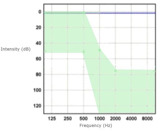
Figure 1: Preoperative audiogram. Indication thresholds are presented as a green zone. Please click here to view a larger version of this figure.
2. Perioperative preparations in the operating room (OR)
- Ensure that there is sufficient space between the patient's face and the draping. In addition, check that communication with the patient is possible with their contralateral hearing aid (HA). Connect the HA wirelessly with a remote microphone given to the nurse. For the HA, use a long sound tube to prevent unpleasant feedback.
- Alternatively, if the patient does not have the option for a contralateral HA, use a tablet computer to provide them with written communication. Test the communication with the patient.
- Administer antibiotics and dexamethasone (e.g., 1.5 g of cefuroxime and 0.1 mg/kg dexamethasone) intravenously before the skin incision.
- Insert the ECochG stimulator earphone into the patient's external ear canal prior to the incision. Seal the external ear canal in the earphone with bone wax (Figure 2).
NOTE: Some manufacturers provide foam earphones in sterile packaging. Alternatively, the earphone can be inserted prior to the draping, and the tube can be fed under the draping to the ECochG computer. - Place an iodoform sheet over the operation area, and align the earphone and the sound tube away from the incision area. Take care that the iodoform draping does not bend the sound tube during the preparation.
3. Partial mastoidectomy, posterior tympanostomy, and drilling the implant bed with a surgical drill
- Drill the partial mastoidectomy and posterior tympanostomy, and prepare the implant bed.
4. Insertion
- For the ECochG preparation, ensure the presence of a medical physicist/clinical engineer in the OR, referred to here as the ECochG operator.
- Get the equipment ready for conducting the ECochG sound stimulation (see the Table of Materials) and response measurement through the CI; the setup varies slightly depending on the CI manufacturer.
- Connect the sound tube to the stimulator.
- Connect the implant coil and cable to the measuring device.
- Thread the measuring coil and cable into the sterile wrapping.
- Depending on the manufacturer, launch the measurement program from a laptop, or use a tablet-based system.
- The stimulus is a tone burst, and the burst length depends on the stimulus frequency. Select the stimulus frequency (usually, stimuli at the highest preservable frequency of 500 Hz or 1,000 Hz during insertion), the sound pressure to be used, and the stimulus amplitude (e.g., 80-100 dB nHL so that stimulus is well-heard and tolerable) from the ECochG software. Depending on the manufacturer, keep the other measurement parameters at the default values, or change them to the following: a measurement window of 6.5-9.7 ms with a delay up to 2 ms after stimulus onset, and an average of 40-150 recordings per data point. Ideally, repeat the stimulus with alternated polarity and responses with condensation, and subtract the rarefaction polarity to extract the cochlear microphonics component (Figure 3).
- Use the first or the second contact of the electrode for the ECochG measurement. Check the impedance of the measurement channel soon after the contact is in the cochlea and before the first measurement.
- Ask the OR assistant to go to the patient and repeat the instructions to them about the subjective reporting of the changes in the sound stimuli.
- Test the sound stimuli, and confirm from the patient that they can clearly hear the stimuli.
- Advise the patient to inform about any deviations, such as diminishing or disappearances of the perceived stimuli.
- Get the equipment ready for conducting the ECochG sound stimulation (see the Table of Materials) and response measurement through the CI; the setup varies slightly depending on the CI manufacturer.
- Surgeon preparations before insertion
- Take the CI device, and place the receiver/stimulator into the implant bed.
- Open the round window membrane of the cochlea. Make a slit with a hypodermic needle in the lower boundary of the round window, and carefully lift the membrane backward with a microsurgical hook. Flush the round window area with dexamethasone solution.
- Check the alignment of the CI lead wire within the mastoid cavity to avoid any rotational forces on the electrode and intracochlear structures from the lead wire after insertion.
- Obtain a good ergonomic position with the best support possible for the hands to facilitate a slow and controlled insertion.
- Start of the insertion
- Have the surgeon inform the team that the insertion is starting, and make sure that everything is ready.
- Take the electrode with the insertion forceps, and start the insertion of the electrode into the cochlea through the round window.
- At the beginning of the insertion, first insert the electrode just inside the cochlea (one or two contacts inside the cochlea). Have the ECochG operator measure the impedance after the insertion of the first and/or second contact. If the impedance is very high, change the measuring channel.
- Have the ECochG operator start the measurement after the impedance measurements. Ensure the electrode is progressed very slowly with constant feedback from the person observing the ECochG responses (Figure 4).
NOTE: Depending on the manufacturer, there might also be an option that the recording device provides auditory feedback-a tone that becomes louder with an increasing response. However, this tone along with other noise in the OR may disturb the patient's subjective evaluation of the perceived stimulus. - Have the surgeon continue the insertion with 1-2 mm advancements (one electrode). Between these advancements, obtain feedback from the patient about the loudness of the perceived sound stimuli.
- If the ECochG responses become weaker or the patient reports a decrease in the perceived loudness of the sound stimuli, discontinue the insertion.
- Wait for 30-60 s, and then inquire about the stimuli from the patient. Simultaneously communicate with the ECochG operator about the current state of the ECochG monitoring (CM response).
- If either of the responses (the ECochG or subjective responses) do not recover when the electrode is motionless, draw the electrode back gradually by one to two contacts at a time (the value of ~2 mm is dependent on which brand of electrode is being used), and wait for the response each time. Draw the electrode back just enough until the responses have recovered.
- If the ECochG responses or the perceived loudness do not increase again after adjusting the insertion, but the desired IDA (270°-300°) (based on preoperative measurements and the number of electrodes inside the cochlea) has been achieved, discontinue the insertion. The situation is interpreted as a completed partial insertion.
- If the responses increase again, continue the insertion as described above (steps 4.4.5-4.4.7).
- Seal the round window with fat tissue gathered from the postauricular incision.
- In addition, use fibrin glue and bone dust to seal the groove for the lead wire between the receiver/stimulator and the mastoid cavity.
- Fix the lead wire inside the mastoid cavity with bone dust and fibrin glue.
- Close the wound with surgical sutures. Monitor the patient after the operation during an overnight stay in the ward.
5. Postoperative care
- Refer the patient to cone-beam computed tomography (CBCT) for postoperative imaging of the implantation.
- Verify the electrode's placement from the CBCT images.
- Open the CBCT images, and choose MPR from the dropdown list.
- Obtain the cochlear view in the MPR.
- Verify the IDA and the location of the electrode by scrolling through the images in the MPR. Check the number of intracochlear electrodes from the cochlear view to guide the fitting of the CI, especially in cases where there is a partial insertion, and identify the stimulating electrodes.
- Verify the electrode's placement from the CBCT images.
- Refer the patient for a postoperative audiogram to measure the residual hearing on the first postoperative day after the surgery and 1 month later. Measure the residual hearing routinely during the fitting follow-ups.
NOTE: The 1 month follow-up is more reliable than the first postoperative day in terms of the hearing preservation results.
Access restricted. Please log in or start a trial to view this content.
Results
Both subjective monitoring and ECochG may help prevent the occurrence of insertion trauma and, thus, provide better hearing preservation results postoperatively. In the audiogram, a decrease in the PTA(125-500 Hz) within 15 dB of the preoperative hearing levels is considered to represent preserved residual hearing and, thus, a positive result after surgery. A negative result is the loss of residual hearing: a PTA(125-500 Hz) change of over 30 dB from the p...
Access restricted. Please log in or start a trial to view this content.
Discussion
The optimal utilization of cochlear implantation is important for the effective management of hearing loss. The expansion of the indications for CI rehabilitation has created a grey zone where individualized decisions must be made regarding the rehabilitation modalities used. Nowadays, for patients with high-frequency hearing loss, there is a trend toward the provision of CIs. However, residual hearing in the low frequencies cannot be preserved in every patient. Cochlear monitoring via ECochG has been proposed a...
Access restricted. Please log in or start a trial to view this content.
Disclosures
The authors report no conflicts of interest.
Acknowledgements
Aarno Dietz has received grants from the Academy of Finland (Grant number 333525) and from the North Savo Regional Grant. Pia Linder has received a grant from the Finnish Government research funding (Grant number 5551877). Matti Iso-Mustajärvi has received grants from the Finnish Government research funding (Grant number 5551876), the Instrumentarium Science Foundation, the North Savo Regional Grant, and The Finnish Society of Ear Surgery.
Access restricted. Please log in or start a trial to view this content.
Materials
| Name | Company | Catalog Number | Comments |
|---|---|---|---|
| EarPhone and sound tube | AB/Cochlear/Medel | Usually provided by the company in sterile packages, can be inserted in ear without sterility issues | |
| Ecocgh program | AB/Cochlear/Medel | AB and Medel provides software for clinical use. Also Cochlear has software for ECochG, atleas for research purposes. | |
| Equipments for Cochlear implantation | Basic setup and instrumentation for Cochlear implantation, not spesific to the ECoGh or Subjective hearing monitoring | ||
| Laptop/tablet | AB/Cochlear/Medel | AB has tablet consept as "AIM" for intraoperative ECochG measuring. It is provided by the company. Cochlear and Medelare operated with laptop | |
| Sound Processor | AB/Cochlear/Medel | Also company-specific, need for the connection to the electrode and measuring during the insertion |
References
- Aschendorff, A., Kromeier, J., Klenzner, T., Laszig, R. Quality control after insertion of the nucleus contour and contour advance electrode in adults. Ear and Hearing. 28 (2), 75-79 (2007).
- Holden, L. K., et al. Factors affecting open-set word recognition in adults with cochlear implants. Ear and Hearing. 34 (3), 342-360 (2013).
- Gantz, B. J., Turner, C., Gfeller, K. E., Lowder, M. W. Preservation of hearing in cochlear implant surgery: Advantages of combined electrical and acoustical speech processing. Laryngoscope. 115 (5), 796-802 (2005).
- Lenarz, T., et al. European multi-centre study of the Nucleus Hybrid L24 cochlear implant. International Journal of Audiology. 52 (12), 838-848 (2013).
- Iso-Mustajärvi, M., Sipari, S., Löppönen, H., Dietz, A. Preservation of residual hearing after cochlear implant surgery with slim modiolar electrode. European Archives of Oto-Rhino-Laryngology. 277 (2), 367-375 (2020).
- Roland, J. T., Gantz, B. J., Waltzman, S. B., Parkinson, A. J. Multicenter clinical trial group. United States multicenter clinical trial of the cochlear nucleus hybrid implant system. Laryngoscope. 126 (1), 175-181 (2016).
- Roland, J. T., Gantz, B. J., Waltzman, S. B., Parkinson, A. J. Long-term outcomes of cochlear implantation in patients with high-frequency hearing loss. Laryngoscope. 128 (8), 1939-1945 (2018).
- Lenarz, T., Timm, M. E., Salcher, R., Büchner, A. Individual hearing preservation cochlear implantation using the concept of partial insertion. Otology & Neurotology. 40 (3), 326-335 (2019).
- Calloway, N. H., et al. Intracochlear electrocochleography during cochlear implantation. Otology & Neurotology. 35 (8), 1451-1457 (2014).
- Trecca, E. M., et al. Electrocochleography and cochlear implantation: A systematic review. Otology & Neurotology. 41 (7), 864-878 (2020).
- Mandalà, M., Colletti, L., Tonoli, G., Colletti, V. Electrocochleography during cochlear implantation for hearing preservation. Otolaryngology-Head and Neck Surgery. 146 (5), 774-781 (2012).
- Kim, J. S. Electrocochleography in cochlear implant users with residual acoustic hearing: A systematic review. International Journal of Environmental Research and Public Health. 17 (19), 7043(2020).
- Alzahrani, M., Martin, F., Bobillier, C., Robier, A., Lescanne, E. Combined local anesthesia and monitored anesthesia care for cochlear implantation. European Annals of Otorhinolaryngology, Head and Neck Diseases. 131 (4), 261-262 (2014).
- Dietz, A., Lenarz, T. Cochlear implantation under local anesthesia in 117 cases: Patients' subjective experience and outcomes. European Archives of Oto-Rhino-Laryngology. 279 (7), 3379-3385 (2021).
- Dietz, A., Wüstefeld, M., Niskanen, M., Löppönen, H. Cochlear implant surgery in the elderly: the feasibility of a modified suprameatal approach under local anesthesia. Otology & Neurotology. 37 (5), 487-491 (2016).
- Hamerschmidt, R., Moreira, A. T. R., Wiemes, G. R. M., Tenório, S. B., Tâmbara, E. M. Cochlear implant surgery with local anesthesia and sedation: comparison with general anesthesia. Otology & Neurotology. 34 (1), 75-78 (2013).
- Strøm, C., Rasmussen, L. S. Challenges in anesthesia for elderly. Singapore Dental Journal. 35, 23-29 (2014).
- Kecskeméti, N., et al. Cochlear implantation under local anesthesia: A possible alternative for elderly patients. European Archives of Oto-Rhino-Laryngology. 276 (6), 1643-1647 (2019).
- Shabashev, S., Fouad, Y., Huncke, T. K., Roland, J. T. Cochlear implantation under conscious sedation with local anesthesia; Safety, efficacy, costs and satisfaction. Cochlear Implants International. 18 (6), 297-303 (2017).
- Linder, P., Iso-Mustajärvi, M., Dietz, A. A comparison of ECochG with the subjective sound perception during cochlear implantation under local anesthesia-A case series study. Otology & Neurotology. 43 (5), 540-547 (2022).
- Escudé, B., et al. The size of the cochlea and predictions of insertion depth angles for cochlear implant electrodes. Audiology and Neurotology. 11 (1), 27-33 (2006).
- Gantz, B., et al. Outcomes of adolescents with a short electrode cochlear implant with preserved residual hearing. Otology & Neurotology. 37 (2), 118-125 (2016).
- Suhling, M. -C., et al. The impact of electrode array length on hearing preservation in cochlear implantation. Otology & Neurotology. 37 (8), 1006-1015 (2016).
Access restricted. Please log in or start a trial to view this content.
Reprints and Permissions
This article has been published
Video Coming Soon