Splenic artery aneurysms (SAA) are recognized as the most common type of visceral artery aneurysms, comprising approximately 60% of such cases. Historically, the management of SAA has included open surgical, endovascular, or non-operative management. This paper describes a robotic-assisted laparoscopic surgical technique for splenic artery aneurysm resection.
Method Article
Robotic Assisted Laparoscopic Splenic Artery Aneurysm Resection
In This Article
Summary
Abstract
Splenic artery aneurysms (SAA) are the most common visceral aneurysm and may develop significant risk for rupture and life-threatening complications when more than 3 cm, in pregnancy, or if resembling pseudoaneurysms. Traditional treatment options of open surgical repair and endovascular repair have been extensively studied, but the role of robotic-assisted laparoscopic surgery in definitively managing SAA is emerging as a promising minimally invasive alternative in select pathology. This case aims to present the technical details, challenges, and outcomes of an SAA in a young woman treated with robotic-assisted laparoscopic resection, highlighting the feasibility and efficacy of this approach. A 38-year-old woman with no significant past medical history was incidentally found to have a distal third splenic artery aneurysm measuring 2.2 cm in diameter and was referred to a vascular surgery clinic for further evaluation. Although she was not pregnant at the time, she was undergoing fertility treatments with plans for assisted pregnancy. She requested definitive aneurysm treatment, wished to avoid multiple interventions, and intended to become pregnant via IVF within the coming year, making repeated axial imaging unsuitable for follow-up. Imaging evaluation revealed the aneurysm sac contained one prominent feeding artery and two highly tortuous draining branches. After providing informed consent, the patient underwent successful robotic-assisted laparoscopic excision of the splenic artery aneurysm. Robotic-assisted laparoscopic surgery represents a valuable approach for the definitive management of distal splenic artery aneurysms or aneurysms involving highly tortuous vessels, conditions historically requiring open surgical ligation, resection, or vascular reconstruction. This paper illustrates how challenging aneurysms can be precisely visualized, dissected, and managed using robotic-assisted laparoscopy with the DaVinci surgical system, highlighting key minimally invasive techniques for optimal exposure and vascular control in splenic artery aneurysm repair.
Introduction
Splenic artery aneurysms (SAA) are recognized as the most common type of visceral artery aneurysms (VAAs), comprising approximately 60% of such cases1. These aneurysms can carry a significant risk of rupture when meeting size criteria (more than 3 cm) or in pregnancy with an increased plasma volume and cardiac output, leading to potentially life-threatening hemorrhage2. Historically, the management of SAA has included open surgical repair and conservative management (non-operative management for patients not meeting size criteria, pseudoaneurysm morphology, or not young fertile women)3,4. Over the past decade, endovascular (EV) approaches have been increasingly utilized due to their less invasive nature and favorable short-term outcomes compared to open repair5.
Recent studies comparing endovascular and open-surgical approaches for SAA management have shown promising results for endovascular techniques. Endovascular repair is associated with lower complication rates, shorter hospital stays, and reduced resource utilization compared to open surgery5,6. A meta-analysis found significantly lower overall mortality rates with endovascular repair7. However, randomized trials showed similar splenectomy rates and technical options for both approaches, with laparoscopy offering shorter procedures and lower morbidity8. While endovascular repair demonstrates advantages in perioperative outcomes9, the need for reintervention after endovascular repair remains a concern, with freedom from reintervention at 3 years postoperatively reported as 82.4%, mostly due to sac reperfusion6. These findings suggest that endovascular repair may be a preferred initial strategy for SAA management, but further comparative studies are needed.
Robotic-assisted and laparoscopic surgery have emerged as viable minimally invasive options for treating SAA. These approaches demonstrate comparable operative times, low open conversion rates, and reduced morbidity compared to open surgery2. Robotic-assisted laparoscopic surgery allows for complex procedures, including aneurysm excision with arterial reconstruction, and shows promising mid-term results4,9. A randomized study comparing open and laparoscopic SAA management found that laparoscopy permitted multiple technical options without increasing splenectomy rates while reducing postoperative complications and hospital stay8. However, laparoscopic arterial anastomoses showed poor long-term results. Complex vascular reconstructions have now been demonstrated in the transplant literature and by international vascular surgeons on the DaVinci Xi platform, emphasizing the increased dexterity and wristed instrument of the robotic platform, a prior limitation of laparoscopic surgery10. Both robotic and laparoscopic approaches result in shorter hospital stays and lower overall morbidity rates compared to open surgery2,8. These minimally invasive techniques represent important advancements in SAA treatment, offering tailored options for patients, particularly those at high surgical risk.
Despite these advancements, the rarity of SAA has precluded the conduct of large randomized controlled trials, resulting in a reliance on case reports and series to inform clinical practice4. Existing literature indicates that while endovascular approaches may offer superior short-term results, they also require more reinterventions compared to open surgery, which is associated with fewer late complications5. Moreover, splenic infarction and pancreatitis are noted as common complications in minimally invasive approaches11.
By examining this case, we aim to provide further insights into the technical details of robotic-assisted laparoscopic surgery for SAA by providing a comprehensive protocol. This paper and accompanying video will serve as a template to instruct robotic instrument selection, port placement and DaVinci Xi robot docking, gastropexy for visualization, exposure of a distal splenic artery aneurysm, vascular control of feeding and draining arteries, and ligation of a splenic artery aneurysm.
CASE PRESENTATION:
A 38-year-old Caucasian female presented to the vascular surgery clinic for evaluation after an incidental finding of a distal splenic artery aneurysm found on an MRI performed for evaluation of adrenal glands. She had a surgical history of prior C-sections and no other significant medical history. Her home medications included Aspirin 81 mg daily, folic acid, fish oil, and prenatal vitamins. She was undergoing fertility treatments with plans for in vitro fertilization this year. She drank alcohol socially and denied tobacco and drug use. In regard to the SAA, she was asymptomatic, and on review of her imaging, there were no other visceral aneurysms identified. The physical exam was unremarkable other than a well-healed Pfannenstiel incision.
Diagnosis, assessment, and plan
The decision was made to obtain new contrasted CT imaging to investigate the anatomy of the splenic artery aneurysm and allow for surgical planning. CTA Abdomen/pelvis showed a 2.2 cm x 1.9 cm distal 1/3 splenic artery aneurysm with two draining branches and one feeding artery. All arteries showed high levels of tortuosity (Figure 1A-B). The patient was counseled about the recommendation for repair, given her plan for the upcoming pregnancy, which would place her at increased risk for an aneurysm rupture. She agreed to surgical repair but requested to avoid the endovascular approach as she did not want to have increased exposure to radiation in her operation and follow-up course, given her fertility treatments and plans for upcoming assisted pregnancy. The options of open surgery through midline laparotomy with a plan for splenic artery aneurysm repair versus robotic-assisted laparoscopic splenic artery aneurysm resection and possible repair were described to her. She opted for the minimally invasive robotic-assisted approach. Risks and consequences, including but not limited to perioperative bleeding, perioperative pancreatitis, splenic artery occlusion and infarct of the spleen, secondary infections, the need for blood transfusions, splenectomy, and the need for post-splenectomy vaccines, were discussed with the patient in detail. All questions were answered, and she wished to proceed.
Protocol
The patient provided informed consent to the report of her case details and imaging studies. This study was conducted, and the protocol was written in accordance with the ethical guidelines and regulations of the Houston Methodist Hospital Ethics Committee.
1. Pre-operative planning
- Obtain complete written informed consent from the patient. Consent should discuss the following: Robotic-assisted laparoscopic splenic artery aneurysm ligation and/or resection, splenic artery reconstruction, conversion to open splenic artery aneurysm resection (laparotomy), splenectomy (total or partial), pancreatic injury repair, bowel injury repair, blood transfusion, placement of surgical drains, post-splenectomy vaccines.
- Use a proper timeout procedure to identify the correct patient, the correct procedure, and the operating surgeon.
- Take the patient to the operating room, place them in a supine position, and administer general endotracheal anesthesia. Secure the patient using a bean bag or gel pads.
- Prepare the surgical site with an antiseptic solution (2% chlorhexidine or betadine solution) and drape in a standard sterile fashion (with the patient exposed from pubic symphysis to xiphoid process). Give 2 g Ancef intravenously prior to induction for antibiotic prophylaxis.
2. Laparoscopic and robotic entry
- Mark the four robotic port sites at 8 cm intervals in a horizontal line approximately 14 cm inferior to the xiphoid process. Make a horizontal 8 mm incision in the skin on the left paramedian region and use an optical trocar to gain access to the peritoneal cavity using the standard technique (Figure 2).
- Establish pneumoperitoneum. Under laparoscopic visualization,place three additional 8 mm ports in the port sites marked, and place an extra 5 mm port in the right subcostal region for a left liver retractor using standard technique and fix it to the bed. Tilt the bed in reverse Trendelenburg (15°-20°) to facilitate better visualization of the upper abdomen.
- Place a 12 mm assistant port inferiorly and in between the supraumbilical and left paramedian port (Figure 2).
- Remove the 12 mm port and use a PMI device and 0 Vicryl suture to pre-close the fascial defect by placing a suture through the abdominal fascia, but do not tie it at this point. Then, reinsert the 12 mm assistant port.
3. Robotic docking and setup
- Dock the DaVinci Xi robot from the patient's right side using the standard technique. Align the boom arm parallel to the patient's midline. Press the Port Assignment button on the touchscreen monitor and select Four-arm Setup. Ensure the camera boom is centered over the planned camera port (port #2) using the laser-assisted crosshairs to assist.
- Attach the robotic arms to the ports: Robot arm 1 (left lateral port) to fenestrated bipolar grasper, arm 2 (midline port) to the camera, arm 3 (right lateral port) to the vessel sealer, arm 4 (far right lateral port) to Cadiere forceps.
- Perform exposure of the splenic artery as described below.
- Using a bipolar fenestrated grasper, a vessel sealer, and Cadiere forceps, perform dissection along the greater curvature of the stomach to divide the anterior leaflet of the greater omentum and enter the lesser sac. Perform further dissection superiorly and inferiorly, superiorly up to the takeoff of the short gastric arteries, which should be kept intact, and inferiorly to the inferior portion of the greater curvature.
- Use tacking sutures with 2-0 Vicryl V-Loc sutures to tack up the stomach to the anterior abdominal wall.
- After opening the lesser sac, the posterior gastric fat pad was visualized. A pulsating mass was seen; use it as a landmark for where to open the fat pad the visualize the splenic artery. Identify the splenic artery at the superior border of the pancreas (Figure 1D). Perform dissection with a fenestrated bipolar grasper and monopolar hook around the proximal splenic artery and obtain proximal control.
4. Vascular control and ligation
- Control the artery with a vessel loop and a 1 cm red rubber catheter to make a modified Rumel technique. Clip the vessel loops to secure them in place. Next, carry out dissection around the inflow artery and the outflow arteries of the aneurysm using a fenestrated grasper and hook electrocautery.
- Encircle the arterial inflow and outflow branches and obtain vascular control with vessel loops. Occlude the feeding artery and check Firefly fluorescence for adequate perfusion of the spleen.
- Perform careful inspection of the distal outflow branches. This showed three branches, two of which were large, and one was small. Both the large branches were extremely thin walled, which would then be a risk for anastomotic suture dehiscence and breakdown with pseudoaneurysm formation and, hence, not worth the risk of primary repair. Therefore, the decision was made to ligate the artery and resect the aneurysm.
- Clip all the arterial inflow and outflow branches and divide them with the vessel sealer using the standard technique (Figure 1E).
5. Removal of aneurysm specimen, hemostasis, and closure
- Dissect the aneurysm from the retroperitoneum with a hook electrocautery using standard technique. Maintain hemostasis with a fenestrated bipolar grasper. Excise the aneurysm completely and place in an Endo Catch retrieval bag (Figure 1C).
- Irrigate the area with normal saline till the returning fluid is clear. Secure hemostasis. Remove the stray sutures on the stomach for the gastropexy. Remove the liver retractor from the right subcostal port.
- Remove the ports under visualization. Secure hemostasis to the port sites. Remove the Endo Catch bag with the specimen from the 12 mm assist port. Discontinue pneumoperitoneum completely.
- Tie down the Vicryl sutures previously placed in the fascia at the 12 mm port site. Irrigate all the incisions with normal saline and close the remaining 8 mm and 5 mm port sites with 3-0 PDS deep dermal simple interrupted suture. Extubate the patient and move to the recovery room.
6. Post-operative Care
- For pain control, use the following regimen post-operatively. Administer Tylenol 650 mg per oral (PO) every 6 h, methocarbamol 500 mg PO 3x daily, gabapentin 200 mg PO 2x daily, lidocaine 4% patch placed on abdomen daily, and tramadol 50 mg PO every 6 h as needed for severe pain (pain score 7-10).
- Administer intravenous (IV) Zofran 4 mg as needed every 8 h and IV promethazine 6.25 mg every 6 h as needed for nausea.
- Anticipate a hospital stay of 2-3 days. Here, the patient stayed a total of 4 days before discharging home due to some persistent post-operative nausea that improved with administration of 4 mg IV Zofran and 6.25 mg IV promethazine.
- Ask the patient to follow up at the clinic at 1 month post-operatively. Perform a physical examination and abdominal ultrasound exam, with the ultrasound confirming the proximal and mid-splenic artery without residual aneurysm or formation of new pseudoaneurysm. Perform a repeat splenic ultrasound at 7 months after surgery with normal arterial and venous waveforms confirming adequate perfusion to the spleen.
Results
The procedure was carried out by a vascular surgeon trained in robotic surgery. The surgical team consisted of the Attending surgeon, a Circulator nurse, a Scrub nurse, and a Surgical Resident. The total operative time was 233 min. The estimated blood loss was 20 mL. There were no intraoperative complications or transfusions. The patient was transferred from the recovery room to the surgical floor with normal range hemodynamics and controlled pain.
From a technical perspective, these results validate the operative workflow described, including port placement, gastropexy for visualization, precise vascular dissection, and ligation techniques. The successful identification and preservation of short gastric arterial collaterals, verified intraoperatively with FireFly fluorescence imaging, ensured adequate splenic perfusion post-ligation, reducing the risk of splenic infarction or the need for arterial reconstruction. Additionally, the absence of pseudoaneurysm formation or need for reintervention at the 1-month and 7-month ultrasound follow-ups provides supporting evidence for the durability of this surgical technique (Figure 3). This was determined from the normal arterial and venous waveforms seen on the splenic ultrasound showing adequate perfusion of the spleen, and the healthy appearing proximal and mid segment splenic artery near the operative site where the ligation took place.
Post-operative care
On the postoperative day (POD) 0, the pain was well controlled, but the patient developed nausea and vomiting. The patient remained on nothing per oral (NPO) overnight after surgery and received IV 4mg Zofran and 6.25mg Phenergan. On POD 1 the patient required IV pain medication but was comfortable with mild nausea that improved with additional Zofran and Phenergan. On POD 2, the patient tolerated a soft diet, walked 200 feet in the hospital unit, and received prophylactic splenectomy vaccines. On POD 3, the patient no longer required IV pain medication was tolerating a full diet and had regular bowel function. The patient was discharged home early morning on POD 4 in good condition. The patient was seen in the clinic at 2 months follow-up. A physical examination and abdominal ultrasound exam were performed, with the ultrasound confirming the patent proximal and mid-splenic artery without an aneurysm. The patient had returned to daily activities without difficulty and had no signs of surgical site complications. The patient had a repeat splenic ultrasound 8 months after surgery with normal arterial and venous waveforms.
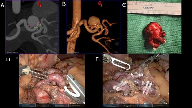
Figure 1: Pre-operative and intra-operative imaging of splenic artery aneurysm. (A) Computed tomography angiography (CTA) Imaging of Splenic artery aneurysm (SAA). Contrasted imaging of the mid-distal highly tortuous SAA. (B) 3D-Reconstruction of SAA. 3D reconstruction highlighting tortuous branching pattern of SAA and multiple draining branches present. (C) Excised SAA. Excised SAA on the back table at the completion of the case. (D) Robotic View of SAA. Initial dissection of the SAA prior to vascular control and ligation of SAA branches. (E) Ligation of SAA. Picture of SAA with clips applied to each branch prior to excision from the patient. Please click here to view a larger version of this figure.
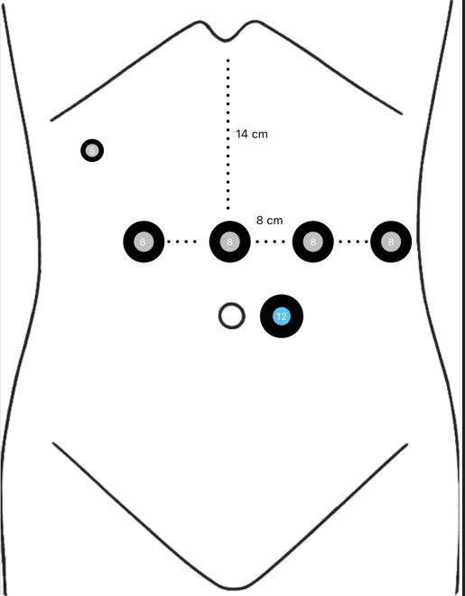
Figure 2: Anatomical landmarks and port placement. Four robotic port sites at 8 cm intervals in a horizontal line approximately 14 cm inferior to the xiphoid process. A 5 mm port in the right subcostal region for a left liver retractor. A 12 mm assistant port inferiorly and in between the supraumbilical and left paramedian port. Please click here to view a larger version of this figure.
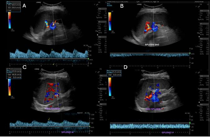
Figure 3: Post-operative splenic ultrasound. (A) The 1-month splenic artery waveforms. (B) The 1-month splenic vein waveforms. (C) The 7-month splenic artery waveforms. Both (A and C) show a low resistance and monophasic waveform with a sharp systolic upstroke. This signifies continuous forward flow through the cardiac cycle and lack of turbulent flow. (D) The 7-month splenic vein waveforms. Both (B and D) show continuous, low-velocity flow directed towards the portal vein. Please click here to view a larger version of this figure.
Discussion
Laparoscopic and robotic treatments of visceral artery aneurysms (VAA) are described in the literature12,13,14,15. Still, they historically are limited in daily clinical practice mainly due to limited expertise amongst vascular surgeons and the facility cost of the surgical robots. In addition, endovascular repair continues to represent first-choice management in most cases16. This combination of factors makes evidence concerning the robotic-assisted laparoscopic approach for VAAs limited. Prior to 2010, there were only seven cases of robotically treated VAAs published in the literature. Between 2011 and 2019, a systematic review found only 20 patients and just 27 more between 2020 and 202417. The use of robotic techniques in the treatment of VAAs is increasing at a rapid pace, but the only available data regarding its outcomes remains limited to case reports and case series.
Although other case reports describing similar cases already exist, this study's value lies in its detailed description of the operative technique, including a video presentation-and in its comprehensive protocol for peri-operative patient care.
One of the most important advantages of robotic assisted laparoscopic surgery (RALS) is the extremely low rate of peri-operative complications, thanks to the minimally invasive approach. Many reviews have shown significantly decreased cardiac, respiratory, thromboembolic, and infections compared to open surgery for VAAs. Moreover, the robotic-assisted approach is characterized by early recovery of oral feeding and short hospital stay17. These added benefits might counterbalance the additional cost of the robotic equipment; however, we are unable to provide a detailed cost analysis for this case.
While there are many benefits to the RALS approach, possible disadvantages are the operative time and periods of organ ischemia if performing a vascular reconstruction. There is no information based on large clinical trials concerning the operative time and ischemia times of open surgery of VAAs, but those of RALS may be slightly longer18,19. This may be due to the learning curve and could be liable to improve in the future. However, on review of early data, such longer times did not cause significant adverse clinical outcomes. In our case, the total operative time was 233 min. This is longer than the average reported time in the 2020 review2 (150 ± 87.7 min) but within one standard deviation. The presented case involved complex anatomy with an extra draining artery branch that needed to be controlled and ligated. This was also the first robotic splenic artery surgery in our institution; thus, the relatively longer operative time was needed to instruct assistants in the operating room. We were able to reduce the operative time in the following 3 cases, with the latest one consisting of aneurysm resection and arterial repair (130 min and 188 min for ligation, and 256 min for arterial reconstruction with end-to-end anastomosis).
Early data indicate satisfactory rates of intraoperative conversion to laparoscopic or open surgery (5.6%). Among VAAs, splenic artery aneurysms (SAA) appear particularly susceptible to conversion, primarily due to dense tissue adhesions that complicate vascular control; this was identified as the most frequent reason for procedural failure17. Consequently, patients with hostile abdominal anatomy may carry a higher risk of conversion, emphasizing the importance of careful patient selection to reduce this risk. Patients eligible for RALS should be informed about the possibility of conversion, similar to other minimally invasive procedures such as endovascular or laparoscopic treatments. However, robotic repair offers an advantage over endovascular approaches, as it does not carry the risk of aneurysm reperfusion and rupture, a significant limitation associated with endovascular treatment.
The robotic-assisted laparoscopic splenic artery aneurysm (SAA) resection involves several critical steps to ensure safe and effective aneurysm management. First, pneumoperitoneum is established, and the DaVinci Xi robot is docked to four abdominal ports, with an additional 12 mm assistant port for instrument exchange and a 5 mm port for liver retractor placement. After docking, the greater omentum is dissected from the greater curvature of the stomach, exposing the posterior gastric fat pad and creating access to the lesser sac. The splenic artery is identified along the superior border of the pancreas, and meticulous dissection is performed to isolate the aneurysm and its vascular branches. The inflow and outflow vessels are encircled with vessel loops for vascular control, with the option of using a modified Rumel tourniquet technique for proximal occlusion. Intraoperative FireFly fluorescence imaging is utilized to assess splenic perfusion through the short gastric arteries before proceeding with arterial ligation and aneurysm excision. The aneurysm is resected, the surgical field is irrigated, and hemostasis is secured. Finally, ports are removed under direct visualization, the fascial defects are closed, and the patient is carefully monitored postoperatively for early recovery and return to normal function.
Recommended modifications to the technique are the use of robotic bulldog clamps (Klein Surgical) for vascular control which decreases operating time compared to vessel loop and Rumel tourniquet4. Another modification would be performing an end-to-end anastomosis to reconstruct the splenic artery in patients with questionable collateral blood supply to the spleen, and that would not tolerate splenectomy/splenic embolization.
A future application of this technique would be the expanded use in other visceral artery aneurysms (hepatic, renal, superior mesenteric, and celiac artery aneurysms). Other applications would be performing robotic-assisted arterial bypass and reconstruction procedures, performing hybrid endovascular-robotic techniques, and adopting these skills to perform vascular repairs for iatrogenic injuries when vascular surgeons are consulted intra-operatively.
In conclusion, RALS offers several advantages, although its applicability remains limited to specific situations. Currently, there is neither consensus nor clear evidence to guide optimal patient selection, and defined inclusion and exclusion criteria for RALS candidates are still lacking. In cases of splenic artery aneurysm (SAA), RALS might be preferred over endovascular approaches for aneurysms located at the splenic hilum, particularly when embolization or endograft placement risks sacrificing collateral vessels, potentially leading to splenic infarction or post-embolization syndrome. Additionally, younger patients at higher risk for long-term reintervention after endovascular treatment could also benefit from robotic-assisted procedures.
Disclosures
The authors have nothing to disclose
Acknowledgements
The authors have no acknowledgments.
Materials
| Name | Company | Catalog Number | Comments |
|---|---|---|---|
| 2-0 Vicryl V-Lock Suture | VLOCM0115 | ||
| 8m endoscope plus, 30 degree | Intuitive Surgical | 470057 | |
| 8mm blunt obturator | Intuitive Surgical | 470008 | |
| 8mm cannula | Intuitive Surgical | 470002 | |
| BD Bard All-Purpose Red Rubber Unisex, 16FR, Straight | Bard | 277716 | |
| Bipolar energy instrument cord | Intuitive Surgical | 470384 | |
| Cadiere Forceps | Intuitive Surgical | 471049 | |
| Da Vinci Xi surgical robot | Intuitive Surgical | 380662-21 | Robotic assisted surgical platform |
| Endo catch specimen retrieval bag | Medtronic | 173050G | |
| Fenestrated Bipolar Forceps | Intuitive Surgical | 471205 | |
| Hot Shears (Monopolar Curved Scissors) | Intuitive Surgical | 470179 | |
| Laparoscopic 5mm x 100mm Trocar | Ethicon | 2B12LT | |
| Laparoscopic Suction Irrigator | Pilling | 728145 | |
| Large Clip Applier | Intuitive Surgical | 470230 | |
| Large SutureCut Needle Driver | Intuitive Surgical | 471296 | |
| Monopolar energy instrument cord | Intuitive Surgical | 470383 | |
| Permanent Monopolar Cautery Hook | Intuitive Surgical | 470183 | |
| Snake Liver Retractor 5mm 80mm End | Artisan Medical Devices | SNK-100 | |
| Tip-Up Fenestrated Grasper | Intuitive Surgical | 470347 | |
| Vessel Loop McKesson Mini Red Silicone Radiopaque | McKesson | 487858 | |
| Vessel Sealer Extend | Intuitive Surgical | 480422 |
References
- Ibrahim, F., Dunn, J., Rundback, J., Pellerito, J., Galmer, A. Visceral artery aneurysms: diagnosis, surveillance, and treatment. Curr Treat Options Cardiovasc Med. 20 (12), 97(2018).
- Ossola, P., Mascioli, F., Coletta, D. Laparoscopic and robotic surgery for splenic artery aneurysm: a systematic review. Ann Vasc Surg. 68, 527-535 (2020).
- Antoniou, G. A., et al. Clinical applications of robotic technology in vascular and endovascular surgery. J Vasc Surg. 53 (2), 493-499 (2011).
- Marone, E. M., et al. Robotic treatment of complex splenic artery aneurysms with deep hilar location: technical insights and midterm results. Ann Vasc Surg. 68, 50-56 (2020).
- Sticco, A., et al. A comparison of open and endovascular treatment strategies for the management of splenic artery aneurysms. Vascular. 24 (5), 487-491 (2016).
- Zhu, C., et al. Endovascular and surgical management of intact splenic artery aneurysm. Ann Vasc Surg. 57, 75-82 (2019).
- Damara, F., Pratama, D. Endovascular and open surgical management in splenic artery aneurysm: an updated meta-analysis of comparative studies. J Vasc Surg. 75 (6), E114-E115 (2022).
- Giulianotti, P. C., et al. Robot-assisted treatment of splenic artery aneurysms. Ann Vasc Surg. 25 (3), 377-383 (2011).
- Tiberio, G. A. M., et al. Prospective randomized comparison of open versus laparoscopic management of splenic artery aneurysms: a 10-year study. Surg Endosc. , (2012).
- tádler, P., Dvořáček, L., Vitásek, P., Matouš, P. The application of robotic surgery in vascular medicine. Innovations. 7 (4), 247-253 (2012).
- Madoff, D. C., et al. Splenic arterial interventions: anatomy, indications, technical considerations, and potential complications. Radiographics. 25 (suppl 1), S191-S211 (2005).
- Grandhomme, J., et al. Robotic surgery for in situ renal artery aneurysm repair: technical note and literature review about a mini-invasive alternative. Ann Vasc Surg. 74, 526.e7-526.e12 (2021).
- Wu, Q., et al. Robot-assisted laparoscopic retroperitoneal renal artery aneurysm repair: a rare case report and literature review. Urol Int. 106 (12), 1298-1303 (2022).
- Ceccarelli, G., Gusai, G., Rondelli, F., Balestra, F., De Rosa, M. Video-robotic aneurysmectomy for splenic artery aneurysm: case report and literature review. Minim Invasive Ther Allied Technol. 29 (4), 244-249 (2020).
- Wei, H. B., et al. Robot-assisted laparoscopic reconstructed management of multiple aneurysms in renal artery primary bifurcations: a case report and literature review. BMC Urol. 17 (1), 96(2017).
- Long, K., et al. Complete robotic repair of a renal artery aneurysm. J Vasc Surg Cases Innov Tech. 3 (4), 225-227 (2017).
- Rinaldi, L. F., Brioschi, C., Marone, E. M. Robotic surgery for elective repair of visceral and renal artery aneurysms: a systematic review. J Clin Med. 13 (12), 3385(2024).
- Barrionuevo, P., et al. A systematic review and meta-analysis of the management of visceral artery aneurysms. J Vasc Surg. 70 (5), 1694-1699 (2019).
- Chaer, R. A., et al. The society for vascular surgery clinical practice guidelines on the management of visceral aneurysms. J Vasc Surg. 72 (1S), 3S-39S (2020).
Reprints and Permissions
This article has been published
Video Coming Soon