Method Article

Rapid Identification of Neisseria gonorrhoeae, Chlamydia trachomatis, and Ureaplasma urealyticum by RNA Amplification Lateral Flow Assay

DOI:

10.3791/68413

June 20th, 2025

In This Article

Summary

Loading...
$$\rightleftharpoonup{xx}$$ $$\longleftharp{xx}$$, $$\longrightharp{xx}$$,

Here, we present a protocol demonstrating RNA amplification lateral flow assay and evaluate the efficacy for rapidly detecting Neisseria gonorrhoeae, Chlamydia trachomatis, and Ureaplasmaurealyticum.

Abstract

Loading...
$$\rightleftharpoonup{xx}$$ $$\longleftharp{xx}$$, $$\longrightharp{xx}$$,

Neisseria gonorrhoeae (NG), Chlamydia trachomatis (CT), and Ureaplasma urealyticum (UU) are common pathogens associated with sexually transmitted diseases (STDs). NG is the causative agent of gonorrhea, which can result in various disorders of the genitourinary system, including epididymitis, prostatitis, cervicitis, and infertility. CT is a microorganism responsible for non-gonococcal urethritis and pelvic inflammatory disease. UU is associated with non-gonococcal urethritis, cervicitis, prostatitis, and infertility. Current molecular technologies for the analysis of NG, CT, and UU are often complex and time-consuming. Rapid molecular testing for these pathogens may facilitate the early diagnosis of STDs. The RNA amplification lateral flow assay (RGT) is a method that does not require nucleic acid extraction. In this process, samples are lysed to release nucleic acid fragments, which are then amplified through reverse transcription and transcription. The amplified RNA product is recognized and captured by specific probes, forming an RNA-detection probe-gold probe complex that can be immobilized on a nitrocellulose membrane via lateral flow to produce visible bands. The entire procedure takes less than 1 h. The aim of this study was to evaluate the efficacy of RGT for the rapid detection of NG, CT, and UU. A total of 1,416 samples were included. The consistency of NG, CT, and UU when analyzed by RGT compared to the polymerase chain reaction (PCR) fluorescence probing method was 99.03% (307/310), 99.38% (159/160), and 99.02% (303/306), respectively. The consistency of NG, CT, and UU when analyzed by RGT compared to gene sequencing was 99.17% (238/240), 98.95% (188/190), and 98.30% (173/176), respectively. Compared with the pathogen isolation culture method, the detection rates for NG, CT, and UU assays were 100.00% (9/9, 17/17, 8/8). The high sensitivity and specificity, ease of use, and reduced time requirements make this assay ideal for the rapid and accurate detection of NG, CT, and UU.

Introduction

Loading...
$$\rightleftharpoonup{xx}$$ $$\longleftharp{xx}$$, $$\longrightharp{xx}$$,

Sexually transmitted infections (STIs) represent significant global public health challenges, leading to serious genital and reproductive morbidity and mortality1. Neisseria gonorrhoeae (NG) is the causative agent of gonorrhea, with approximately 87 million new cases reported annually worldwide2. This pathogen primarily induces inflammation by invading the columnar epithelium of the urogenital tract3. Chlamydia trachomatis (CT), another microorganism, is responsible for non-gonococcal urethritis and pelvic infections, accounting for around 130 million new cases each year4. Ureaplasma urealyticum (UU), the simplest and smallest self-replicating cell, readily invades damaged cervical tissue and is associated with non-gonococcal urethritis, cervicitis, prostatitis, and infertility5. All of these infections are often asymptomatic in both female and male patients, yet they can lead to severe complications if not treated promptly. Maternal infections can result in serious adverse pregnancy outcomes, including stillbirth, miscarriage, preterm birth, and direct fetal infection6. Therefore, the development of a rapid, sensitive, specific, and user-friendly method for pathogen detection is of paramount importance.

Rapid and accurate detection of pathogens is critical for effective disease diagnosis and management. Traditional culture methods, which require the isolation, culture, and identification of pathogens, have significant disadvantages, including being time-consuming and having low sensitivity. Although gene sequencing boasts a high accuracy rate, it necessitates a complex environment and equipment, making it costly and also time-consuming. Conventional molecular technologies for the analysis of NG, CT, and UU have been widely accepted; however, these methods are often complicated and time-consuming, requiring sophisticated instrumentation, trained technicians, and extended processing times. Therefore, there is a pressing need for a simple and reliable molecular diagnostic method to address the limitations of conventional approaches. The RNA amplification lateral flow assay (RGT) offers a rapid, sensitive, and specific alternative without the need for complex procedures7,8. In this method, samples are directly lysed to release nucleic acid fragments without nucleic acid extraction, and these fragments are amplified through reverse transcription and transcription. The amplified RNA product is then recognized and captured by specific probes, forming an RNA-detection probe-gold probe complex, which can be immobilized on a nitrocellulose membrane via lateral flow to create visible bands9. The entire process takes less than 1 h, making it highly suitable for emergency laboratory tests.

In this study, we recruited 1,416 samples to evaluate the use of RGT for the detection of NG, CT, and UU. Samples were analyzed using RGT, PCR fluorescence probing, gene sequencing, and pathogen isolation culture methods. The results indicated that the RGT method was accurate and consistent with conventional diagnostic methods. In conclusion, the high sensitivity and specificity, ease of use, and reduced time requirements make this assay ideal for the rapid and accurate detection of NG, CT, and UU, suggesting its promising application in the preliminary medical diagnosis of sexually transmitted diseases.

Protocol

Loading...
$$\rightleftharpoonup{xx}$$ $$\longleftharp{xx}$$, $$\longrightharp{xx}$$,

This study was conducted in accordance with the ethical guidelines and approval granted by the Institutional Review Board of the First People's Hospital of Foshan, Guangdong Province, China (Ethical Review Document No. LR (2025) 136). A total of 1,416 genital swab specimens (716 from female patients, 700 from male patients) were prospectively collected from individuals clinically suspected of STDs who had not undergone antibiotic therapy within the preceding 14 days (Supplementary Table 1). For each participant, paired swabs were obtained simultaneously and analyzed in parallel: one by the test method (RGT) and the other by reference method (PCR fluorescence probing, gene sequencing, and pathogen isolating culture, respectively). Inter-method agreement was quantified using Cohen's Kappa coefficient (κ), with interpretation guided by Landis and Koch's criteria.

NOTE: The assay protocol for CT and UU follows identical procedural steps to those described for NG, with modifications limited to pathogen-specific reagents. Detail reagent formulations are provided in step 2.1 (Supplementary Table 2, Supplementary Table 3, and Supplementary Table 4).

1. Sample collection

  1. Applicable specimen type: Collect genital swabs (female cervical swab, male urethral swab).
  2. Specimen collection
    1. Insert the genital swab into the male urethral orifice for 2-3 cm or into the female cervical orifice for approximately 1- 2 cm.
    2. Rotate once and hold for 10 s.
    3. Remove the swab and insert it into the collection tube containing 1 mL of normal saline.
    4. Break off and discard the tail of the swab, then tighten the tube cap.
    5. Record the collection time and conduct the test promptly.
  3. Specimen storage and transportation
    1. Transport specimens at room temperature (RT) within 4 h or at 2-8 °C with an ice pack within 24 h.
    2. Store the remaining cell pellets of processed specimens at -20 °C for 3 months or -80 °C for up to 6 months if the tests cannot be conducted in a timely manner.

2. Preparation

  1. Reagents
    1. For reagents of NG nucleic acid assay, refer to Supplementary Table 2.
    2. For reagents of CT nucleic acid assay, refer to Supplementary Table 3.
    3. For reagents of UU nucleic acid assay, refer to Supplementary Table 4.
    4. Storage: Store all reagents at temperatures ranging from -40 °C to -15 °C and the detection cards at temperatures between 2 °C and 30 °C.
      NOTE: The amplification buffer and detection solution should be equilibrated to RT before use, based on the daily detection requirements. After reaching RT, they should be stored at 4 °C and can be used for the next detection. Avoid repeated freezing and thawing. The amplification enzyme should be stored at -20 °C except during the experimental process. An increase in storage temperature can lead to a decline or loss of enzyme activity.
  2. Experimental environment requirements
    1. Conduct the experiment in four physically separated zones:
      1. Reagent preparation area: Maintain at 22 ± 1°C with positive air pressure (High-efficiency particulate air (HEPA)-filtered airflow rate ≥ 0.3 m/s). Store pre-aliquoted reagents in UV-sterilized cabinets.
      2. Sample preparation area: Perform all specimen handling in a Class II biological safety cabinet pre-treated with 70% ethanol.
      3. Amplification area: Operate thermal cyclers on anti-vibration tables.
      4. Product analysis area: Use a UV-sterilized bench (30 min exposure pre/post use).
    2. Regulate personnel and airflow in each section to prevent cross-contamination as follows:
      1. Maintain unidirectional airflow from reagent preparation to product analysis zones.
      2. Ensure negative pressure in the sample preparation area (-5 Pa relative to adjacent zones).
      3. Restrict personnel movement; decontaminate gloves with 75% ethanol before zone transitions.
      4. Install airlock doors with interlock systems (minimum 30 s delay between openings).
    3. Use zone-specific consumables (e.g., centrifuge tubes, pipette tips, etc.) and discard consumables after a single use.
    4. Maintain cleanliness and quality control:
      1. Clean benches daily with 10% sodium hypochlorite followed by 75% ethanol or distilled water.
      2. Validate consumable sterility via endotoxin testing (Limulus amebocyte lysate assay, <0.25 EU/mL).
      3. Monitor air quality weekly using settle plates (Tryptic soy agar [TSA] agar, ≤5 colony-forming unit (CFU)/plate after 4 h exposure).
  3. Personnel qualifications
    1. Possess certificates or qualifications related to PCR technology to ensure the accuracy and reliability of this experiment.

3. Lyse the cells to extract RNA

  1. Specimen processing
    1. Vortex the specimen collection tube at 2,600 times/min for 10 s to dislodge the cells from the swab head into the solution.
    2. Transfer 1 mL of the solution to a 1.5 mL microcentrifuge tube using a calibrated pipette set to 1000 µL.
    3. Centrifuge at 12,000 × g for 2 min at 25 °C.
    4. Aspirate 950 µL of the supernatant without disturbing the pellet. Resuspend the pellets in 25 µL of residual solution by pipetting up and down 10 times using a 200 µL calibrated pipette set to 50 µL.
  2. Lysis of specimen and quality control materials
    1. Pre-equilibrate cell lysis buffer and negative control at 25 °C for 15 min.
    2. Centrifuge the quality control materials at 1000 × g for 10 s at 25 °C to ensure that all powders settle at the bottom of the tube.
    3. Add 30 µL of lysis buffer to the quality control materials and 20 µL to the specimens.
    4. Incubate for 10 min at 25 °C.
      NOTE: When using the cell lysis buffer, it is essential to mix it thoroughly. Mix by pipetting 15 times (200 µL tip) and add both the particles and the liquid to the precipitate. Handle positive control in a UV-treated Class II biosafety cabinet followed by 10% sodium hypochlorite decontamination.

4. RNA isothermal amplification

  1. Pre-equilibration of the amplification buffer
    1. Thaw the amplification buffer and equilibrate at 25 °C ± 1 °C for 15 min.
  2. Control setup
    1. Include both positive and negative control in each experimental run.
  3. Tube labeling
    1. Label amplification tubes according to the number of specimens.
  4. Reaction assembly
    1. Pipette 17 µL of amplification buffer into each tube using a calibrated 20 µL pipette. Add 2 µL of the specimen or control lysate directly into the buffer droplet.
      NOTE: Ensure the lysate enters the buffer without contacting the tube wall (briefly centrifuge at 500 × g for 5 s if necessary). Immediately cap tubes to prevent aerosol contamination.
  5. Thermal activation
    1. Incubate tubes in a preheated thermal cycler under the following program:
      Step 1: 95 °C for 2 min (denaturation)
      ​Step 2: 42 °C for 2 min (primer annealing).
  6. Enzymatic amplification
    1. Add 1 µL of amplification enzyme to each tube and mix by flicking the tube 5 times followed by brief centrifugation (500 × g for 5 s). Transfer the tubes to a second preheated thermal cycler at 42 °C for 45 min immediately.
      NOTE: Use separate incubators to avoid temperature fluctuations, one set to 95 °C and the other to 42 °C. For multi-sample runs, maintain the remaining amplification tubes at 42 °C while adding the amplification enzyme to the last amplification tube.

5. Lateral flow assay

  1. Detection solution pre-equilibration
    1. Transfer the detection solution to the incubator at 42 °C for 10 min.
  2. Homogenization
    1. Vortex the detection solution at 2,600 times/min for 15 s until the color is uniform.
  3. Temperature maintenance
    1. Keep the detection solution at 25 °C ± 2 °C for ≤30 min prior to use.
  4. Reaction initiation
    1. Add 30 µL of detection solution to each amplification tube using a calibrated 20-200 µL pipette.
      NOTE: Pipette tip must contact the tube wall at a 45° angle during dispensing to prevent bubble formation.
  5. Hybridization
    1. Mix the solution by pipetting up and down 5 times and incubate in a preheated incubator at 53 °C for 10 min.
  6. Assay device preparation
    1. Remove the detection cards from foil pouches.
      NOTE: Avoid touching the sample application zone.
  7. Sample application
    1. Transfer the entire mixture to the specimen port of the detection card using a 20-200 µL pipette.
    2. Immediately place the detection card in a 42 °C incubator for 15 min.

6. Result analysis

  1. Controlled environment for readout
    1. Maintain the detection cards in a preheated incubator at 53 °C during visual interpretation to stabilize signal intensity.
  2. Visual interpretation protocol
    1. Read the test results directly by visual inspection of the bands on the detection cards within 10-20 min.
      NOTE: The result becomes invalid after 20 min.
  3. For the interpretation matrix for RGT result interpretation, refer to Supplementary Table 5.
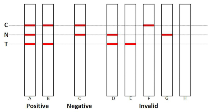
    NOTE: Visual interpretation of the result is shown in Figure 1.

Results

Loading...
$$\rightleftharpoonup{xx}$$ $$\longleftharp{xx}$$, $$\longrightharp{xx}$$,

Consistency of the RGT and PCR fluorescence probing methods
A total of 310 samples were analyzed for NG, consisting of 161 females and 149 males. The positive coincidence rate was 98.78% (95%CI: 93.39%-99.97%), while the negative coincidence rate was 99.12% (95% CI: 96.87%-99.89%). The overall coincidence rate was 99.03% (95% CI: 97.20%-99.80%). The Kappa consistency value was 0.975 (95% CI: 0.947-1.003, Z = 17.171, P < 0.05) (Table 1). There were three samples with inconsistent results: one sample tested positive using the PCR fluorescence probing method but negative with RGT, and the gene sequencing review confirmed a negative result. Among the two samples that tested RGT-positive but PCR-negative, gene sequencing confirmed one true positive (RGT result concordant with sequencing) and one false positive (RGT-positive but sequencing-negative).

For the CT analysis, a total of 160 samples were included, consisting of 80 females and 80 males. The positive coincidence rate was 100.00% (95%CI: 93.02%-100.00%). The negative coincidence rate was 99.08% (95%CI: 94.99%-99.98%). The overall coincidence rate was 99.38% (95% CI: 96.57%-99.98%). The Kappa consistency value was 0.986 (95% CI: 0.958-1.014, Z = 12.469, P < 0.05) (Table 2). There was one sample with inconsistent results; it was negative according to the PCR fluorescence probing method but positive with the RGT. The gene sequencing review confirmed a negative result.

For UU, a total of 306 samples were analyzed, consisting of 154 females and 152 males. The positive coincidence rate was 98.25% (95% CI: 93.81%-99.79%), while the negative coincidence rate was 99.48% (95%CI: 97.13%-99.99%). The overall coincidence rate was 99.02% (95% CI: 97.16%-99.80%). The Kappa consistency value was 0.979 (95% CI: 0.955-1.003, Z = 17.129, P < 0.05) (Table 3). There were three samples with inconsistent results: two samples tested positive using the PCR fluorescence probing method but negative with the rapid diagnostic test (RGT). Gene sequencing revealed one of these samples as positive and the other as negative. Additionally, one sample was negative with the PCR fluorescence probing method but positive with the RGT, and gene sequencing confirmed this sample as positive.

Consistency of RGT and gene sequencing
A total of 240 samples were analyzed for NG, consisting of 121 females and 119 males. The positive coincidence rate was 98.57% (95% CI: 92.30%-99.96%), while the negative coincidence rate was 99.41% (95% CI: 96.77%-99.99%). The overall coincidence rate was 99.17% (95% CI: 97.02%-99.90%). The Kappa consistency value was 0.980 (95% CI: 0.952-1.008, Z = 15.180, P < 0.05) (Table 4). There were two samples with inconsistent results: one sample tested positive by gene sequencing but negative by RGT, and another sample tested negative by gene sequencing but positive by RGT.

For the CT analysis, a total of 190 samples were included, consisting of 95 females and 95 males. The positive coincidence rate was 100.00% (95% CI: 94.31%-99.81%). The negative coincidence rate was 98.43% (95% CI: 94.43%-99.81%). The overall coincidence rate was 98.95% (95% CI: 96.25%-99.87%). The Kappa consistency value was 0.976 (95% CI: 0.944-1.009, Z = 13.463, P < 0.05) (Table 5). There were 2 samples with inconsistent results; these samples were negative by gene sequencing but positive by RGT.

For UU, a total of 176 samples were analyzed, consisting of 90 females and 86 males. The positive coincidence rate was 98.70% (95% CI: 92.98%-99.97%). The negative coincidence rate was 97.98% (95% CI: 92.89%-99.75%). The overall coincidence rate was 98.30% (95% CI: 95.10%-99.65%). The Kappa consistency value was 0.965 (95% CI: 0.927-1.004, Z = 12.809, P < 0.05) (Table 6). There were three samples with inconsistent results: one sample was positive by gene sequencing but negative by RGT, while two samples were negative by gene sequencing but positive by RGT.

Consistency of RGT and pathogen isolating culture method
A total of 34 positive samples were analyzed regarding the consistency between the RGT and the pathogen isolation culture method, consisting of 17 females and 17 males. RGT detected 9 cases of NG positive, 17 cases of CT positive, and 8 cases of UU positive, with all positive rates reaching 100.00%.

Lateral flow assay results diagram, positive, negative, and invalid test lines illustrated.
Figure 1: Interpretation criteria for lateral flow assay results. (A,B) A valid positive result is defined by the presence of bands on both the Control line (C) and Test line (T), irrespective of the visibility of the Internal Control line (N). (B) The absence of an N-line band with a visible T-line may indicate competitive inhibition of 18S rRNA amplification due to high target RNA concentrations. (C) A valid negative result requires concurrent visibility of bands on the C-line and N-line, with no T-line signal. (D-H) Invalid results: (D, E, G, H) Complete absence of the C-line band, regardless of N/T-line visibility. (F) The presence of a C-line band with the simultaneous absence of both N-line and T-line signals suggests specimen contamination with amplification inhibitors or improper collection/storage. Please click here to view a larger version of this figure.

positive coincidence rate98.78% (93.39%–99.97%)
RGT  ResultsPCR Fluorescence Probing ResultsTotalnegative coincidence rate99.12% (96.87%–99.89%)
Positive (+)Negative (-)positive predictive value97.59% (91.57%–99.71%)
Positive (+)81283negative predictive value99.56% (97.57%–99.99%)
Negative (-)1226227total coincidence rate99.03% (97.20%–99.80%)
Total82228310kappa value0.975(0.947–1.003)
Z value17.171

Table 1: Comparative analysis of Neisseria gonorrhoeae (NG) detection between RNA amplification lateral flow assay (RGT) and PCR fluorescence probing (n = 310). Definition of positivity: RGT: Visible band on both Control (C) and Test (T) lines; PCR: Cycle threshold (Ct) <35.0 with S-shaped amplification curve. Statistical analysis: Sensitivity: 98.78% (81/82, 95% CI: 93.39-99.97%); Specificity: 99.12% (226/228, 95% CI: 96.87-99.89%); Cohen's Kappa (κ): 0.975 (95% CI: 0.947-1.003), indicating excellent agreement (Landis & Koch criteria: κ >0.80). Error reporting: Confidence intervals calculated using the Wilson score method.

positive coincidence rate100.00% (93.02%–100.00%)
RGT  ResultsPCR Fluorescence Probing ResultsTotalnegative coincidence rate99.08% (94.99%–99.98%)
Positive (+)Negative (-)positive predictive value98.08% (89.74%–99.95%)
Positive (+)51152negative predictive value100.00% (96.64%–100.00%)
Negative (-)0108108total coincidence rate99.38% (96.57%–99.98%)
Total51109160kappa value0.986(0.958–1.014)
Z value12.469

Table 2: Comparative analysis of Chlamydia trachomatis (CT) detection between RNA amplification lateral flow assay (RGT) and PCR fluorescence probing (n = 160). Definition of positivity: RGT: Visible band on both Control (C) and Test (T) lines; PCR: Cycle threshold (Ct) <35.0 with S-shaped amplification curve. Statistical analysis: Sensitivity: 100.00% (51/51, 95% CI: 93.02-100.00%); Specificity: 99.08% (108/109, 95% CI: 94.99-99.98%); Cohen's Kappa (κ): 0.986 (95% CI: 0.958-1.014), indicating excellent agreement (Landis & Koch criteria: κ >0.80). Error reporting: Confidence intervals calculated using the Wilson score method.

positive coincidence rate98.25% (93.81%–99.79%)
RGT  ResultsPCR Fluorescence Probing ResultsTotalnegative coincidence rate99.48% (97.13%–99.99%)
Positive (+)Negative (-)positive predictive value99.12% (95.17%–99.98%)
Positive (+)1121113negative predictive value98.96% (96.31%–99.87%)
Negative (-)2191193total coincidence rate99.02% (97.16%–99.80%)
Total114192306kappa value0.979(0.955–1.003)
Z value17.129

Table 3: Comparative analysis of Ureaplasma urealyticum (UU) detection between RNA amplification lateral flow assay (RGT) and PCR fluorescence probing (n = 306). Definition of positivity: RGT: Visible band on both Control (C) and Test (T) lines; PCR: Cycle threshold (Ct) <35.0 with S-shaped amplification curve. Statistical analysis: Sensitivity: 98.25% (112/114, 95% CI: 93.81-99.79%); Specificity: 99.48% (191/192, 95% CI: 97.13-99.99%); Cohen's Kappa (κ): 0.979 (95% CI: 0.955-1.003), indicating excellent agreement (Landis & Koch criteria: κ >0.80). Error reporting: Confidence intervals calculated using the Wilson score method.

positive coincidence rate98.57% (92.30%–99.96%)
RGT  Results Gene Sequencing ResultsTotalnegative coincidence rate99.41% (96.77%–99.99%)
Positive (+)Negative (-)positive predictive value98.57% (92.30%–99.96%)
Positive (+)69170negative predictive value99.41% (96.77%–99.99%)
Negative (-)1169170total coincidence rate99.17% (97.02%–99.90%)
Total70170240kappa value0.980(0.952–1.008)
Z value15.180

Table 4: Comparative analysis of Neisseria gonorrhoeae (NG) detection between RNA amplification lateral flow assay (RGT) and gene sequencing (n = 240). Definition of positivity: RGT: Visible band on both Control (C) and Test (T) lines; PCR: Cycle threshold (Ct) <35.0 with S-shaped amplification curve. Statistical analysis: Sensitivity: 98.57% (69/70, 95% CI: 92.30-99.96%); Specificity: 99.41% (169/170, 95% CI: 96.77-99.99%); Cohen's Kappa (κ): 0.980(95% CI: 0.952-1.008), indicating excellent agreement (Landis & Koch criteria: κ >0.80). Error reporting: Confidence intervals calculated using the Wilson score method.

positive coincidence rate100.00% (94.31%–100.00%)
RGT  Results Gene Sequencing ResultsTotalnegative coincidence rate98.43% (94.43%–99.81%)
Positive (+)Negative (-)positive predictive value96.92% (89.32%–99.63%)
Positive (+)63265negative predictive value100.00% (97.09%–100.00%)
Negative (-)0125125total coincidence rate98.95% (96.25%–99.87%)
Total63127190kappa value0.976(0.944–1.009)
Z value13.463

Table 5: Comparative analysis of Chlamydia trachomatis (CT) detection between RNA amplification lateral flow assay (RGT) and gene sequencing (n = 190). Definition of positivity: RGT: Visible band on both Control (C) and Test (T) lines; PCR: Cycle threshold (Ct) <35.0 with S-shaped amplification curve. Statistical analysis: Sensitivity: 100.00% (63/63, 95% CI: 94.31-100.00%); Specificity: 98.43% (125/127, 95% CI: 94.43-99.81%); Cohen's Kappa (κ): 0.976 (95% CI: 0.944-1.009), indicating excellent agreement (Landis & Koch criteria: κ >0.80). Error reporting: Confidence intervals calculated using the Wilson score method.

positive coincidence rate98.70% (92.98%–99.97%)
RGT  Results Gene Sequencing ResultsTotalnegative coincidence rate97.98% (92.89%–99.75%)
Positive (+)Negative (-)positive predictive value97.44% (91.04%–99.69%)
Positive (+)76278negative predictive value98.98% (94.45%–99.97%)
Negative (-)19798total coincidence rate98.30% (95.10%–99.65%)
Total7799176kappa value0.965(0.927–1.004)
Z value12.809

Table 6: Comparative analysis of Ureaplasma urealyticum (UU) detection between RNA amplification lateral flow assay (RGT) and gene sequencing (n = 176). Definition of positivity: RGT: Visible band on both Control (C) and Test (T) lines; PCR: Cycle threshold (Ct) <35.0 with S-shaped amplification curve. Statistical analysis: Sensitivity: 98.70% (76/77, 95% CI: 92.98-99.97%); Specificity: 97.98% (97/99, 95% CI: 92.89-99.75%); Cohen's Kappa (κ): 0.965 (95% CI: 0.927-1.004), indicating excellent agreement (Landis & Koch criteria: κ >0.80). Error reporting: Confidence intervals calculated using the Wilson score method.

Supplementary Table 1: Distribution of clinical samples analyzed by RNA amplification lateral flow assay (RGT) compared to reference methods, stratified by pathogen and sex. Abbreviations: RGT: RNA amplification lateral flow assay; NG: Neisseria gonorrhoeae; CT: Chlamydia trachomatis; UU: Ureaplasma urealyticum. Sample criteria: All specimens were collected from patients clinically suspected of sexually transmitted infections (STIs) who had not received antibiotic therapy within 14 days prior to sampling. Sex distribution: Female: 50.6% (716/1,416); Male: 49.4% (700/1,416). Reference method details: PCR positivity threshold: Cycle threshold (Ct) <35.0. Pathogen isolation culture: Performed adherence to the specified culture protocols or guidelines for distinct pathogens.  Please click here to download this table.

Supplementary Table 2: Reagents of NG nucleic acid assay Please click here to download this table.

Supplementary Table 3: Reagents of CT nucleic acid assay. Please click here to download this table.

Supplementary Table 4: Reagents of UU nucleic acid assay. Please click here to download this table.

Supplementary Table 5: Interpretation matrix for RGT result interpretation. C-line: Nitrocellulose-immobilized IgG control; N-line: 18S rRNA internal control probe; T-line: Pathogen-specific RNA detection probe; +: Visible band; -: No band. Please click here to download this table.

Discussion

Loading...
$$\rightleftharpoonup{xx}$$ $$\longleftharp{xx}$$, $$\longrightharp{xx}$$,

In this study, RGT was employed to detect STI pathogens, including NG, CT, and UU. When performed under stringent operational protocols, the RGT assay demonstrated strong concordance with established diagnostic methods such as PCR fluorescence probing, gene sequencing, and pathogen isolation culture techniques. The sensitivity and specificity of the RGT assay were comparable to or exceeded those of previously reported methodologies, as evidenced by comparative analyses with established benchmarks10,11.

The reliability of the RGT methodology hinges on several critical protocol steps. Sample homogenization must be performed at 2,600 times/min for 10 s to ensure complete cell detachment from swabs. Proper homogenization of the amplification buffer and detection solution through gentle agitation followed by prompt centrifugation is essential to achieve homogeneity and prevent diminished amplification efficiency. The introduction of air bubbles during reagent transfer must be meticulously avoided, as such artifacts may compromise reaction reproducibility. Owing to the minimal volumetric requirement (1 µL), precise pipetting of the amplification enzyme using low-retention tips to the microchip's reaction chamber is imperative to ensure optimal substrate contact. Furthermore, the presence of persistent insoluble particulate matter within the detection solution post-reconstitution necessitates reagent discard, as residual precipitates may interfere with optical readouts or reaction kinetics.

For the RGT assay, pathogens are capable of semi-quantitative detection of pathogens. The detection limitations of NG, CT, and UU for this assay are 1 × 10³ CFU/mL, 1 × 10³ infectious units [IFU]/mL, and 1 × 10⁴ color-changing units [CCU]/mL, respectively. For samples with extremely low concentrations, the detection rate of RGT assay may be lower compared to some highly sensitive molecular technologies12,13.

The RGT method demonstrates distinct advantages relative to established and emerging diagnostic platforms. Compared to PCR and nucleic acid amplification tests (NAATs), which remain benchmark methodologies for sensitivity and quantitative precision, RGT circumvents dependencies on thermocycling equipment, purified nucleic acid templates, and specialized technical expertise. This enables its application in resource-limited settings, as the method operates efficiently under low-temperature isothermal conditions (37-42 °C) and tolerates crude sample inputs, such as direct clinical swab extracts, thereby facilitating rapid diagnostics in decentralized clinical or field environments10,14. Such operational flexibility positions RGT as a pragmatic alternative for regions lacking access to conventional NAAT infrastructure15.

Traditional culture-based approaches for pathogens like NG and CT, while historically foundational, are constrained by prolonged incubation periods (3-7 days) and variable sensitivity. In contrast, RGT delivers results within <2 h while preserving diagnostic specificity comparable to culture methods11. Emerging CRISPR-based diagnostic platforms, despite their high specificity, frequently require labor-intensive pre-amplification steps16,17. RGT addresses this limitation by consolidating nucleic acid amplification and detection within a unified microfluidic system, significantly reducing procedural complexity and manual handling. The results of this study also indicate that RGT was a rapid, sensitive, and specific method for NG, CT, and UU detection. Collectively, these attributes position RGT as a hybrid solution bridging the gap between low-sensitivity rapid assays and high-complexity molecular techniques, effectively addressing unmet needs in scenarios requiring both rapid turnaround and analytical rigor.

NAAT methods targeting microbial DNA or RNA in clinical specimens are currently regarded as the gold standard for diagnosing numerous STIs18. Molecular biology testing primarily encompasses two major categories: DNA and RNA. In this study, the RGT method was employed to detect RNA in reproductive tract samples, which leveraged the inherent advantages of RNA-based detection. Given the presence of multiple RNA copies in non-viral pathogenic microbial cells, this technique demonstrates superior sensitivity and accuracy compared to DNA-targeted PCR and other conventional methods19. The RGT method is applicable to various sample types, including urine specimens, and exhibits excellent consistency across samples obtained from different anatomical sites. In male patients, studies have demonstrated nearly perfect agreement between urine and urethral swab results using this method20. Furthermore, since RNA is exclusively present in viable pathogenic microorganisms21, RNA-based testing provides more timely and clinically relevant data for therapeutic efficacy evaluation compared to DNA testing, thereby aligning with the principles of precision medicine.

Consequently, the RGT methodology demonstrates substantial potential for resolving pivotal challenges in diverse healthcare and research applications. This innovative approach offers significant advantages in clinical diagnostics and therapeutic monitoring, particularly through its capacity to detect viable pathogens with enhanced sensitivity and specificity and its adaptability to various biological specimens, which positions it as a valuable tool for advancing precision medicine initiatives.

Disclosures

Loading...
$$\rightleftharpoonup{xx}$$ $$\longleftharp{xx}$$, $$\longrightharp{xx}$$,

The authors have no conflicts of interest to disclose.

Acknowledgements

Loading...
$$\rightleftharpoonup{xx}$$ $$\longleftharp{xx}$$, $$\longrightharp{xx}$$,

We thank Wuhan Zhongzhi Bio-technologies Inc. for the reagents and technical guidance offered for this study.

Materials

List of materials used in this article
NameCompanyCatalog NumberComments
10% (w/v)  sodium hypochloritePreparation in the laboratory.Preparation in the laboratory.Diluted bleach solution for decontamination and inactivation of pathogens on equipment or surfaces.
75% (v/v) ethanolAnte Food Co., Ltd (Anhui,China)http://www.antegroup.com/product_detail_cn/id/81.htmlAqueous ethanol solution used for surface disinfection
Adjustable-volume Pipette (0.5–10 μL)DLAB Scientific Co., Ltd.7010101004Single-channel micropipette for accurate handling of microliter-scale volumes.
Adjustable-volume Pipette (100–1000 μL)DLAB Scientific Co., Ltd.7010101014Single-channel pipette calibrated for large-volume liquid handling.
Adjustable-volume Pipette (10–100 μL)DLAB Scientific Co., Ltd.7010101008Single-channel pipette optimized for intermediate volumes, commonly used in molecular biology protocols.
Adjustable-volume Pipette (20–200 μL)DLAB Scientific Co., Ltd.7010101009Single-channel pipette for medium-volume transfers, suitable for precise aliquoting of reagents.
Biosafety cabinet (HF Safe-1200)HealForce Bio-Meditech Holdings Limited (Hong Kong, China)HF Safe-1200A Class II biosafety cabinet ensuring personnel, sample, and environmental protection during handling of pathogenic microorganisms or cell cultures.
Centrifuge TubesHaozhi Medical Devices Co., Ltd. (Guangzhou, China)Guangzhou Yuexiu Medical Device Filing No. 20210414For nucleic acid extraction, cell pelleting, and other laboratory procedures.
Chamydia trachomatis (CT) Nucleic Acid Assay (RNA Amplification-Lateral Flow Assay)Wuhan Zhongzhi Biotechnologies INC.(Wuhan, China)A.10.309-STD-RG-002For qualitative detection of Chamydia trachomatis nucleic acid RNA in genital and urinary tract swab samples.
CO2 IncubatorThermo Fisher Scientific(Asheville, USA) LLCSN:300504406Used for cell culture, providing a stable environment with controlled temperature (37°C), humidity, and CO2 concentration (typically 5%) to ensure optimal cell growth.
Culture Plate Corning Incorporated (Corning, USA)50 mm ´ 9 mmFor adherent cell culture and expansion.
Detection Kit for Nucleic Acid to Chamydia trachomatis (CT) (PCR-Fluorescence Probing)Fosun Diagnostics (Shanghai, China) Co., LtdChina Medical Device Registration Certificate No. 20143402234For qualitative detection of nucleic acid of Chamydia trachomatis in genital and urinary tract swab samples.
Detection Kit for Nucleic Acid to Neisseria Gonorrhoeae (NG)(PCR-Fluorescence Probing)Fosun Diagnostics (Shanghai, China) Co., LtdChina Medical Device Registration Certificate No. 20173401080For qualitative detection of nucleic acid of Neisseria gonorrhoeae in genital and urinary tract swab samples.
Detection Kit for Nucleic Acid to Ureaplasma Urealyticum (UU) (PCR-Fluorescence Probing)Fosun Diagnostics (Shanghai, China) Co., LtdChina Medical Device Registration Certificate No. 20173400536For qualitative detection of nucleic acid of Ureaplasma Urealyticum in genital and urinary tract swab samples.
DMEM mediumThermo Fisher Scientific Inc.(Waltham, USA)11965092For Chlamydia trachomatis culture.
Filter Pipette Tips (10 μL)Guangzhou Surbiopure Biotechnology Co., Ltd (Guangzhou, China)TF-10-BXLFor precise pipetting in the experiments.
Filter Pipette Tips (200 μL)Guangzhou Surbiopure Biotechnology Co., Ltd (Guangzhou, China)TF-200-BXLFor precise pipetting in the experiments.
Genital swabHaozhi Medical Devices Co., Ltd. (Guangzhou, China)Hz112Sterile cotton-tipped swabs for collecting clinical specimens from genital mucosal surfaces.
Gibco Fetal Bovine Serum (FBS)Thermo Fisher Scientific Inc. (Waltham, USA)10270106For Chlamydia trachomatis culture.
Glass slidesHaozhi Medical Devices Co., Ltd. (Guangzhou, China)Guangzhou Yuexiu Medical Device Filing No. 20210414Standard glass slides with anti-fade treatment for preparing cell smears or tissue sections, compatible with Gram staining protocols.
Goldenstar RT6 cDNA Synthesis Kit Ver2Beijing Tsingke Biotech Co., Ltd (Beijing, China)TSK302SFor the synthesis of the first strand of cDNA in the first step of the two-step RT-PCR. It contains all the reagents required for the synthesis of cDNA from RNA.
Gram Stain KitBaso Diagnostics,Inc. (Zhuhai, China)BA4012A staining kit containing crystal violet, iodine, decolorizer, and safranin for differentiating Neisseria Gonorrhoeae. 
IBM SPSS Statistics (V25.0)International Business Machines Corporation (New York, USA)Version 25.0For data analysis.
Infrared sterilizerHuaxixinrui (Qingdao) Analytical Instruments Co., Ltd.MJ-200WFor instant decontamination of inoculation loops or forceps, minimizing cross-contamination in biosafety cabinets.
Inoculation loopBKMAM Biotechnology Co., Ltd.(Hunan, China)110306002For microbial sample transfer, streaking, or inoculation.
Inverted Microscope (IX-51)Olympus Corporation (Tokyo, Japan)Asset Code of Labortory:  71807101For observing adherent cells or live-cell dynamics in culture dishes.
MALDI Biotyper automated microbial mass spectrometry systemBruker Daltonics GmbH & Co. KG (Bremen, Germany)National Medical Device Import Registration No. 20182222017A MALDI-TOF mass spectrometry-based system for rapid microbial identification via protein profiling, enabling species identification of bacteria and fungi in clinical microbiology laboratories.
McCoy cell linesPreserved in the laboratory.Preserved in the laboratory.For Chlamydia trachomatis culture.
Microplate Thermostatic Incubator Leopard70-4 (4 Plate Capacity)Leaptec Scientific Instruments (Beijing) Co., Ltd.Leopard70-4A microplate incubator accommodating up to 4 standard microplates, with a temperature range from ambient to 70°C,  used for the incubation steps.
Microscope (OLYMPUS CX43)Olympus Corporation (Tokyo, Japan)https://lifescience.evidentscientific.com.cn/zh/microscopes/upright/cx43-33/For clinical sample observation (e.g., cell morphology, stained specimens).
Mini Metal Bath MiniG-R100Leaptec Scientific Instruments (Beijing) Co., Ltd.MiniG-R100For rapid temperature regulation during the experiment, such as heating, incubating, or terminating reactions in small-volume samples.
Neisseria gonorrhoeae (NG) Nucleic Acid Assay (RNA Amplification-Lateral Flow Assay)Wuhan Zhongzhi Biotechnologies INC.(Wuhan, China)A.10.309-STD-RG-001For qualitative detection of Neisseria gonorrhoeae nucleic acid RNA in genital and urinary tract swab samples.
Nucleic Acid Extraction ReagentWuhan Zhongzhi Biotechnologies INC.(Wuhan, China)A.10.105-SC-001For the extraction, enrichment and purification of nucleic acids.
Specimen Transport MediumQIAGEN N.V. (Venlo, Netherlands)5128-1220This medium is specifically formulated for the collection, preservation, and transport of clinical specimens, ensuring the viability of Chlamydia trachomatis and Neisseria gonorrhoeae for subsequent diagnostic testing.
Thermostatic CentrifugeBeckman Coulter, Inc. (Brea, USA)SN: MRZ21L037For cell, microbial, or nucleic acid pelleting.
TM mediumShanghai Yihua Medical Science & Technology Co., Ltd (Shanghai, China)Shanghai Pu Medical Device Filing No. 20140081TM medium can be used for the isolation, cultivation and identification of Neisseria gonorrhoeae from clinical samples to assist in the diagnosis of gonorrhea.
Trypsin-EDTA (0.25%)Thermo Fisher Scientific Inc.(Waltham, USA)25200072For gentle dissociation of adherent cells during subculturing or experimental procedures.
Ureaplasma urealyticum (UU) Nucleic Acid Assay (RNA Amplification-Lateral Flow Assay)Wuhan Zhongzhi Biotechnologies INC.(Wuhan, China)A.10.309-STD-RG-003For qualitative detection of Ureaplasma Urealyticum nucleic acid RNA in genital and urinary tract swab samples.
Ureaplasma urealyticum and Mycoplasma hominis Selective Isolation Culture, Identification and Drug Sensitivity KitHebei Zhongaisheng Biotechnology Co., Ltd.(Hebei, China)Hebei Medical Device Registration No. 20152400168For the selective isolation, culture, identification, and drug sensitivity determination of Ureaplasma urealyticum and Mycoplasma hominis.
Vortex mixerXinkang Medical Instrument Co. Ltd (Jiangsu, China)XK80-ALaboratory device for homogenizing liquid samples by rapid agitation.

References

Loading...
$$\rightleftharpoonup{xx}$$ $$\longleftharp{xx}$$, $$\longrightharp{xx}$$,
  1. Clinical updates in sexually transmitted infections, 2024. J Womens Health (Larchmt). 33 (6), 827-837 (2024).">Hufstetler, K., Llata, E., Miele, K., Quilter, L. A. S. Clinical updates in sexually transmitted infections, 2024. J Womens Health (Larchmt). 33 (6), 827-837 (2024).
  2. Molecular regulatory mechanisms drive emergent pathogenetic properties of Neisseria gonorrhoeae. Microorganisms. 10 (5), 922(2022).">Sunkavalli, A., McClure, R., Genco, C. Molecular regulatory mechanisms drive emergent pathogenetic properties of Neisseria gonorrhoeae. Microorganisms. 10 (5), 922(2022).
  3. Neisseria gonorrhoeae host adaptation and pathogenesis. Nat Rev Microbiol. 16 (4), 226-240 (2018).">Quillin, S. J., Seifert, H. S. Neisseria gonorrhoeae host adaptation and pathogenesis. Nat Rev Microbiol. 16 (4), 226-240 (2018).
  4. Chlamydia, gonorrhoea, trichomoniasis, and syphilis: global prevalence and incidence estimates, 2016. Bull World Health Organ. 97 (8), 548-562 (2016).">Rowley, J., et al. Chlamydia, gonorrhoea, trichomoniasis, and syphilis: global prevalence and incidence estimates, 2016. Bull World Health Organ. 97 (8), 548-562 (2016).
  5. Intrauterine detection of Ureaplasma species after vaginal colonization in pregnancy and neonatal outcome. Neonatology. 121 (2), 187-194 (2024).">Rittenschober-Boehm, J., et al. Intrauterine detection of Ureaplasma species after vaginal colonization in pregnancy and neonatal outcome. Neonatology. 121 (2), 187-194 (2024).
  6. Analysis of Ureaplasma urealyticum, Chlamydia trachomatis, Mycoplasma genitalium and Neisseria gonorrhoeae infections among obstetrics and gynecological outpatients in southwest China: a retrospective study. BMC Infect Dis. 22 (1), 283(2022).">Liu, T., et al. Analysis of Ureaplasma urealyticum, Chlamydia trachomatis, Mycoplasma genitalium and Neisseria gonorrhoeae infections among obstetrics and gynecological outpatients in southwest China: a retrospective study. BMC Infect Dis. 22 (1), 283(2022).
  7. Visualized lateral flow assay for dual viral RNA fragment detection. Anal Chem. 95 (30), 11187-11192 (2023).">Song, J., Zhang, L., Zeng, L., Xu, X. Visualized lateral flow assay for dual viral RNA fragment detection. Anal Chem. 95 (30), 11187-11192 (2023).
  8. A rapid RNA extraction-free lateral flow assay for molecular point-of-care detection of SARS-CoV-2 augmented by chemical probes. Biosens Bioelectron. 200, 113900(2002).">Dighe, K., Moitra, P., Alafeef, M., Gunaseelan, N., Pan, D. A rapid RNA extraction-free lateral flow assay for molecular point-of-care detection of SARS-CoV-2 augmented by chemical probes. Biosens Bioelectron. 200, 113900(2002).
  9. Salmonella detection using 16S ribosomal DNA/RNA probe-gold nanoparticles and lateral flow immunoassay. Food Chem. 141 (3), 2526-2532 (2013).">Liu, C. C., Yeung, C. Y., Chen, P. H., Yeh, M. K., Hou, S. Y. Salmonella detection using 16S ribosomal DNA/RNA probe-gold nanoparticles and lateral flow immunoassay. Food Chem. 141 (3), 2526-2532 (2013).
  10. Clinical performance of four multiplex real-time PCR kits detecting urogenital and sexually transmitted pathogens. Clin Microbiol Infect. 28 (5), 733.e7-733.e13 (2022).">Pereyre, S., Caméléna, F., Hénin, N., Berçot, B., Bébéar, C. Clinical performance of four multiplex real-time PCR kits detecting urogenital and sexually transmitted pathogens. Clin Microbiol Infect. 28 (5), 733.e7-733.e13 (2022).
  11. Performance of a single-use, rapid, point-of-care PCR device for the detection of Neisseria gonorrhoeae, Chlamydia trachomatis, and Trichomonas vaginalis: a cross-sectional study. Lancet Infect Dis. 21 (5), 668-676 (2021).">Morris, S. R., Bristow, C. C., Wierzbicki, M. R., et al. Performance of a single-use, rapid, point-of-care PCR device for the detection of Neisseria gonorrhoeae, Chlamydia trachomatis, and Trichomonas vaginalis: a cross-sectional study. Lancet Infect Dis. 21 (5), 668-676 (2021).
  12. Analytical and clinical validation of a multiplex PCR assay for detection of Neisseria gonorrhoeae and Chlamydia trachomatis including simultaneous LGV serotyping on an automated high-throughput PCR system. Microbiol Spectr. 12 (3), e0275623(2024).">Pflüger, L. S., Nörz, D., Grunwald, M., et al. Analytical and clinical validation of a multiplex PCR assay for detection of Neisseria gonorrhoeae and Chlamydia trachomatis including simultaneous LGV serotyping on an automated high-throughput PCR system. Microbiol Spectr. 12 (3), e0275623(2024).
  13. Droplet digital PCR (ddPCR) for the detection and quantification of Ureaplasma spp. BMC Infect Dis. 21 (1), 804(2021).">Huang, Y., et al. Droplet digital PCR (ddPCR) for the detection and quantification of Ureaplasma spp. BMC Infect Dis. 21 (1), 804(2021).
  14. Comparison of analytical sensitivity of DNA-based and RNA-based nucleic acid amplification tests for reproductive tract infection pathogens: implications for clinical applications. Microbiol Spectr. 11 (5), e0149723(2023).">Ma, Y., et al. Comparison of analytical sensitivity of DNA-based and RNA-based nucleic acid amplification tests for reproductive tract infection pathogens: implications for clinical applications. Microbiol Spectr. 11 (5), e0149723(2023).
  15. Multicenter comparison of nucleic acid amplification tests for the diagnosis of rectal and oropharyngeal Chlamydia trachomatis and Neisseria gonorrhoeae infections. J Clin Microbiol. 60 (1), e0136321(2022).">Van Der Pol, B., et al. Multicenter comparison of nucleic acid amplification tests for the diagnosis of rectal and oropharyngeal Chlamydia trachomatis and Neisseria gonorrhoeae infections. J Clin Microbiol. 60 (1), e0136321(2022).
  16. An isothermal CRISPR-based diagnostic assay for Neisseria gonorrhoeae and Chlamydia trachomatis detection. Microbiol Spectr. 11 (6), e0046423(2023).">Luo, H., et al. An isothermal CRISPR-based diagnostic assay for Neisseria gonorrhoeae and Chlamydia trachomatis detection. Microbiol Spectr. 11 (6), e0046423(2023).
  17. Point-of-care detection of Neisseria gonorrhoeae based on RPA-CRISPR/Cas12a. AMB Express. 13 (1), 50(2023).">Tu, Q., et al. Point-of-care detection of Neisseria gonorrhoeae based on RPA-CRISPR/Cas12a. AMB Express. 13 (1), 50(2023).
  18. Current and future trends in the laboratory diagnosis of sexually transmitted infections. Int J Environ Res Public Health. 18 (3), 1038(2021).">Caruso, G., Giammanco, A., Virruso, R., Fasciana, T. Current and future trends in the laboratory diagnosis of sexually transmitted infections. Int J Environ Res Public Health. 18 (3), 1038(2021).
  19. Comparison of an rRNA-based and DNA-based nucleic acid amplification test for the detection of Chlamydia trachomatis in trachoma. Br J Ophthalmol. 91 (3), 293-295 (2007).">Yang, J. L., et al. Comparison of an rRNA-based and DNA-based nucleic acid amplification test for the detection of Chlamydia trachomatis in trachoma. Br J Ophthalmol. 91 (3), 293-295 (2007).
  20. Real-time fluorescence constant-temperature simultaneous amplification and testing of nucleic acid for detection of Mycoplasma genitalium infection in the genitourinary tract. Zhonghua Nan Ke Xue. 25 (6), Chinese 535-538 (2019).">Luo, Z. G., Li, R. P., Wang, Y. B., Shang, X. J., Zhu, J. Y. Real-time fluorescence constant-temperature simultaneous amplification and testing of nucleic acid for detection of Mycoplasma genitalium infection in the genitourinary tract. Zhonghua Nan Ke Xue. 25 (6), Chinese 535-538 (2019).
  21. Light-up RNA aptamer signaling-CRISPR-Cas13a-based mix-and-read assays for profiling viable pathogenic bacteria. Biosens Bioelectron. 176, 112906(2021).">Zhang, T., et al. Light-up RNA aptamer signaling-CRISPR-Cas13a-based mix-and-read assays for profiling viable pathogenic bacteria. Biosens Bioelectron. 176, 112906(2021).

Reprints and Permissions

Request permission to reuse the text or figures of this JoVE article

Request Permission

Tags

Rapid Molecular TestingSexually Transmitted DiseasesPathogen DetectionReverse TranscriptionGene Sequencing
Video Coming Soon

Related Articles