This protocol describes a low-cost production and efficient validation method for glue-based TMA construction, providing a convenient pathological diagnosis platform for tumor and disease research.
Method Article
Glue-Based Tissue Microarray Construction for Tumor and Disease Research
Junwen Cai1 , Ying Zheng2 , Wenguang He3 , Yuanyuan Zhang1 , Xiang Wang4 , Wubi Zhou3 , Jingjing Dai1
1Department of Medical Laboratory, The Affiliated Huai'an No.1 People's Hospital of Nanjing Medical University, 2Department of Anesthesiology, The Affiliated Huai'an No.1 People's Hospital of Nanjing Medical University, 3Department of Pathology, The Affiliated Huai'an No.1 People's Hospital of Nanjing Medical University, 4Department of Pediatrics, The Affiliated Huai'an No.1 People's Hospital of Nanjing Medical University
Opens in a new tab
In This Article
Summary
Loading...
$$\rightleftharpoonup{xx}$$
$$\longleftharp{xx}$$,
$$\longrightharp{xx}$$,
Abstract
Loading...
$$\rightleftharpoonup{xx}$$
$$\longleftharp{xx}$$,
$$\longrightharp{xx}$$,
Tissue microarray technology (TMA) is a high-throughput platform for the simultaneous detection and analysis of multiple tissue samples, facilitating efficient tumor and disease biomarker research. However, conventional TMA construction methods often face limitations such as operational complexity, time-consuming procedures, and variable accuracy. A glue-based TMA construction method was developed to overcome these challenges, offering improved tissue core fixation and enhanced structural stability. Systematic validation included histological evaluation (HE staining), immunohistochemical profiling of target proteins, and fluorescence in situ hybridization (FISH) analysis. Results demonstrated that the glue-based method maintained excellent slice integrity, improved signal-to-noise ratio, and ensured consistent batch-to-batch reproducibility. Although the approach is limited to manual operation, it presents a reliable and cost-effective option for moderate-throughput TMA production. This method is particularly suited to research environments that value flexibility and sample diversity over large-scale automation, expanding the utility of TMA in academic and diagnostic settings.
Introduction
Loading...
$$\rightleftharpoonup{xx}$$
$$\longleftharp{xx}$$,
$$\longrightharp{xx}$$,
Tissue Microarray (TMA) is a high-throughput technique for analyzing tissue samples1,2,3. Multiple donor tissue cores are obtained, and donor paraffin blocks are transferred into recipient tissue blocks for simultaneous differential and comparative molecular analysis under theoretically the same performance conditions2,4. The classic way to construct a tissue microarray (TMA) is to use a hole punch to extract tissue cores from a donor tissue sample and arrange them sequentially into a recipient paraffin block. This method is suitable for donor paraffin blocks of similar depth and allows for the efficient analysis of multiple samples on a single slice, greatly improving the efficiency of the study and the consistency of data5,6,7. Nevertheless, this method still possesses certain limitations, including inadequate handling of the tissue core during the embedding process, which may result in inconsistent staining of samples in subsequent analyses8.
The second method of TMA construction is the tape method9,10. This method inverts the construction process by casting the block around inverted upright cores that, upon completion, are flush with the top of the TMA, irrespective of core length11,12. However, this method requires ensuring the proper placement of the tissue core and the effectiveness of the tape, and also limits the number of samples that can be processed compared to traditional techniques.
This study proposes an innovative glue method for constructing TMA, aiming to solve problems such as the insufficient stability of tissue cores and complex operation that exist in traditional techniques13. This method uses glue to precisely fix multiple tissue cores together and has the advantages of simple operation and strong sample firmness. Compared with the traditional sectioning or tape methods, the glue method can increase the retention rate of tissue cores and reduce costs. This method is applicable to clinical studies with a medium sample size and is particularly suitable for research designs that require flexible adjustment of the experimental plan. However, it should be noted that the processing capacity of this method does not meet the demands of ultra-high throughput9. Meanwhile, in the actual application process, a complete set of standardized operation norms and quality control systems must be established. Operators need to receive systematic training and pass professional assessments to ensure the standardization of technical operations and the repeatability of results. Comprehensive analysis indicates that this technology achieves a good balance among operational flexibility, cost-effectiveness, and technical reliability, and is particularly suitable for research scenarios where resources are limited but quality control still needs to be guaranteed.
Protocol
Loading...
$$\rightleftharpoonup{xx}$$
$$\longleftharp{xx}$$,
$$\longrightharp{xx}$$,
All donor blocks were obtained from archival pathological specimens collected between 2016 and 2018 at the Affiliated Huai'an No.1 People's Hospital of Nanjing Medical University. The samples were deidentified prior to use and processed in compliance with approved protocols (the ethics committee of the Affiliated Huai'an No.1 People's Hospital of Nanjing Medical University, KY-2024-250-01).
1. Assessment and tagging of donor tissue
- Select tissue blocks with a thickness of ≥5 mm and an area of ≥15 mm × 15 mm. Ensure a 2 mm safety margin at the edges, and exclude regions exhibiting necrosis, hemorrhage, or calcification to maintain tissue integrity.
- Target area identification and marking
- Evaluate the H&E-stained sections using an optical microscope. Locate the target area through low-power scanning, then switch to high-power to confirm the cellular morphological characteristics. Mark the target area boundaries on the slides.
- Map the coordinates of the selected area to the surface of the paraffin block to complete the marking conversion.
2. Tissue core extraction
- Puncture preparation and sample pretreatment
- Select a 2 mm diameter hand-held hole puncher and confirm the smoothness of its cutting edge and ease of operation.
- Place the donor paraffin block on a cold platform at 4 °C for 15 min.
- Align the needle vertically with the marked point, and apply steady rotational pressure to reach the limiting depth. Avoid tilting or vibrating during the process7 (Figure 1A).
- Gently rotate the needle counterclockwise (≤2 rotations/s) to withdraw it. Carefully eject the tissue core from the needle and transfer it into an individual well of a pre-chilled 96-well plate. Record the core location in each well (Figure 1B).
NOTE: If the bottom of the tissue core is uneven, level it with a blade. After trimming, recheck the integrity. - Pre-record all donor numbers and the corresponding ELISA plate well positions (A1-H12) in the registration form. During the operation, recheck to ensure strict correspondence between donor numbers and well positions.
- Immediately inspect the cores for integrity. Discard cores that are fractured, bent, or exhibit diameter deviations >0.1 mm.
NOTE: Ensure core length consistency (coefficient of variation, CV ≤5%) and avoid surface scratches or compression artifacts.
3. Tissue core fixation
- Use a waterproof marker to draw a grid directly on the mold surface, ensuring clear and evenly spaced lines.
- Use a standard mold with an effective bottom area of 3.0 cm × 2.5 cm, and reserve a blank boundary area of 1.5 mm on each side.
- Use a 0.5 mm fine-point waterproof marker to draw nine equally spaced parallel lines horizontally and vertically, forming a 9 × 9 positioning grid (81 intersection points in total).
NOTE: Before starting, clean the mold interior with a xylene-soaked cotton swab to remove residual paraffin, which may cause delamination. During the drawing process, use a steel ruler to ensure uniform line thickness and clearly distinguishable intersection points.
- Use a pipette to draw 5 µL of glue and gently place it at the center of the pre-labeled slide to form a single droplet. Immediately use fine tweezers to vertically pick up the tissue core at a 90° angle and slowly press it vertically into the glue drop (Figure 1C).
- Insert the cores into the corresponding grid intersections on the mold using tweezers. Apply gentle pressure for 5 s to ensure secure adhesion (Figure 1D).
- If the tissue core is displaced, fine-tune and reset it immediately using fine tweezers before the glue solidifies (within 30 s). Discard solidified, misaligned cores to maintain preparation quality while preserving precious samples to the greatest extent.
4. Cassette installation and paraffin embedding
- Vertically place the tissue cassette over the steel mold, ensuring contact with the core tips without applying compression. Secure the cassette using magnetic clamps at all four corners, and seal the seams with molten paraffin.
- Infuse molten paraffin at a 45° angle in a zigzag pattern, maintaining a flow rate of 1 mL/s. Ensure the paraffin level exceeds the core tips by 2 mm (Figure 1E).
- Rapidly cool the block on a 4 °C cold plate for 5 min to form a rigid support layer. Transfer the block to a 22 °C environment for 20 min to reduce internal stress. Finally, use a 40 °C heat gun to gently smooth the surface and eliminate shrinkage marks.
- Slightly heat the paraffin to soften it, then carefully cut along the cassette edges to loosen the block. Gently lift the cassette and remove any residual paraffin to ensure the integrity of the tissue cores (Figure 1F).
Results
Loading...
$$\rightleftharpoonup{xx}$$
$$\longleftharp{xx}$$,
$$\longrightharp{xx}$$,
In the present study, high-quality tissue microarrays were constructed using the glue method. To verify the effectiveness of the method, a series of experiments were performed, including H&E staining, immunohistochemical detection of specific proteins, and fluorescence in situ hybridization (FISH) analysis. A critical component of the construction process is the presence of tissue core dots at the expected positions and distances apart from one another, which is assessed by visual inspection. Visual inspection of the TMA blocks shows that the cores are present and regularly spaced in each TMA (Figure 2A). H&E staining showed that all tissue cores were neatly arranged, with uniform spacing, consistent size, and abundant target tissues (Figure 2B). The immunohistochemical staining results demonstrated robust expression of 10 prostate cancer-related proteins (ZCCHC24, SMAD9, TARBP1, CDHR4, CRTAC1, DNASE1L3, GPR146, IGSF10, ITIH1, and CDK1) in the TMAs, showing consistent staining intensity and minimal background interference (Figure 3). In addition, the FISH analysis showed a clear probe signal against a clean background. In clear tumor areas, identify at least two infiltrating cancer regions and randomly count GSP HER2 (red) and CSP 17 (green) signals in at least 20 infiltrating cancer cells. Record the total number of GSP HER2 and CSP 17 signals as 90 and 40, respectively. When the HER2/CSP 17 ratio is ≥2.0, it is HER2 gene amplification (Figure 4).
The TMA constructed using the glue method was validated by H&E staining, immunohistochemical detection of specific proteins, and FISH analysis. These results demonstrate that the glue method provides a reliable means for systematic analysis of tumor tissue samples, demonstrating its potential for tumor feature recognition and research.
To verify the consistency of immunohistochemical scoring between tissue microarrays (TMA) and conventional large wax block sections, two pathologists conducted independent double-blind reading of 80 samples and scored TMA and conventional large wax block sections, respectively. All samples were evaluated, and the tissue core loss rate was 10%. Statistical analysis showed that there was no significant difference in the scores between the two, indicating that the TMA constructed by the glue method could completely retain the antigenic characteristics of the original tissue and had good technical stability (Table 1).

Figure 1: Construction of tissue microarrays using the glue method. (A) Donor blocks are punched to extract cores from tumor-rich areas identified through H&E staining and pathologist review. (B) Tissue cores are pre-loaded into the collection plate. (C) Each tissue core is vertically dipped into an appropriate amount of glue. (D) Cores are inserted into the acceptor wax block in numerical order at the corresponding grid intersections. (E) Molten paraffin is gently poured into a steel mold to surround and submerge the upright cores positioned beneath the cassette. (F) The mold is gently removed after paraffin solidification. Please click here to view a larger version of this figure.
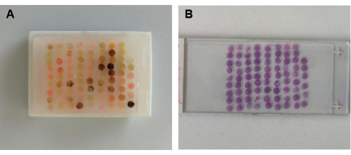
Figure 2: H&E staining of donor blocks. (A) TMA was successfully constructed using the glue method. (B) Corresponding H&E-stained section from the constructed TMA. Please click here to view a larger version of this figure.

Figure 3: Immunohistochemical (IHC) analysis of target protein expression in TMA sections. Full-face TMA sections were evaluated by IHC for expression of 10 prostate cancer-associated proteins: ZCCHC24 (A), SMAD9 (B), TARBP1 (C), CDHR4 (D), CRTAC1 (E), DNASE1L3 (F), GPR146 (G), IGSF10 (H), ITIH1 (I), and CDK1 (J) in formalin-fixed, paraffin-embedded (FFPE) TMA sections. Please click here to view a larger version of this figure.

Figure 4: Fluorescence in situ hybridization (FISH) analysis of target gene expression in TMA sections. (A) Negative FISH result. (B) Positive FISH result showing HER2 (red), CSP17 (green), and nuclei stained with DAPI (blue), imaged at 1000× magnification. Please click here to view a larger version of this figure.
Table 1: Double-blind scoring of tissue sections by pathologists. Two pathologists independently performed double-blind evaluations of 80 samples, scoring both the TMAs and corresponding conventional large wax block sections. Please click here to download this Table.
Table 2: Cost comparison of TMA construction methods. Comparison of the cost of TMA chips generated using the glue method in this study versus chips produced using conventional methods. Please click here to download this Table.
Discussion
Loading...
$$\rightleftharpoonup{xx}$$
$$\longleftharp{xx}$$,
$$\longrightharp{xx}$$,
As an innovative method for constructing tissue microarrays (TMA), the glue method has shown significant advantages due to its ease of construction procedures and cost-effectiveness. Compared to the traditional needle array method, which relies on sophisticated instruments14,15, the glue method enables sample fixation by glueing, which can be done with only basic tools, greatly reducing the technical threshold. In contrast to the traditional embedding method, the glueing method is fixed by local dispensing, which significantly shortens the localization time of the tissue core and avoids the destruction of sensitive antigens by high temperatures16,17(Table 2).
However, in actual construction procedures, there are many complex and delicate steps. These steps not only consume a lot of time, but also put forward extremely high requirements for the technical level and experience of the operators. From the selection and marking of donor tissues to the extraction, fixation, and paraffin embedding of tissue cores, each step requires precise construction procedures and strict quality control18,19. Common technical problems during histological sectioning and paraffin embedding include core displacement of tissue blocks, glue overflow, and malfunctions in sectioning operations. Core displacement of tissue blocks is often caused by inaccurate positioning during embedding or the mold not being placed horizontally. This can be prevented by promptly adjusting the tissue position with preheated tweezers. If displacement has already occurred, secondary correction is required. The problem of glue overflow is usually caused by improper control of glue volume. It is recommended to use a micropipette to precisely control the glue volume of 5 µL. The phenomenon of sticking to the blade during the slicing process requires regular cleaning of the blade and maintaining the environmental humidity. To ensure the quality of the experiment, preventive measures should be emphasized, such as ensuring that the tissue is fully dehydrated and maintaining a stable wax temperature. At the same time, an operation log should be established to record problems and solutions. When necessary, equipment calibration or consumables should be checked or replaced. These targeted measures can effectively enhance the quality of slices and the efficiency of experiments.
The potential of the glue method is not limited to traditional pathology but can also be extended to the emerging field of multi-omics research. When combined with spatial transcriptomics, TMA can accurately locate specific cell regions in the tumor microenvironment, reveal tumor heterogeneity and immune cell infiltration properties20, and provide new ideas for the screening of immunotherapy targets20,21,22.
Disclosures
Loading...
$$\rightleftharpoonup{xx}$$
$$\longleftharp{xx}$$,
$$\longrightharp{xx}$$,
The authors have nothing to disclose.
Acknowledgements
Loading...
$$\rightleftharpoonup{xx}$$
$$\longleftharp{xx}$$,
$$\longrightharp{xx}$$,
Thank you to the team members for their support and contribution to this experiment.
Materials
| Name | Company | Catalog Number | Comments |
|---|---|---|---|
| Breast cancer HER2 Detection kit | Anbiping | 2502001 | Breast cancer HER2 Detection kit |
| CDHR4 antibody | Abcam | ab166914 | CDHR4 antibody |
| CDK1 antibody | Abcam | ab265590 | CDK1 antibody |
| CRTAC1 antibody | Abcam | ab254691 | CRTAC1 antibody |
| DNASE1L3 antibody | Abcam | ab203669 | DNASE1L3 antibody |
| Embedding machine | P.S.J MEDICAL | BM450A | Embedding machine |
| Fully automatic tissue dehydrator | Leica Biosystems | ASP3005 | Fully automatic tissue dehydrator |
| Glass microscope slides | Citotest | 250124A1 | Glass microscope slides |
| Glue | TIZO | 200 | Glue |
| GPR146 antibody | Abcam | ab117104 | GPR146 antibody |
| IGSF10 antibody | Abcam | ab197671 | IGSF10 antibody |
| ITIH1 antibody | Abcam | ab233032 | ITIH1 antibody |
| Low Profile Microtome Blades | Thermo Fisher | 3052835 | Low Profile Microtome Blades |
| Marker pen | Deli | SK109 | Marker pen |
| Microtome | Leica Biosystems | HistoCore BIOCUT | Microtome |
| Paraffin wax | Solarbio | YA0012 | Paraffin wax |
| SMAD9 antibody | Abcam | ab262940 | SMAD9 antibody |
| TARBP1 antibody | Abcam | ab115896 | TARBP1 antibody |
| ZCCHC24 antibody | Abcam | ab88756 | ZCCHC24 antibody |
References
Loading...
$$\rightleftharpoonup{xx}$$
$$\longleftharp{xx}$$,
$$\longrightharp{xx}$$,
- Oral Biol Mol Tech Appl. Seymour, G. J., Cullinan, M. P., Heng, N. C. K. , 429-438 (2017).">Bingle, L., Fonseca, F. P., Farthing, P. M. Constructing tissue microarrays: Protocols and methods considering potential advantages and disadvantages for downstream use. Oral Biol Mol Tech Appl. Seymour, G. J., Cullinan, M. P., Heng, N. C. K. , 429-438 (2017).
- Tissue microarray analysis for epithelial membrane protein-2 as a novel biomarker for gliomas. Brain Tumor Pathol. 35 (1), 1-9 (2018).">Chung, L. K., et al. Tissue microarray analysis for epithelial membrane protein-2 as a novel biomarker for gliomas. Brain Tumor Pathol. 35 (1), 1-9 (2018).
- Tissue tablet method: An efficient tissue banking procedure applicable to both molecular analysis and frozen tissue microarray. Hum Pathol. 45 (1), 143-152 (2014).">Torata, N., et al. Tissue tablet method: An efficient tissue banking procedure applicable to both molecular analysis and frozen tissue microarray. Hum Pathol. 45 (1), 143-152 (2014).
- Biobank Methods Protocols. Yong, W. H. , 313-323 (2019).">Koo, M., Squires, J. M., Ying, D., Huang, J. Making a tissue microarray. Biobank Methods Protocols. Yong, W. H. , 313-323 (2019).
- Amplification of tissue by construction of tissue microarrays. Exp Mol Pathol. 70 (3), 255-264 (2001).">Rimm, D. L., Camp, R. L., Charette, L. A., Olsen, D. A., Provost, E. Amplification of tissue by construction of tissue microarrays. Exp Mol Pathol. 70 (3), 255-264 (2001).
- Simple inexpensive, and precise paraffin tissue microarrays constructed with a conventional microcompound table and a drill grinder. Am J Clin Pathol. 126 (3), 342-348 (2006).">Vogel, U. F., Bueltmann, B. D. Simple inexpensive, and precise paraffin tissue microarrays constructed with a conventional microcompound table and a drill grinder. Am J Clin Pathol. 126 (3), 342-348 (2006).
- TMA for all: A new method for the construction of tissue microarrays without recipient paraffin block using custom-built needles. Diagn Pathol. 1 (1), 14(2006).">Pires, A. R. C., Andreiuolo, F. M., de Souza, S. R. TMA for all: A new method for the construction of tissue microarrays without recipient paraffin block using custom-built needles. Diagn Pathol. 1 (1), 14(2006).
- Next-generation tissue microarray (ngTMA) increases the quality of biomarker studies: An example using CD3, CD8, and CD45RO in the tumor microenvironment of six different solid tumor types. J Transl Med. 11 (1), 104(2013).">Zlobec, I., Koelzer, V. H., Dawson, H., Perren, A., Lugli, A. Next-generation tissue microarray (ngTMA) increases the quality of biomarker studies: An example using CD3, CD8, and CD45RO in the tumor microenvironment of six different solid tumor types. J Transl Med. 11 (1), 104(2013).
- Recommendations for tissue microarray construction and quality assurance. Appl Immunohistochem Mol Morphol. 28 (4), 325-330 (2020).">Glinsmann-Gibson, B., et al. Recommendations for tissue microarray construction and quality assurance. Appl Immunohistochem Mol Morphol. 28 (4), 325-330 (2020).
- Constructing tissue microarrays without prefabricating recipient blocks: A novel approach. Am J Clin Pathol. 124 (1), 103-107 (2005).">Chen, N., Zhou, Q. Constructing tissue microarrays without prefabricating recipient blocks: A novel approach. Am J Clin Pathol. 124 (1), 103-107 (2005).
- Hypodermic needle without recipient paraffin block technique. Methods Mol Biol. 664, 53-61 (2010).">Pires, A. R. C., de Souza, S. R. Hypodermic needle without recipient paraffin block technique. Methods Mol Biol. 664, 53-61 (2010).
- Application of a novel and low-cost technique to construct paraffin tissue microarrays out of paraffinised needle biopsy specimens from patients with breast cancer. J Clin Pathol. 63 (7), 640-643 (2010).">Vogel, U. F., Bültmann, B. Application of a novel and low-cost technique to construct paraffin tissue microarrays out of paraffinised needle biopsy specimens from patients with breast cancer. J Clin Pathol. 63 (7), 640-643 (2010).
- Constructing high-density tissue microarrays with a novel method and a self-made tissue-arraying instrument. Pathol Res Pract. 245, 154430(2023).">Qin, P., Li, L., Zhao, L., Bian, P., Xiong, Z. Constructing high-density tissue microarrays with a novel method and a self-made tissue-arraying instrument. Pathol Res Pract. 245, 154430(2023).
- A next-generation tissue microarray (ngTMA) protocol for biomarker studies. J Vis Exp. (91), e51893(2014).">Zlobec, I., Suter, G., Perren, A., Lugli, A. A next-generation tissue microarray (ngTMA) protocol for biomarker studies. J Vis Exp. (91), e51893(2014).
- Tissue microarray. J Invest Dermatol. 134 (9), 1-4 (2014).">Barrette, K., van den Oord, J. J., Garmyn, M. Tissue microarray. J Invest Dermatol. 134 (9), 1-4 (2014).
- Optimization of tissue microarrays from banked human formalin-fixed paraffin embedded tissues in the cancer research setting. Biopreserv Biobank. 17 (5), 452-457 (2019).">Sexton, T., Kucera, G. L., Levine, E. A., Watabe, K., O'Neill, S. S. Optimization of tissue microarrays from banked human formalin-fixed paraffin embedded tissues in the cancer research setting. Biopreserv Biobank. 17 (5), 452-457 (2019).
- Optimization of tissue microarray technique for breast cancer patients: A short communication. Ann Med Surg. 85 (10), 5299-5303 (2023).">Harfouch, R. M., et al. Optimization of tissue microarray technique for breast cancer patients: A short communication. Ann Med Surg. 85 (10), 5299-5303 (2023).
- Construction of high-density tissue microarrays at low cost by using self-made manual microarray kits and recipient paraffin blocks. Korean J Pathol. 46 (6), 562-568 (2012).">Choi, C. H., et al. Construction of high-density tissue microarrays at low cost by using self-made manual microarray kits and recipient paraffin blocks. Korean J Pathol. 46 (6), 562-568 (2012).
- In-house manual construction of high-density and high-quality tissue microarrays by using homemade recipient agarose-paraffin blocks. Korean J Pathol. 47 (3), 238-244 (2013).">Kim, K. H., et al. In-house manual construction of high-density and high-quality tissue microarrays by using homemade recipient agarose-paraffin blocks. Korean J Pathol. 47 (3), 238-244 (2013).
- Integrating microarray-based spatial transcriptomics and single-cell RNA-seq reveals tissue architecture in pancreatic ductal adenocarcinomas. Nat Biotechnol. 38 (3), 333-342 (2020).">Moncada, R., et al. Integrating microarray-based spatial transcriptomics and single-cell RNA-seq reveals tissue architecture in pancreatic ductal adenocarcinomas. Nat Biotechnol. 38 (3), 333-342 (2020).
- Topographic analysis of pancreatic cancer by TMA and digital spatial profiling reveals biological complexity with potential therapeutic implications. Sci Rep. 14 (1), 11361(2024).">Bingham, V., et al. Topographic analysis of pancreatic cancer by TMA and digital spatial profiling reveals biological complexity with potential therapeutic implications. Sci Rep. 14 (1), 11361(2024).
- MALDI IMS and cancer tissue microarrays. Adv Cancer Res. 134, 173-200 (2017).">Casadonte, R., Longuespée, R., Kriegsmann, J., Kriegsmann, M. MALDI IMS and cancer tissue microarrays. Adv Cancer Res. 134, 173-200 (2017).
Reprints and Permissions
Request permission to reuse the text or figures of this JoVE article
Request Permission