Despite being widely used in gene delivery, recombinant adeno-associated viral (rAAV) vectors face notable production challenges. This protocol outlines microscale and miniscale production methods using both adherent and suspension cells. The methods streamline vector generation, purification, and optimization, thereby accelerating preclinical research and facilitating progress within the gene therapy pipeline.
Method Article
Scaling Down for Big Impact: Streamlined High-throughput Recombinant Adeno-associated Virus Production
In This Article
Summary
Abstract
In recent years, recombinant adeno-associated viral (rAAV) vectors have emerged as a leading platform for gene therapy applications. However, the cumbersome production and purification processes remain significant bottlenecks in drug development. High-throughput, small-scale rAAV production without labor-intensive and time-consuming purification procedures offers a valuable alternative strategy to accelerate preclinical research and early-stage therapeutic screening. This approach enables the rapid generation of diverse rAAV variants or therapeutic constructs with sufficient quality for initial in vitro and in vivo evaluation. This protocol introduces two methods distinguished by cell type and production scale: microscale and miniscale rAAV production. Microscale production is carried out in a 6-well format using adherent human embryonic kidney (HEK) 293T cells (micro-6-well), while miniscale production utilizes suspension cells in 24-well plates (mini-HT24). Vectors generated through these methods undergo downstream analyses to optimize cell culturing conditions and/or are used to assess the biological activity of transgenes. Key analytical readouts include vector genome titration (vg/mL), capsid titer determination (vp/mL), western blot analysis, and cell transduction assays. Additionally, vectors produced at the miniscale and semi-purified using affinity resins are compared with midiscale preparations purified via cesium chloride density gradients. The streamlined methods described here demonstrate significant potential for refining cell culture parameters, identifying lead candidates during screenings (i.e., optimized transgene cassettes or superior AAV capsids), and enhancing transgene expression. Collectively, the presented workflows support the establishment of a robust and efficient platform for iterative development in the gene therapy pipeline that can be readily implemented in any research lab.
Introduction
Drug development is a complex and resource-intensive process that requires integrating high-quality data from various stages, including target identification, preclinical validation, and clinical trials1. In response to growing demands for faster timelines and reduced costs2, the pharmaceutical industry has increasingly adopted high-throughput (HT) small-scale assays in combination with advanced artificial intelligence (AI) as a cornerstone of modern development strategies3,4,5. HT assays allow for the rapid screening and evaluation of large numbers of drug candidates or assay variables at a fraction of the time and cost compared to traditional methods5,6.
In gene therapy applications, particularly those utilizing adeno-associated viral (AAV) vectors, the high cost of goods and lengthy manufacturing timelines pose significant challenges7. The development process begins with producing the plasmid material required for transfection, followed by cell growth to high densities sufficient for viral vector production. After transfection, an incubation period of two to five days is needed before harvesting, and the process concludes with purification and batch characterization8,9. Collectively, this workflow can take up to a month and is typically constrained to a limited number of preparations that can run in parallel when using affinity columns or gradient ultracentrifugation9,10,11,12.
Recognizing the need for small, mini-, or microscale AAV production protocols, the field has adapted several approaches to produce recombinant AAV (rAAV) in small lab-scale setups. These approaches typically involve using multiple 15 cm dishes for adherent cells13,14 or 50-2000 mL15 shake flasks for suspension cultures. In the past, AAV purification methods, such as gradient ultracentrifugation (e.g., iodixanol16, cesium chloride (CsCl)17, or sucrose gradients18), were employed. This inexpensive and serotype-independent method was highly suitable for producing rAAV at lab scale, but its labor-intensive nature and limited ultracentrifuge capacity make it less conducive to high-throughput applications19,20.
The ongoing demand for higher throughput led to the exploration of alternative approaches to downscale rAAV production. Using adherent cells, formats such as 6-well13,21,22, 96-well23, and even 384-well plates24 have been investigated. More recently, suspension cells have been adopted, given their improved scalability and the comparable, or even superior potency of vector preparations8.
However, high-throughput production of rAAV in suspension cells with culture volumes less than 15 mL remains a specialized area, with limited published protocols7,25,26. Most studies focus on larger volumes to achieve sufficient yields for downstream applications8,9,27, but as with adherent cells, miniscale formats offer significant reductions in resource consumption and processing time, thus meeting a critical need in the field. Importantly, mini- and microscale protocols often utilize "crude lysates," or unpurified cell culture harvests containing vector particles, for downstream characterization and analyses.
This study outlines two protocols utilizing both adherent and suspension producer cells (Figure 1). Crude rAAV lysates produced from adherent HEK293T cells are used directly for subsequent applications, such as transduction of target cells. In contrast, rAAVs generated in suspension cells undergo further processing via high-throughput affinity-resin purification. These methods were specifically developed to improve yield, assess vector quality, and monitor transgene expression, all while minimizing the consumption of expensive reagents and materials. The results presented here show that both crude lysates (from adherent cells) and affinity-purified material (from suspension cells) are suitable, depending on the scope of the application. Furthermore, rAAV obtained using affinity resins was directly compared to counterparts purified using traditional cesium chloride ultracentrifugation in terms of yield, purity, and transduction efficiency in human-derived neuronal co-cultures.
The adoption of high-throughput, small-scale assays in gene therapy drug development represents an indispensable advancement, accelerating innovation and bridging the gap between scientific discovery and therapeutic reality. These methods not only reduce costs and timelines but also enable the parallel testing of numerous conditions, ultimately contributing to the efficient development of transformative therapies.
Protocol
1. Micro-6-well production of rAAV vectors in adherent cells
NOTE: The micro-6-well production of rAAV is an adapted version from a previously published protocol by Fakhiri et al.21.
- Seeding and transfection of HEK293T cells
- Thaw a vial of HEK293T cells28 in a water bath at 37 °C.
- Transfer cells to 30 mL of adherent cell culture media in T175 flasks and maintain in Dulbecco's Modified Eagle Medium (DMEM) + 10% Fetal Calf Serum (FCS), at 37 °C and 5% CO2.
- Split HEK293T every 2-3 days: Aspirate cell culture medium and carefully add 10 mL of phosphate buffered saline (PBS) to wash the cells.
NOTE: Only cell passages below passage number 15 were used in this protocol to ensure the integrity and health of the cells, which directly affects rAAV production. - Add 2 mL of 0.025% Trypsin/Ethylenediaminetetraacetic acid (EDTA) to the cells and incubate for 2 min at 37 °C (or until cells detach).
- Stop the reaction by adding 20 mL of fresh medium and suspending the cells in the medium using a 25 mL pipette. Depending on confluency, cells can be diluted 1:10 or 1:20 in 24 mL of DMEM high glucose medium. To gain a sufficient cell density for transfection, seed 7 × 10⁶ HEK293T cells in a T175 flask.
- After 72 h at 37 °C, harvest the cells by trypsinization (as described in step 1.1.3-1.1.5), resuspend in 20 mL of medium, and count the cells.
NOTE: Cells can be counted using various methods, depending on the desired accuracy, throughput, and available equipment. Mainly, two approaches are used: manual counting (Neubauer chamber) and automated cell counters. This protocol employs a fully automated, image-based cell analyzer to measure critical cell parameters such as viability, cell density, and aggregation rate. - Seed cells at a density of 0.5 × 10⁶ cells/well in a 6-well plate. To distribute cells evenly, gently shake the plate and place it into the CO2 incubator.
- After 24 h, cells reach a confluence of 60-70% and can be triple-transfected.
- For each well, mix 90 µL of serum-free medium with a total of 2.6 µg plasmid DNA, including pTransgene, pRep/Cap and pHelper in a 1:1:1 molar ratio.
- In a second tube, add 5 µL of Polyethylenimine (PEI) to 90 µL of serum-free medium just before transfection.
- After combining the two solutions, vortex the mixture and incubate at room temperature (RT) for 10 min.
- Add the transfection mix dropwise to each well. Gently shake the plates before placing them back into the incubator.
NOTE: A medium change after transfection can be performed, but it was not applied as a standard practice, as the cells maintained good viability throughout the experiment.
- Harvest and lysis of cells
- 72 h post-transfection, carefully aspirate the cell medium from the plate(s).
- Detach Cells by adding 500 µL of PBS into each well, and carefully pipette up and down.
- Transfer the cell suspension into 1.5 mL microcentrifuge tubes.
- Measure cell viability and density manually or automatically by following the manufacturer's instructions.
- Transfer 50 µL from the suspension into a 96 U-bottom plate to determine transfection efficiency by quantifying reporter-positive populations via flow cytometry.
NOTE: It is always recommended to perform a transfection control in parallel, i.e., a transfection of 1-2 wells with a transgene carrying a fluorescent protein (e.g., green fluorescent protein (GFP), mCherry, etc.) to monitor the transfection efficiency on the plate. Wells with non-transduced cells serve as a negative control. - Centrifuge the remaining cell suspension at 800 × g for 10 min at RT to pellet cells, which contain the main part of the produced vectors.
- Discard supernatant and resuspend cells in 100 µL of PBS. The cell suspension can be stored at -80 °C.
NOTE: To obtain a crude lysate from the harvested cells, use one of the two approaches described below. - In the first approach, cells can be subjected to five freeze-thaw cycles, each 5 min, using a 37 °C prewarmed water bath and liquid nitrogen. Briefly vortex the samples between the cycles for 2-3 s.
- The second and mainly used option in this protocol is sonication. Here, transfer the cell suspension to thin-walled polymerase chain reaction (PCR) plates or tubes and place at 4 °C in a water bath sonicator.
- Sonicate samples for a total of 3 min with 30 s on-off-increments at 25% amplitude (intensity).
- After sonication, pellet the cells using a tabletop centrifuge (16,100 × g for 10 min at 4 °C) to remove cell debris and obtain a cell-free vector suspension.
- Transfer supernatants to 1.5 mL tubes and pipette 10 µL of each sample into a low-binding 96-well plate for digital droplet (dd)PCR or digital (d)PCR analysis.
- Store the tubes and 96-well plates at -80 °C until further use.
- Transduction of HEK293A cells using crude lysates
NOTE: HEK293A cells may not be suitable for all AAV serotypes. To identify an appropriate cell line, please consult databases such as SPIRIT29 (see Table of Materials for a link to the SPIRIT database) or relevant original research articles30,31.- Seed HEK293A cells in three 96-well plates one day prior to transduction at densities of 1.5 × 10⁶ cells per plate.
- The next day, mix 10 µL of the produced vectors with different volumes of PBS to generate 1:2, 1:10, and 1:100 dilutions.
- Using a multichannel pipette, transduce the cells in 96-well plates in technical triplicates.
- Incubate cells with the rAAVs without medium exchange for 72 h. After that, perform the downstream experiments depending on the transgene expressed, such as protein level measurements, flow cytometry analysis for fluorescent protein expression, knockdown/ knockout analyses, etc.
- For standard flow cytometry as performed in this protocol, aspirate the medium and detach the cells by adding 25 µL of 0.025% Trypsin/EDTA.
- Incubate at 37 °C for 3 min.
- Stop the reaction by adding 175 µL of 1% bovine serum albumin (BSA) in PBS into each well.
- Use 100 µL of the cell suspension for flow cytometry analysis.
2. Mini-HT24 production of rAAV vectors in suspension cells
NOTE: In the mini-HT24 setup, in contrast to the micro-6-well production described earlier, purified preparations of rAAV vectors are generated using suspension cells. Suspension cultures yield higher amounts of rAAV due to the increased cell densities that can be achieved within the same culture volume. In this protocol, a 24-deep well format was employed. Crude cell lysates are purified in high-throughput using AAVX affinity resins with the aim of generating cleaner products required for certain applications where crude lysates are not suitable (see Discussion section).
- Cultivation and triple-transfection of suspension cells in 24-deep well plates
- Thaw a vial of suspension Viral Production Cells (VPC) in a water bath at 37 °C. Passage cells less than 30 times to guarantee the consistent quality of the produced vectors.
- Transfer cells to 30 mL of suspension culture media in a shake flask and culture them for 2-3 days at maintenance culture conditions: 37 °C, 8% CO2, 95% humidity, and 140 rpm (shaking platform radius 25 mm).
- Determine viable cell density after 2-3 days (see NOTE at step 1.1.6). Continue with subculturing and expanding until the desired number of viable cells required for AAV production is reached.
NOTE Subculture cells at a viable density of > 4 × 10⁶ cells/mL and < 6 × 10⁶ cells/mL or according to the manufacturer's instructions based on the individual cell line used. Seed at a viable density of 6 × 10⁵ cells/mL for 3 days, split or 3 × 10⁵ cells/mL for 4 days. Split according to the manufacturer's instructions. - Seed 3 × 10⁶ cells/mL in 2.5 mL of each well of the 24-deep well plate and cover the plate with a sterile membrane lid.
- Prepare the triple-transfection mix for each rAAV construct as follows:
- Prepare DNA-Mix of the production plasmids (pHelper, pRep/Capsid, pTransgene) in a 1:1:1 molar ratio at a final mass of 600 ng DNA per 1 × 10⁶ viable cells.
- Prepare the mix in Suspension Culture Medium at a final volume of 2.5% of the culture volume to be transfected.
- Prepare the PEI-Mix in Suspension Culture Medium at a final volume of 2.5% of the culture volume to be transfected. Use a final mass of 1 ng PEI per 1 ng of DNA.
- Combine both DNA-Mix and PEI-Mix by transferring the entire volume of the individual PEI-Mix to the corresponding DNA-Mix, resulting in a transfection mix volume of 5% of the culture volume.
- Mix by pipetting up and down 10 times.
NOTE: Do not vortex the transfection mix after the incubation time to avoid disruption of PEI-DNA complexes that have been formed during incubation32. - Incubate at RT for 10 min.
- Add the transfection mixes drop-wise to the corresponding wells while shaking the deep-well plate carefully.
- Seal the plate using a sterile breathable rayon film and cover it additionally with the corresponding membrane lid to minimize evaporation.
- Make sure that the plate is placed on a suitable deep-well shaker tray and fix it tightly.
- Incubate the cells at 37 °C, 8% CO2, 95% humidity and 225 rpm for 72 h.
NOTE: Different deep-well plate formats require individual assessment of shaking speed to enable efficient gas exchange and culturing of cells in a homogenous suspension without cells settling to the bottom over time. This requires initial testing dependent on the individual laboratory equipment and setup, as well as the cell lines used for production. Especially consider shaking radius, plate format, and well bottom shape to keep the cell suspension homogeneous and to avoid cells settling to the bottom.
- Harvest and lysis
- Harvest cells by transferring the full culture volume of each well to individually labeled 5 mL tubes, and centrifuge at 800 × g for 20 min at 4 °C.
- Aspirate the supernatant and resuspend the pellet in 550 µL of Dulbecco's (D)PBS and transfer the suspension to 1.8-2.0 mL screw cap tube to avoid aerosols and drops when opening the tubes.
NOTE: Pellets can also be stored at this point to proceed with the purification process later. Store the Dulbecco's phosphate-buffered saline (DPBS) resuspended pellets at -80 °C and make sure to consider this during the freeze-thaw cycle steps. - Prepare lysates by placing the tubes in liquid nitrogen for approximately 2 min, then thawing them in a 37 °C water bath. Repeat this cycle 3-5x.
- Centrifuge lysates for 5 min at 1000 × g at 4 °C. Cover the centrifuge buckets with aerosol-tight lids.
- Carefully remove tubes from the centrifuge.
NOTE: Lysates need to be clear and free of visible debris or turbidity to avoid clogging the resin used in the purification process. Avoid shaking or disturbing the pellets. Repeat the centrifugation step in case the supernatants are not clear. Consider a higher centrifugation speed or longer duration in case of highly concentrated samples or samples with large amounts of debris.
- Affinity resin-based high-throughput rAAV purification
- In a beaker, prepare the Elution Buffer by adding 720 mg of citric acid to 50 mL of ultrapure water to reach a final concentration of 75 mM citric acid.
- Stir the Buffer until the powder has fully dissolved.
NOTE: Ensure that the well volume is sufficient to accommodate the buffer volumes and pipette tips without spilling or risking cross-contamination of neighboring wells. - Adjust pH to 3.0 by dropwise adding 1 M hydrochloric acid (HCl) while continuously stirring the buffer and measuring with a pH electrode.
- Run sterile filtration on the buffer using a pore size of 0.22 µm.
- Prepare columns of a 96 deep-well plate with a well volume of 2-2.2 mL for purification according to the scheme described in Table 1.
NOTE: The samples here were purified using an electronic multichannel pipette setup. The process is compatible with automated liquid handling systems and can be scaled up for higher sample throughput. - Run the purification in a biosafety cabinet on RT using parameters shown in the purification scheme (see Table 1) or optimize parameters according to individual requirements, depending on resin or sample volume, expected titer, etc.
- After Elution, add ¼ elution volume of Neutralization Buffer to each sample.
- Mix by pipetting 80% of the volume 10x up and down.
- Prepare the dialysis plates by removing the membrane cassettes and adding 1600 µL of Dialysis Buffer to each well.
NOTE: Make sure not to get the membranes in contact with liquid before performing the dialysis, as re-drying can impair membrane integrity and function. - Transfer the full sample volume (300 µL maximum) to a dialysis cassette with a suitable volume format and membrane pore size (≥ 20,000 molecular-weight cutoff (MWCO)). Make sure the membranes are homogeneously loaded and no air bubbles are present.
- Place the cassettes in the loaded wells of the dialysis plate and seal the plate with a sealing foil.
- Transfer the dialysis plate to a shaking platform and incubate at 300 rpm, 4 °C for 30 min.
- Replace dialysis buffer and repeat incubation at 300 rpm, 4 °C for 30 min.
- Repeat Buffer exchange and incubation 3x in total.
- Transfer the full volume of the dialyzed samples to labeled storage tubes and store them at 4 °C if they are subsequently used for characterization or other experiments.
- Store at -80 °C for long-term storage. Avoid freeze thaw cycles as this can impact potency of the AAVs, and prepare small volume aliquots if needed.
- Titration of rAAV genomic titer from crude lysates and resin-based purification using digital PCR (dPCR)
- To determine the genomic titer of the crude lysates, i.e., viral genomes per mL (vg/mL), thaw the harvested vector samples at RT and then pipette 10 µL into a tube.
NOTE: Several methods are available to determine the viral genome (vg/mL) titer of a vector preparation, with absolute quantification typically relying on two main approaches: the droplet or emulsion-based method (ddPCR), and the microplate-based method, represented by digital PCR (dPCR)33,34. In addition to these digital methods, absolute quantification can also be achieved using quantitative PCR (qPCR). While qPCR is traditionally a relative quantification method, it can be adapted for absolute quantification by using a plasmid standard curve. This involves a plasmid containing the target sequence at a known concentration, which serves as a reference to extrapolate the viral genome titer21,33. Although qPCR is faster and less expensive than ddPCR or dPCR, it is less precise and relies heavily on the accuracy of the plasmid standard16. Regardless of the quantification method, samples have to be pre-treated to release the viral genomes from the particles. This protocol follows a previously described method by Dobnik, D. et al35. - Treat rAAV samples with a nuclease at 37 °C for 30 min to degrade free plasmid DNA from the transfection process.
NOTE: Optional, depending on the enzyme used: heat samples to 95 °C for 15 min to inactivate the enzyme and lyse the capsid of the vector. - After cooling to RT, add Proteinase K for 15 min to digest capsid fragments.
- For inactivation of Proteinase K, heat samples again to 95 °C for 15 min.
- Prepare the ddPCR or dPCR master mixes by following the manufacturer's instructions.
- Use labeled DNA probes as they provide a more specific signal than intercalating dyes. Design probes to bind "centrally" in the viral genome, i.e., in the transgene (see NOTE), rather than the promoter or polyA tail, and select a fluorophore like hexachloro-6-carboxyfluorescein (HEX) or 6-carboxyfluorescein (FAM) that is compatible with the respective PCR system.
NOTE : Recently, multiplex ddPCR or dPCR, employing primer/probe sets targeting different regions of the viral genome, has been utilized to enhance the accuracy of titer determination36,37. This approach enables the quantification of intact genomes by applying formulas outlined in the referenced studies or using integrated software solutions. - Conduct a 10-fold serial dilution of the post-processed samples in ddH2O using low DNA-binding 96-well plates. Finally, transfer 5.5 µL (for ddPCR) or 2 µL (for dPCR) of the template (dilutions 104- and 105) to the ddPCR 96-well plate, which already contains the ddPCR/dPCR Master Mix including primer/probes. Adjust the dilution range based on the production efficiency of individual constructs to ensure measurements fall within the linear detection range.
NOTE: As dPCR/ddPCR quantification is a highly sensitive method, 104- and 105-dilutions of the samples are needed. - Follow the steps described below for ddPCR.
- For PCR amplification, follow the manufacturer's instructions for optimizing assay conditions, including adjustments to annealing and amplification times based on the primer/probe set(s) used. The standard cycling protocol used in this study was as follows: initial denaturation at 9 °C for 10 min (ramping °C/s); 35 cycles of denaturation at 9 °C for 30 s and amplification at 6 °C for 60 s; followed by a final elongation step at 9 °C for 10 min (ramping °C/s).
- After PCR amplification of the rAAV viral genome targets, transfer the plate to the droplet reader and follow the manufacturer's instructions to measure the fluorescence intensity in each well. The droplet reader enables absolute quantification of viral genome copy numbers pe µL reaction.
- Follow the steps described below for dPCR.
- As described for the ddPCR-based quantification, follow the manufacturer's instructions for optimizing assay conditions.
- As a starting point, follow the pre-set thermal profile on the dPCR device, which includes an initial denaturation step at 9 °C for 2 min, followed by 40 cycles of denaturation at 9 °C for 15 s and annealing/extension at 5 °C for 30 s.
NOTE: This protocol was carried out using the recommended ramp rates and reaction volumes specified by the manufacturer.
- Analyze ddPCR and dPCR data using various software solutions, typically provided by the instrument manufacturer (e.g., QX manager and QIAcuity Software Suite 1.2, respectively). For both methods, select only sample dilutions with an adequate ratio of positive to negative partitions/droplets for quantification.
NOTE: For determination of rAAV particle titers (vp/mL; vp = viral particles) from crude lysates and AAVX resin-purified material, an Enzyme-linked Immunosorbent Assay (ELISA) can be used., Various commercially available standard sandwich ELISA kits for AAV quantification are available (the kits used in this work are listed in the Table of Materials). Perform the assay according to the manufacturer's manual(s). Ensure that the ELISA kit used is compatible with the AAV serotype being produced.
- To determine the genomic titer of the crude lysates, i.e., viral genomes per mL (vg/mL), thaw the harvested vector samples at RT and then pipette 10 µL into a tube.
- Analysis of VP ratio from purified rAAVs
NOTE: To determine the viral capsid protein (VP) ratio of each sample from the individual wells of the 24 deep-well setup, either standard western blot or automated capillary-based protocols can be performed.- To detect AAV2 capsid proteins VP1, VP2, and VP3, use a primary anti-AAV VP1/VP2/VP3 monoclonal antibody. Make sure the primary antibody works with the respective serotype or capsid variant.
- Use suitable positive controls, such as recombinant VP1, VP2, and VP3 or an AAV reference standard.
- Prepare the positive control at a molecular ratio of 1:1:10 (VP1:VP2:VP3) to mimic the expected VP ratio. This also serves as a reference for VP ratio quantification using signal area integration.
- Select a capillary kit with a suitable range that encompasses the capsid proteins to be detected, including the smallest (VP3 with ~ 60 kDa) and the largest (VP1 with ~ 82 kDa). This can vary depending on serotype and capsid variant used.
- Prepare the reagents and biotinylated ladder provided in the kit according to the manufacturer's instructions.
- Prepare a dilution series of the samples (either based on the genomic titer starting from 1 × 10¹² vg/mL to 1 × 10¹⁰ vg/mL or 1:2 dilutions starting from the undiluted sample to 1:64) in the provided dilution buffer of the kit to ensure a good detection range for each sample.
- Dilute primary antibody in provided antibody dilution buffer according to manufacturer's instructions or dependent on previous testing. Start with 1:100.
- Use a corresponding detection kit dependent on the source of the primary antibody.
- Load the plate according to the scheme provided with the kit.
- Make sure to fill empty columns with water or sample dilution buffer as well to not damage the instrument capillaries by running dry.
- Cover the plate with a sealing foil and centrifuge at 1200 × g, 5 min, at 4 °C.
- Transfer plate and capillaries to the instrument and use pre-set standard conditions for the run.
- Analyse the run using a data analysis software, such as Compass (version 6.3.0)38.
NOTE: To analyze particle integrity, monodispersity, and purity negative staining transmission electron microscopy (ns-TEM) is recommended. TEM analysis can be performed as described by Steininger et. al25. However, other methods, such as Mass photometry39, SEC-MALS40 and sliver staining41 can also provide complementary readouts to assess the quality of rAAV preparations.
- Transduction with purified viral particles
NOTE: To transduce cells with purified viral vectors, a multiplicity of infection (MOI) must be determined. MOI is defined as the number of particle-containing genomes added per cell. It is important to note that this is a theoretical value; the actual number of rAAV genomes delivered per cell depends on the capsid's ability to successfully transduce the target cells. Therefore, the optimal MOI must always be determined experimentally. In this section, the goal was to compare two viral vector stocks, which were produced using different purification methods (cesium chloride ultracentrifugation and AAVX affinity resin). To this end, human-derived neuronal co-cultures were transduced with the two preparations, and transduction efficiency was assessed using fluorescence microscopy.- Coat a 96-well black optical bottom plate with 0.1% (Poly-L-Ornithine) PLO overnight at 4 °C or incubate at 37 °C for ≥ 1 h.
- On the day of seeding, aspirate the PLO coating reagent and coat with Reduced Growth Factor Basement Membrane Matrix and incubate at 37 °C for ≥1 h.
- Culture motor neurons with astrocytes at a 1:6 ratio or per suppliers recommendation.
- At 2 days in vitro (DIV), transduce the co-culture at an MOI of 50,000.
- Dethaw viral aliquots at 4 °C.
- Prepare transduction media (viral vectors at desired MOI based on viral genomes per cell + cell culture media in appropriate volumes to substitute 50% of the culture media per well).
- Perform 50% media exchange with transduction media.
- At 5 DIV, perform full media exchange with regular culture media.
- Maintain cells with regular culture media exchange every 2-3 days or per the supplier's recommendation.
- Monitor expression levels with a live imaging microscope equipped with correct excitation for reporter transgene. For human-derived neurons plateau expression is observed between 10-14 days after transduction (see Figure 4A).
| Step | Buffer/Material | Well volume [µL] | Working Volume [µL] | Cycles | Flow Rate [µL/min] |
| 1. Equilibration | Equilibration/ Wash I Buffer | 1100 | 950 | 2 | 500 |
| 2.Loading/ Capturing | Sample Supernatant | 550 | 500 | 4 | |
| 3. Wash I | Equilibration/ Wash I Buffer | 1100 | 950 | 2 | |
| 4. Wash II | Wash II Buffer | 1100 | 950 | 2 | |
| 5. Elution | Elution Buffer | 70 µL (20 µL Resin tips) 250 µL (80 µL Resin tips) | 60 µL (20 µL Resin tips) 240 µL (80 µL Resin tips) | 4 | |
| 6. Neutralization | Neutralization Buffer | Add ¼ of Elution volume directly to each sample 70 µL eluted sample → Add 17.5 µL 250 µL eluted sample → Add 62.5 µL | - | - | |
Table 1: Plate layout and purification parameters for affinity resin purification of rAAVs.
Results
In recent years, the demand for lab-scale AAV vector production has increased significantly, leading to the establishment of numerous contract research organizations offering this as a service42. Given the high costs and delayed timelines often associated with outsourcing, in-house rAAV production remains an attractive option. In particular, high-throughput protocols that minimize material consumption and working hours while integrating purification steps are highly beneficial in the drug development pipeline.
This protocol outlines two high-throughput setups for rAAV production: a micro-6-well approach using adherent cells and a mini-HT24 protocol for suspension cells (see overview in Figure 1). The micro-6-well setup utilizes adherent HEK293T cells to produce rAAV in a 6-well format. This material was used to evaluate the effects of two commercially available PEI transfection reagents and sodium chloride (NaCl) on rAAV2 production (Figure 2A). Here, different parameters were monitored at the same time: (i) viability of the producer cell and transfection efficiency using a GFP transgene (Figure 2B and Supplementary Figure 1), and (iii) percentage of GFP-positive (GFP+) cells after transduction with crude lysates (Figure 2C). The transduction experiment served as an inexpensive and quick indirect readout of vector titers and functionalities. Interestingly, lower ratios of transfection reagent to DNA were sufficient to promote efficient vector production (Figure 2B) while maintaining good cell viability. Moreover, the addition of 150 mM NaCl appeared beneficial, particularly when using the PEIpro reagent. Performing this experiment at a microscale offered several advantages: (i) a fast workflow, with cell seeding, transfection, and vector harvesting completed within one week; (ii) compatibility with standard downstream analytical assays; and (iii) broad applicability to all AAV serotypes.
For applications requiring larger amounts of rAAV vectors, such as in vivo experiments or in vitro kinetic studies (where a higher or a broader range of MOIs is applied), scaling up is necessary. To establish a mini-HT24 protocol that provides sufficient material for high-throughput purification, suspension cells were used. Compared to adherent cells, suspension cultures offer better scalability and are more space-efficient, as they are not constrained by the surface area. Since suspension cells grow freely in the medium, they can occupy the entire culture volume, making scale-up in three dimensions more straightforward. This enables achieving higher cell densities as compared to layer cultures. Additionally, suspension systems are easier to maintain and to process as no detachment steps are needed during passaging or harvest. The uniformity of rAAV production in the 24-well format was assessed by analyzing three key parameters across the plate (Figure 3): (A) Particle titer (vp/mL); (B) VP ratio in the capsid; (C) Genomic titer (vg/mL) and (D) Empty-to-full ratio.
While some variability was observed across these parameters, it remained largely within a two-fold range, which is within the margin of measurement error. VP protein expression was also evaluated using automated western blot analysis (Figure 3E). Finally, a comparison of total yields obtained from micro-6-well, mini-HT24, and medium-scale (shake flask) production is presented to guide selection of the appropriate scale based on experimental requirements and downstream analytical needs (Figure 3F).
A critical aspect of any AAV purification protocol is the evaluation of vector purity and potency. Here, resin-purified vectors were compared side by side with those purified using a two-step cesium chloride (CsCl)-based ultracentrifugation (UC) method (Figure 4). Comparable potency between the two preparations was observed by fluorescence microscopy, as demonstrated by the successful transduction of motor neurons and astrocytes (Figure 4A). Electron microscopy analysis revealed no significant differences in particle shape or aggregation state. However, there was a slight trend indicating that CsCl purification more consistently yielded cleaner preparations (Figure 4B).
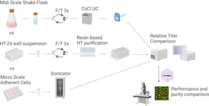
Figure 1: Overview of rAAV production and preparation approaches. This figure illustrates rAAV production methods, ranging from midi-scale shake flasks to mini-HT24 suspension cultures and micro-6-well adherent cultures. These approaches enable rAAV generation at various scales, utilizing different cell lysis and vector purification strategies. Final characterization of rAAVs typically includes qPCR/dPCR for viral genome quantification (vg/mL), ELISA for capsid titer (vp/mL) determination, and cell transduction assays to assess biological activity. Created in BioRender. Ohland, P. (2025) https://BioRender.com/x3sfbdd. Please click here to view a larger version of this figure.

Figure 2: Optimization of transfection protocol for micro-6-well rAAV production. (A) Schematic overview of the experiment with tested parameters and analytical readouts. (B) HEK293T cells were seeded in 6-well plates at a density of 0.5 × 10⁶ cells/well 24 h prior to transfection. Different DNA amounts, PEI types (Pro or Max), ratios, and NaCl concentrations were tested. (C) Percentage of GFP-positive (GFP+) HEK293A cells 72 h post-transduction with equal volumes of lysates from panel A (10 µL each). For simplicity, only the DNA condition that resulted in the highest vector yields (2650 ng) is shown. Please click here to view a larger version of this figure.
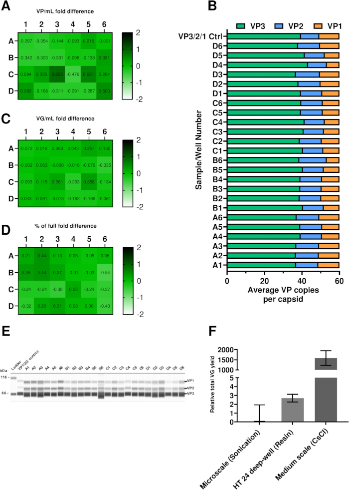
Figure 3: Analytical characterization of rAAV produced in a mini-HT24 setup and titer comparison across different scales. (A) ELISA analysis of purified rAAV capsid titers in each well. Values are represented as variations above the average particle titer of the plate after purification. Expected titers depend on the used AAV capsid and the packaged transgene. Particle titers for different AAV vectors ranged between low 1011 to high 1012 VP/mL. (B) VP-ratio analysis by WES of purified rAAV from the indicated wells. Recombinant AAV2 VP1, VP2, and VP3 were used as positive controls at a molar ratio of 1:1:10. (C) dPCR analysis of purified rAAV from each well. Values are shown as variations above the average genomic titer of the plate after purification. Expected titers depend on the used AAV capsid and the packaged transgene. The genomic titers for different AAV vectors ranged between low 1010 (rAAV2) to high 1011 vg/mL (rAAV9, not shown here). (D) Empty-to-full ratio (E/F) of the indicated wells, based on genomic and particle titer results from panels A and C. Values are normalized to the plate average. Mean E/F in mini-HT24 format for rAAV2 shown is 10.6% ± 3.0% after AAVX affinity resin purification. (E) Capillary western blot analysis of VP proteins in purified rAAV2 samples from each well. (F) Comparison of total viral genome yields considering the final sample volumes and concentration of each method. Data are shown as mean fold difference over the smallest format (micro-6-well), with error bars indicating standard deviation (SD) from biological replicates. For high-throughput 24-deep-well production with AAVX affinity resin purification, SD = ±0.176 (n = 24); for micro-6-well sonication; SD = ±0.349 (n = 6); and for standard medium-scale production with CsCl gradient purification, SD = ±134.70 (n = 2). Please click here to view a larger version of this figure.
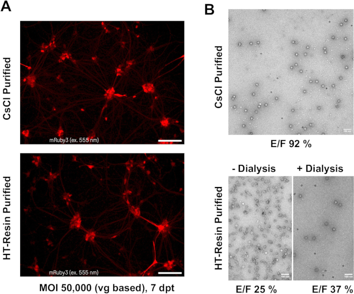
Figure 4: Comparison of potency and purity between high-throughput (HT) resin-purified rAAV and standard CsCl-purified samples. (A) Human-derived motor neurons and astrocytes were transduced with the same rAAV construct expressing fluorescence reporter mRuby3 under the human synapsin promoter as a reporter at an MOI of 50,000. Fluorescence microscopy images were captured on a live imaging microscope seven days post-transduction (DPT). rAAVs were purified using either the standard CsCl gradient method (top) or the HT AAVX affinity resin tip-based approach (bottom). CsCl-purified samples were exposed to one additional freeze-thaw cycle before transduction was performed and compared to resin-purified rAAVs. (B) Transmission electron microscopy (TEM) analysis of rAAV2-serotype-based variants purified via CsCl gradient (top) and HT resin tip methods (bottom). Two batches of HT resin-purified samples of rAAV2 can be compared without dialysis (bottom left) and with dialysis (bottom right). Empty-to Full ratios (E/F) were determined using TEM to be > 90% full for CsCl gradient ultracentrifugation-purified samples and < 40% full for AAX affinity resin-purified samples. Scale bar = 100 nm. Panel B shows representative pictures of purified batches using the indicated methods. Please note that these images do not belong to the same preparations used for transduction experiments shown in panel A. Please click here to view a larger version of this figure.
Supplementary Figure 1:Transfection of HEK293T cells for rAAV2-GFP production using the micro-6-well setup. Various DNA amounts, NaCl concentrations (in mM), PEI products, and PEI-to-DNA ratios were used for transfection (as described in Figure 2). Images were taken 72 h post-transfection using the same exposure time (320 ms). Representative brightfield and fluorescence microscopy images are shown in this figure. Magnification: 10X. Scale bar not shown; image is for illustrative purposes and qualitative assessment only. Please click here to download this File.
Discussion
This protocol paper outlines efficient methods for producing rAAV in high-throughput, small-scale settings utilizing both adherent and suspension cells. These methodologies address the increasing demand for scalable and flexible rAAV production processes that support preclinical research while minimizing time, cost, and resource consumption.
Both adherent and suspension cell systems offer distinct advantages and challenges for small-scale rAAV production. Adherent cell systems, such as HEK293T cells28 grown in multi-well plate formats, are particularly well-suited for early-stage vector design and comparative studies. Their compatibility with established culturing and transfection protocols enables straightforward implementation in most laboratories. For example, Grosse et al. utilized small-scale production in 6-well plates to investigate AAP functionality, a protein crucial for the assembly of the viral capsid22. Drittanti et al. produced rAAV vectors in 96-well plates to assess the kinetics of rAAV replication and test the effects of different Adenovirus helpers on rAAV production23. Notably, in the latter study, a replication assay using specialized HeLa cells expressing AAV Rep and Cap to further amplify the signal was described. More recently, Quan et al. applied a 384-well high-throughput production protocol in the context of a siRNA screen, reporting relative titers24. However, given the limited amount of material, it still remains uncertain how many assays and readouts can be conducted, or whether sufficient MOIs can be achieved in cell transduction experiments beyond the measurement of fluorescent proteins, potentially impacting reproducibility.
In this work, micro-6-well-based production in adherent HEK293T cells was used to optimize culture conditions for rAAV2 (Figure 1 and Figure 2). In line with previous studies and technical reports43,44, lower PEI-to-DNA ratios and the addition of NaCl during transfection positively impacted rAAV production. This was indirectly reflected in the transduction efficiencies of the produced vectors in HEK293A cells. The striking differences observed with the lower PEI:DNA ratios were mainly attributed to cellular stress, as indicated by low cell viability, particularly with PEIpro. Although the effects were less apparent under PEImax conditions, microscopy images revealed cell-free areas in the cellular layer, indicating stress or cell detachment (Supplementary Figure 1).
Importantly, these findings were not directly applicable to vector production in suspension cultures, emphasizing that experimental conditions enhancing rAAV production in adherent HEK293T cells may not always translate to suspension cells, even when the underlying cell type remains the same. This discrepancy could be attributed to differences at the transcriptomic and proteomic levels between these two systems45. Consequently, closer alignment with clinical production processes may necessitate the use of suspension cultures in the early development phases. Building on these observations and the advantage of achieving higher cell densities in suspension, a 24-deep well system was adopted to produce rAAV in miniscale suspension cultures (mini-HT24). This system underwent extensive validation, demonstrating consistent vg/mL and vp/mL titers across the plate, along with uniform VP protein expression and ratios (Figure 3A-E). Indeed, the higher cell densities achieved in the mini-HT24 setup (3 × 10⁶ cells/mL) compared to the micro-6-well format (5 × 10⁵ cells per well) resulted in higher rAAV titers, as shown in Figure 3F. This highlights a key advantage of suspension-based systems, which are not limited by the surface area of the culture vessel. However, implementing such systems requires specialized equipment that may not be available in all laboratories. Finally, it is recommended to further validate identified lead molecules or superior culturing conditions from the mini-HT24 system using minibioreactor systems such as AMBR15 or AMBR250, as these systems have been shown to more closely mimic production conditions in large bioreactors46,47.
The choice of purification level plays a crucial role in determining the suitability of rAAV preparations for specific downstream applications. Crude lysates, derived from both adherent (micro-6-well) and suspension cultures (mini-HT24), provide a rapid and resource-efficient option for preliminary screening assays. These lysates contain a mixture of viral particles, cellular debris, and media components, which is acceptable for applications such as vector genomic/particle titer, transduction efficiency screening, and western blot analysis. Especially applicable in studies addressing fundamental biological questions (as exemplified above in the investigation of AAP functionality), cell culture condition optimization, and preclinical candidate screening and selection. However, the presence of impurities may necessitate higher dilutions, resulting in lower assay sensitivities. Additionally, impurities may negatively affect the viability of sensitive cell types, such as primary cells.
In contrast to crude lysates, AAVX affinity resin-purified preparations, used exclusively in the suspension-based protocol, achieve a higher level of purity by selectively isolating intact capsids. This overcomes the limitations of crude cell lysates, making these preparations more suitable for applications such as in vivo studies, as well as detailed in vitro analyses of transgene expression kinetics, and precise functional readouts, which often require larger amounts of material (see yield comparison in Figure 3F). For in vitro applications, yields of rAAV vectors produced in the mini-HT24 setting were usually sufficient for the majority of read-outs, depending on the number of cells and required MOIs. In contrast, in vivo applications typically require production at midi-shake flask scales or larger, depending on the dose, administration route, and animal model. Despite the increased cost and time required for purification, the consistency and reliability of the resulting material often justify the investment for critical use cases. Notably, the results shown in Figure 4 demonstrate that rAAV vectors purified using affinity resin methods perform comparably to those purified via traditional cesium chloride (CsCl) ultracentrifugation. This is in line with previous observations that empty capsids have either no effect on transduction or may even enhance it, depending on the AAV species present48. It is important to note, however, that only one MOI and one time point were used in this experiment, and optimal conditions need to be determined individually for each cell type. While CsCl purification generally yields a cleaner background, HT resin tip-purified AAVs show greater variability in purity (see TEM images in Figure 4). Collectively, these findings further support the adoption of high-throughput, small-scale workflows.
The use of small-scale affinity resin-filled tips in high-throughput purification provides a practical solution for rapid rAAV screening, but several technical aspects require attention to ensure reliable results. Due to the limited resin bed volume, these tips are particularly prone to clogging, especially when processing crude lysates. Hence, clarification of lysates by high-speed centrifugation is essential to remove cellular debris prior to loading, as insufficient clarification can reduce flow rates and compromise yield. For the mini-HT24 setup, optimizing transfection protocols and maintaining a maximum working volume of 2.5-3 mL per well is recommended. When working with various serotypes or capsid libraries, testing different affinity resin types is advisable, as binding efficiency and recovery may vary depending on capsid structure49. The handheld electronic multichannel pipetting system allows easy integration of this workflow in small labs, offering flexibility without the need for automation. However, manual handling can introduce variability and lot-to-lot differences. For greater consistency, automated liquid handling platforms may be beneficial. Careful attention should be paid to avoid cross-contamination during sample and buffer addition, and the use of appropriate plate formats with adequate well volume is essential to prevent overflow. By addressing these considerations, including sample clarification, protocol optimization, resin selection, and careful pipetting, researchers can minimize technical issues and enhance the robustness and reproducibility of high-throughput, small-scale affinity purification workflows.
In conclusion, the combination of high-throughput, small-scale rAAV production methods with application-specific purification strategies provides a versatile platform for advancing gene therapy research. These protocols address the growing demand for cost-effective and efficient workflows while maintaining the flexibility to meet diverse experimental requirements. By balancing the trade-offs between purity, scalability, and resource consumption, researchers can accelerate preclinical development and facilitate the transition to clinical applications.
Disclosures
The authors declare no competing financial interests.
Acknowledgements
Julia Fakhiri gratefully acknowledges support through a Roche Postdoctoral Fellowship. Electron microscopy data were acquired at the Roche cryo-EM facility Nautilus. We thank Moritz Classen and Lubomir Kovacik for their contributions to the operation and support of the facility.
Materials
| Name | Company | Catalog Number | Comments |
|---|---|---|---|
| 0.9% Non-buffered Saline | B. Braun, Melsungen, Germany | 7647-14-5 | Wash II buffer |
| 1 M Hydrochlocirc acid | Merck Millipore, Burlington, Massachusetts, USA | 1099700050 | pH adjustment of buffers |
| 10x DNase I Buffer | Thermo Fisher Scientific, Waltham, USA | AM8170G | ddPCR/dPCR |
| 1M Tris-HCl, pH 9.0 | Thermo Fisher Scientific, Waltham, USA | J60707.AP | Neutralization buffer |
| 24-deep well microplates | Enzyscreen, Heemstede, Netherlands | CR1424cl | Suspension culture equipment |
| 250 mL Polycarbonate Erlenmeyer Flask with Vent Cap | Corning, New York, NY , USA | 431144 | Suspension culture equipment |
| 66 - 440 kDa separation 8x25 capillary cartridges | BioTechne, Minneapolis, MN, USA | SM-W008 | Automated Western blot kit |
| 96 Well 2.2 mL Polypropylene DeepWell | Thermo Fisher Scientific, Waltham, USA | AB0661 | Sample processing and storage plate |
| 96 Well Black/Clear Bottom Plate, TC Surface | Thermo Fisher Scientific, Waltham, USA | 165305 | Transduction assays |
| AAV2 VP1, recombinant protein | PROGEN, Heidelberg Germany | 640823 | Wes reagent |
| AAV2 VP2, recombinant protein | PROGEN, Heidelberg Germany | 640824 | Wes reagent |
| AAV2 VP3, recombinant protein | PROGEN, Heidelberg Germany | 640825 | Wes reagent |
| AAV2 Xpress ELISA | PROGEN, Heidelberg Germany | PRAAV2XP | Used for HT-resin purified samples. |
| Anti-Mouse Detection Module for Wes | BioTechne, Minneapolis, MN, USA | DM-002 | Automated Western blot kit |
| Assay/Template specific forward primer | Custom | Custom | ddPCR/dPCR |
| Assay/Template specific probe | Custom | Custom | ddPCR/dPCR |
| Assay/Template specific reverse primer | Custom | Custom | ddPCR/dPCR |
| BalanCD HEK293 - Liquid | FUJIFILM IRVINE SCIENTIFIC INC, Santa Ana, CA, United States | NC1192689 | Medium for suspension cell culture |
| Bovine Serum Albumin | SERVA Electrophoresis, Heidelberg, Germany | cBovine Serum Albumin | Supplement for adherent cell medium |
| Breathable rayon film, sterile | VWR International LLC, Radnor, PA, USA | 391-1262 | Suspension culture equipment |
| C1000 Thermocycler | Bio-Rad Laboratories, Hercules, USA | 1851096 | ddPCR |
| CaptureSelec AAVX Ligand Leakage ELISA Kit | Thermo Fisher Scientific, Waltham, USA | 8113522000 | ELISA |
| Cedex HiRes Analyzer | Roche Holding AG, Basel, Switzerland | 5650216001 | Cell counter |
| CELL culture microplate, 96 well | Greiner Bio-One, Kremsmünster, Austria | 655162 | Adeherent cell culture equipment |
| Citric Acid | Sigma-Aldrich, St. Louis, Missouri, United States | 5949-29-1 | Purification reagent; CAUTION |
| CMOS based TEM camera TVIPS XF416 | Tietz Video and Image Processing Systems GmbH, Gilching, Germany | XF416 | Video and Image Processing System |
| Compass software (version 6.3.0) | BioTechne, Minneapolis, MN, USA | WES Software | |
| Costar 6-well Clear TC-treated Multiple Well Plates | Corning, New York, NY , USA | 3516 | Adeherent cell culture equipment |
| ddPCR Droplet Generator Oil | Bio-Rad Laboratories, Hercules, USA | #1864110 | ddPCR reagent |
| ddPCR Droplet Reader Oil | Bio-Rad Laboratories, Hercules, USA | #1863004 | ddPCR reagent |
| ddPCR Supermix for Probes (no dUTP) | Bio-Rad Laboratories, Hercules, USA | #1863023 | ddPCR reagent |
| DMEM, high glucose | Thermo Fisher Scientific, Waltham, USA | 11960044 | Medium for adherent cell culture |
| DMEM, low glucose | Thermo Fisher Scientific, Waltham, USA | 11885084 | Medium for adherent cell culture |
| DNA low bind 96 well plates | Eppendorf, Hamburg, Germany | 30129504 | ddPCR/dPCR |
| DNA low bind tubes | Eppendorf, Hamburg, Germany | 30108051 | ddPCR/dPCR |
| DPBS | Thermo Fisher Scientific, Waltham, USA | 14190094 | Wash buffer |
| EM grids (T600H-Cu 698 l/inch Hex. mesh Thin Bar; EMS) | Electron Microscopy Sciences, Hatfield, PA, USA | FFTH600-Cu-UL | Electron microscopy analysis |
| EVO M5000 Imaging System | Thermo Fisher Scientific, Waltham, USA | AMF5000 | Live imaging microscope for transduction assays |
| EZ Standard Pack 3 | BioTechne, Minneapolis, MN, USA | PS-ST03EZ-8 | Automated Western blot kit |
| Falcon 15mL Conical Centrifuge Tubes | Fisher Scientific, Schwerte, Germany | 11507411 | Sample processing |
| Falcon 50mL Conical Centrifuge Tubes | Fisher Scientific, Schwerte, Germany | 10203001 | Sample processing |
| Geltrex LDEV-Free Reduced Growth Factor Basement Membrane Matrix | Thermo Fisher Scientific, Waltham, USA | A1413201 | Culture plate coating |
| GlutaMAX Supplement (L-Alanyl-L-Glutamine) | Thermo Fisher Scientific, Waltham, USA | 35050061 | Supplement for adherent cell medium |
| HEK293A | Thermo Fisher Scientific, Waltham, USA | R70507 | Transduction assays (research use only) |
| HEK293T | ATCC, Manassas, Virginia, USA | CRL-3216 | Adeherent cell culture, used for production of rAAV (research use only) |
| HEPES solution, 1M | Merck, Darmstadt, Germany | #H0887 | Supplement for adherent cell medium |
| HL-Salt Active Nuclease, SAN (25 U/µL) | ArcticZymes Technologies, Tromsø, Norway | 70910-202 | ddPCR/dPCR |
| HydroSpeed Microplate Washer | Tecan, Männedorf, Switzerland | 30190101 | ELISA |
| iCell Astrocytes Kit, 01434 | FUJIFILM Cellular Dynamics, Inc., Madison, WI, USA | R1092 | Transduction assays |
| iCell Motor Neuron Kit, 01279 | FUJIFILM Cellular Dynamics, Inc., Madison, WI, USA | R1051 | Transduction assays |
| Infinite 200 PRO | Tecan, Männedorf, Switzerland | 30213617 | ELISA |
| JEM-1400Plus is a transmission electron microscope | JEOL Ltd., Akishima, Tokyo, Japan | JEM-1400Plus | transmission electron microscope |
| Low-evaporation sandwich cover with pins | Enzyscreen, Heemstede, Netherlands | CR1224c | Suspension culture equipment |
| Microplate Reader SpectraMax i3 | Molecular Devices, San Jose, CA, USA | i3x | ELISA |
| Mouse anit-AAV2 VP1/VP2/VP3 monoclonal antibody | PROGEN, Heidelberg Germany | 690058 | Wes reagent |
| Nuclease-free water | Roche Diagnostics GmbH, Mannheim, Germany | 3036430103 | ddPCR/dPCR |
| Opti-MEM, Reduced Serum Medium | Thermo Fisher Scientific, Waltham, USA | #31985070 | Medium For PEI-DNA complex formation |
| PBS | Thermo Fisher Scientific, Waltham, USA | #10010023 | Wash buffer |
| PEI-MAX, polyethyleneimine, 1mg/ml stock | Polysciences Europe, Hirschberg an der Bergstraße, Germany | 24765 or 24765 | Transfection reagent |
| PEIpro | Polyplus, Illkirch-Graffenstaden, France | 101000033/10mL | Transfection reagent |
| Pierce Microdialysis Plate, 96 well, 0.3 mL, 20K MWCO | Thermo Fisher Scientific, Waltham, USA | A50472 | Purification equipment |
| Poloxamer 188 | Sigma-Aldrich, St. Louis, Missouri, United States | P5556-100mL | Cell culture reagent; CAUTION |
| Poly-L-Ornithine Solution (0.01%) | Sigma-Aldrich | A-004-M | Transduction assays |
| Proteinase K Solution (20 mg/mL) | Thermo Fisher Scientific, Waltham, USA | 25530049 | ddPCR/dPCR |
| ProteinSimple “Wes” | BioTechne, Minneapolis, MN, USA | 004-600 | Device for autmated Western Blot |
| QIAcuity Eight Platform System | Qiagen, Hilden, Germany | 911052 | dPCR |
| QIAcuity Nanoplate 8.5k 96-well | Qiagen, Hilden, Germany | 250021 | ddPCR/dPCR |
| QIAcuity Probe PCR Kit | Qiagen, Hilden, Germany | 250101, 250102 or 250103 | ddPCR/dPCR |
| QIAcuity Software Suite 1.2 | Qiagen, Hilden, Germany | dPCR Software | |
| QX manager | Bio-Rad Laboratories, Hercules, USA | ddPCR Software | |
| QX200 Droplet Reader | Bio-Rad Laboratories, Hercules, USA | 1864003 | ddPCR |
| Screw cap micro tube, 1.5 ml, PCR Performance Tested | Sarstedt, Nümbrecht, Germany | 72.703.416 | Suspension culture equipment |
| Screw cap, DNA LoBind, 5.0 mL | Eppendorf, Hamburg, Germany | 30122348 | Suspension culture equipment |
| Shaker Incubator | Infors AG, Bottmingen, Switzerland | Multitron | Suspension culture equipment |
| Sodium Pyruvate (100 mM) | Thermo Fisher Scientific, Waltham, USA | 11360070 | Medium for adherent cell culture |
| Sonicator Microplate Horn System | QSonica, Newtown, CT, USA | Q700MPXC | Sonicator used for crude lysate generation |
| SPIRIT | Link to the SPIRIT database: https://spirit.cladiac.com/aav.html | ||
| Steriflip-GP Sterile Centrifuge Tube Top Filter Unit | Merck Millipore, Burlington, Massachusetts, USA | SCGP00525 | Buffer preparation equipment |
| T-175, CELL CULTURE FLASK, 550 ML, 175 CM² | Greiner Bio-One, Kremsmünster, Austria | 660175 | Adeherent cell culture equipment |
| Trypsin/EDTA 0.025% | Thermo Fisher Scientific, Waltham, USA | R001100 | Cell culture reagent |
| Virus Production Cells 2.0 (VPC2.0) | Thermo Fisher Scientific, Waltham, USA | A49784 or A51218 | rAAV production |
| J63596.K2 | Dilute 1:10 for working solution PBST |
References
- Singh, N. Drug discovery and development: Introduction to the general public and patient groups. Front. Drug Discov. 3, 1201419(2023).
- Thakur, G., Mink, S., Bak, H., Tustian, A. D. Improving process efficiency to reduce cost-of-goods per dose in manufacturing of recombinant AAVs. Cell Gene Ther. Insights. 10 (3), 479-499 (2024).
- Reid, C. A., Hörer, M., Mandegar, M. A. Advancing AAV production with high-throughput screening and transcriptomics. Cell Gene Ther. Insights. 10 (6), 821-840 (2024).
- Matsunaga, R., Tsumoto, K. Accelerating antibody discovery and optimization with high-throughput experimentation and machine learning. J. Biomed. Sci. 32 (1), 46(2025).
- Pereira, D. A., Williams, J. A. Origin and evolution of high-throughput screening. Br. J. Pharmacol. 152 (1), 53-61 (2007).
- Aldewachi, H., Al-Zidan, R. N., Conner, M. T., Salman, M. M. High-throughput screening platforms in the discovery of novel drugs for neurodegenerative diseases. Bioengineering. 8 (2), 30(2021).
- Liu, H., et al. Producing high-quantity and high-quality recombinant adeno-associated virus by low-cis triple transfection. Mol. Ther. Methods Clin. Dev. 32 (2), 101230(2024).
- Blessing, D., et al. Scalable production of AAV vectors in orbitally shaken HEK293 cells. Mol. Ther. Methods Clin. Dev. 13, 14-26 (2019).
- Grieger, J. C., Soltys, S. M., Samulski, R. J. Production of recombinant adeno-associated virus vectors using suspension HEK293 cells and continuous harvest of vector from the culture media for GMP FIX and FLT1 clinical vector. Mol. Ther. 24 (2), 287-297 (2016).
- Strobel, B., Miller, F. D., Rist, W., Lamla, T. Comparative analysis of cesium chloride- and iodixanol-based purification of recombinant adeno-associated viral vectors for preclinical applications. Hum. Gene Ther. Methods. 26 (4), 147-157 (2015).
- Fakhiri, J., Nickl, M., Grimm, D. CRISPR gene editing, methods and protocols. Methods Mol. Biol. 1961, 111-126 (2019).
- Strobel, B., et al. Standardized, scalable, and timely flexible adeno-associated virus vector production using frozen high-density HEK-293 cell stocks and CELLdiscs. Hum. Gene Ther. Methods. 30 (1), 23-33 (2019).
- Rumachik, N. G., et al. Methods matter: Standard production platforms for recombinant AAV produce chemically and functionally distinct vectors. Mol. Ther. Methods Clin. Dev. 18, 98-118 (2020).
- Merten, O. W. AAV vector production: State of the art developments and remaining challenges. Cell Gene Ther. Insights. 2 (5), 521-551 (2016).
- Trivedi, P. D., et al. Comparison of highly pure rAAV9 vector stocks produced in suspension by PEI transfection or HSV infection reveals striking quantitative and qualitative differences. Mol. Ther. Methods Clin. Dev. 24, 154-170 (2022).
- Zolotukhin, S., et al. Recombinant adeno-associated virus purification using novel methods improves infectious titer and yield. Gene Ther. 6 (6), 973-985 (1999).
- Wada, M., et al. Large-scale purification of functional AAV particles packaging the full genome using short-term ultracentrifugation with a zonal rotor. Gene Ther. 30 (7-8), 641-648 (2023).
- Kronenberg, S., Böttcher, B., von der Lieth, C. W., Bleker, S., Kleinschmidt, J. A. A conformational change in the adeno-associated virus type 2 capsid leads to the exposure of hidden VP1 N termini. J. Virol. 79 (9), 5296-5303 (2005).
- Strobel, B., Miller, F. D., Rist, W., Lamla, T. Comparative analysis of cesium chloride- and iodixanol-based purification of recombinant adeno-associated viral vectors for preclinical applications. Hum. Gene Ther. Methods. 26 (4), 147-157 (2015).
- Shiraishi, Y., Adachi, T., Cacicedo, J. M., Ido, Y. Development of a high-yield, high-quality purification process for adeno-associated virus vectors that can be used in vivo without ultracentrifugation: Application to a lung endothelial cell-targeted adeno-associated virus. FASEB J. 36 (12), e22653(2022).
- Fakhiri, J., Nickl, M., Grimm, D. CRISPR gene editing, methods and protocols. Methods Mol. Biol. 1961, 111-126 (2019).
- Grosse, S., et al. Relevance of assembly-activating protein for adeno-associated virus vector production and capsid protein stability in mammalian and insect cells. J. Virol. 91 (20), (2017).
- Drittanti, L., Rivet, C., Manceau, P., Danos, O., Vega, M. High throughput production, screening and analysis of adeno-associated viral vectors. Gene Ther. 7 (11), 924-929 (2000).
- Quan, D. N., Shiloach, J. rAAV production and titration at the microscale for high-throughput screening. Hum. Gene Ther. 33 (1-2), 94-102 (2022).
- Steininger, T., et al. Improved recombinant adeno-associated viral vector production via molecular evolution of the viral Rep protein. Int. J. Mol. Sci. 26 (3), 1319(2025).
- Fisher, K., et al. Polo-like kinase inhibitors increase AAV production by halting cell cycle progression. Mol. Ther. Methods Clin. Dev. 33 (1), 101412(2025).
- Chen, K., et al. Advanced biomanufacturing and evaluation of adeno-associated virus. J. Biol. Eng. 18 (1), 15(2024).
- DuBridge, R. B., et al. Analysis of mutation in human cells by using an Epstein-Barr virus shuttle system. Mol. Cell. Biol. 7 (1), 379-387 (1987).
- Börner, K., et al. Pre-arrayed pan-AAV peptide display libraries for rapid single-round screening. Mol. Ther. 28 (4), 1016-1032 (2020).
- Weinmann, J., et al. Identification of broadly applicable adeno-associated virus vectors by systematic comparison of commonly used capsid variants in vitro. Hum. Gene Ther. 33 (21-22), 1197-1212 (2022).
- Westhaus, A., et al. High-throughput in vitro, ex vivo, and in vivo screen of adeno-associated virus vectors based on physical and functional transduction. Hum. Gene Ther. 31 (9-10), 575-589 (2020).
- Yang, S., Shi, H., Chu, X., Zhou, X., Sun, P. A rapid and efficient polyethylenimine-based transfection method to prepare lentiviral or retroviral vectors:useful for making iPS cells and transduction of primary cells. Biotechnol. Lett. 38 (9), 1631-1641 (2016).
- Sanmiguel, J., Gao, G., Vandenberghe, L. H. Adeno-associated virus vectors, design and delivery. Methods Mol. Biol. 1950, 51-83 (2019).
- Wang, G., et al. Assessment of key factors impacting variability in AAV vector genome titration by digital PCR. Int. J. Mol. Sci. 25 (10), 5149(2024).
- Dobnik, D., et al. Accurate quantification and characterization of adeno-associated viral vectors. Front. Microbiol. 10, 1570(2019).
- Duong, T., Firmo, M., Li, C. T., Gu, B., Wang, P. Three-dimensional linkage analysis with digital PCR for genome integrity and identity of recombinant adeno-associated virus. Sci. Rep. 15 (1), 2154(2025).
- Tereshko, L., et al. A novel method for quantitation of AAV genome integrity using duplex digital PCR. PLOS ONE. 18 (12), e0293277(2023).
- Sormunen, A., et al. Comparison of automated and traditional Western blotting methods. Methods Protoc. 6 (2), 43(2023).
- Hiemenz, C., et al. Characterization of virus particles and submicron-sized particulate impurities in recombinant adeno-associated virus drug product. J. Pharm. Sci. 112 (8), 2190-2202 (2023).
- McIntosh, N. L., et al. Comprehensive characterization and quantification of adeno associated vectors by size exclusion chromatography and multi angle light scattering. Sci. Rep. 11 (1), 3012(2021).
- Su, Q., Sena-Esteves, M., Gao, G. Analysis of recombinant adeno-associated virus (rAAV) purity using silver-stained SDS-PAGE. Cold Spring Harb. Protoc. (8), pdb.prot095679(2020).
- Capra, E., Gennari, A., Loche, A., Temps, C. Viral-vector therapies at scale: Today's challenges and future opportunities . , McKinsey & Company. https://www.mckinsey.com/industries/life-sciences/our-insights/viral-vector-therapies-at-scale-todays-challenges-and-future-opportunities (2022).
- Huang, X., et al. AAV2 production with optimized N/P ratio and PEI-mediated transfection results in low toxicity and high titer for in vitro and in vivo applications. J. Virol. Methods. 193 (2), 270-277 (2013).
- Development of a novel cell culture medium for AAV production with multiple HEK293 lineages and process optimization. , Merck. https://www.sigmaaldrich.com/DE/de/technical-documents/technical-article/pharmaceutical-and-biopharmaceutical-manufacturing/gene-therapy-manufacturing/development-of-novel-cell-culture-medium-aav-production (2025).
- Malm, M., et al. Evolution from adherent to suspension: Systems biology of HEK293 cell line development. Sci. Rep. 10 (1), 18996(2020).
- Warr, S. R. C. Animal cell biotechnology, methods and protocols. Methods Mol. Biol. 2095, 43-67 (2019).
- Manahan, M., et al. Scale-down model qualification of ambr 250 high-throughput mini-bioreactor system for two commercial-scale mAb processes. Biotechnol. Prog. 35 (6), e2870(2019).
- Wang, Q., et al. Syngeneic AAV pseudo-particles potentiate gene transduction of AAV vectors. Mol. Ther. Methods Clin. Dev. 4, 149-158 (2017).
- Florea, M., et al. High-efficiency purification of divergent AAV serotypes using AAVX affinity chromatography. Mol. Ther. Methods Clin. Dev. 28, 146-159 (2023).