Stable in vitro amplification of the Primordial Germ Cell-Like Cells (PGCLCs), a pluripotent stem cell-derived surrogate model of the embryonic primordial germ cells, has been facing technical challenges. This protocol supports the long-term expansion of PGCLCs in an in vitro condition without serum or feeder layer cells.
Method Article
Long-term Culture of Human Primordial Germ Cell-like Cells
In This Article
Summary
Abstract
Primordial germ cells (PGCs) are common precursors of all male and female germline cells. PGCs arise in peri-implantation mammalian embryos and become gender-specific germ cell precursors upon sexual differentiation of the gonadal anlage. The in vitro model commonly known as PGC-Like Cells (PGCLCs), generated from human pluripotent stem cells, is a useful surrogate of the human embryonic PGCs, providing a unique opportunity to explore human gametogenesis in vitro. The protocol presented here supports long-term in vitro expansion of human PGCLCs. Freshly isolated PGCLCs are maintained initially on a STO feeder layer and then expanded in a feeder-free condition on basement membrane extract. The long-term culture PGCLCs (LTC-PGCLCs) can be Fluorescence-Activated Cell Sorting (FACS)-enriched as CD38-positive cells and are readily amplified to tens of millions of cells as pure, feeder-free, and serum-free monotonous culture without apparent limitations such as senescence. It is practically feasible to obtain a pure population of more than 1 million human PGCLCs from a few thousand freshly isolated PGCLCs. The human LTC-PGCLC cell culture is a useful and convenient in vitro model to study human germ cell biology and differentiation into the downstream germline cells.
Introduction
Primordial germ cells (PGCs) are the earliest precursors of all germline cells. Human PGCs emerge from posterior epiblast - presumably uncommitted amniotic precursor cells - in early post-implantation, gastrulating embryos at 2-3 weeks of gestation1,2,3,4,5,6,7. For specification of both human and mouse PGCs, the signaling pathways activated by the bone morphogenetic proteins (BMPs) and WNT3 play critical roles3,6. Whereas the induction of three transcription factors, BLIMP1, PRDM14, and TFAP2C, is necessary for the specification of mouse PGCs, in human PGC specification, the role of PRDM14 is diminished. SOX17, which is dispensable for mouse PGC specification but indispensable for human PGC specification, is induced by BMP signaling in a manner dependent exclusively on EOMES or both EOMES and TBXT7,8,9. Human PGCs migrate from the posterior epiblast towards the gonadal anlagen in the genital ridges through the dorsal mesentery at 5-6 weeks of gestation10,11,12,13,14,15,16. The human PGC migration may be driven by chemotaxis, although it awaits experimental confirmation. Transcriptomic profiles of the migratory human PGCs are not identical to those of mouse PGCs at the same stage17,18. For example, human PGCs express DAZL even before their settlement in the gonadal anlagen, whereas mouse PGCs express DAZL only after their gonadal settlement9. Upon initiation of the sex-specific differentiation of the gonadal somatic cells, PGCs also undergo differentiation to prospermatogonia (also known as gonocytes) or oogonia.
Since access to human embryonic PGCs is extremely challenging due to technical and ethical barriers, the use of the PGC-like cells (PGCLCs) derived from pluripotent stem cells is becoming increasingly popular3. Mouse PGCLCs resemble the post-migration stage of PGCs, characterized by advanced global genomic DNA (gDNA) demethylation, and they are capable of generating fertilization-competent sperm and oocytes19,20,21. In contrast, human PGCLCs resemble earlier stages of embryonic PGCs with limited global gDNA demethylation7,22,23,24. Evidence is accumulating that human PGCLCs, despite their relatively immature status, are capable of in vitro differentiation to more advanced stages of germ cells resembling oogonia25,26 or prospermatogonia27.
Since Irie et al. published the first generation of PGCLCs from human pluripotent stem cells in 20157, a number of laboratories have published various protocols for human PGCLC production. The early, standard protocols involve the formation of pluripotent cell aggregates, which are often referred to as embryoid bodies7,22,24,26,28,29. Human PGCLCs can be produced using various aggregate-based protocols that share comparable transcriptomic profiles, all of which strongly express key PGC marker genes such as PRDM1, SOX17, TFAP2C, POU5F1, and NANOS3, whereas expression of the pluripotency marker SOX2 or late-stage PGC marker DDX4 is commonly suppressed24. Successful attempts have also been reported to produce human PGCLCs from monolayer cell cultures for improved scalability and convenience30. Although the yield of PGCLCs is relatively small, the successful production of human PGCLCs from pluripotent stem cell-derived in vitro models of the gastrulation-stage embryos significantly contributes to elucidation of the mechanisms of germline specification and early-stage PGC development31,32,33,34,35,36.
Although yield and quality of human PGCLCs produced using various cell culture methods have been improving, the technical difficulty in cell culture expansion of human PGCLCs poses a significant hurdle to their application for experiments requiring large amounts of homogeneous cell populations, such as chemical or genetic screening, as well as production of advanced stages of germline cells26. Gell et al. reported maintenance of human PGCLCs for up to 21 days on STO feeders in a FR10-based medium containing stem cell factor (SCF), and 2.5% fetal calf serum (FCS); however, PGCLCs tended to diverge from the PGC-like identity to other types of cells that may not be relevant to germline cell development37. Murase et al. repeatedly eliminated such unwanted cells from the culture by FACS at every passaging and showed that human PGCLCs are capable of maintaining their proliferation potential long-term in vitro38. Later, Murase et al. reported successful amplification of human PGCLCs in a medium containing 2.5% FCS39, but their method requires the m220-5 feeder layer cells, which are spontaneously emerged MMC-resistant subclones of the m220 mouse embryonic cells that express the membrane-bound form of mouse SCF from an exogenous vector40. As of today, m220-5 cells have not yet been deposited into a publicly accessible repository. Thus, it remains a major challenge to expand human PGCLC culture in vitro to a large number of homogenous cells, maintaining their PGC-like characteristics in a simple condition that does not require a non-standard feeder cell resource and is free of animal-derived reagents, whose efficiency may vary among different lots.
To overcome the above technical hurdle, a method that enables long-term in vitro expansion of human PGCLCs as a homogeneous cell population, maintaining the PGC-like characteristics in a condition free of serum or feeder layer cells, has been developed41. Because human PGCLCs express telomerase, these cells amplify at a constant rate with no signs of senescence or deviation from other types of cells for at least 150 days. The long-term culture PGCLCs (LTC-PGCLCs) maintain their human PGC-like transcriptomic characteristics, although their global gDNA is modestly demethylated upon repeated passages. LTC-PGCLS are readily converted to pluripotent embryonic germ cell-like cells (EGCLCs), restoring iPSC-like transcriptome and gDNA methylome, although the imprinted allelic restriction of H19 RNA expression is lost. LTC-PGCLCs are also capable of differentiating into DAZL-expressing cells resembling prospermatogonia in the xrTestis xenobiotic organoid culture27. Thus, the human LTC-PGCLC model represents a novel type of stably maintained, pluripotency-relevant cell culture resource that permanently retains the biological characteristics of PGCLCs and is easily expanded in vitro as a stable cell line.
This article describes the method of adapting freshly isolated, FACS-enriched pure population of human PGCLCs to serum-free, long-term maintenance culture through temporal support by the standard STO feeder layer cells and then in a feeder-free condition on Basement Membrane Extract (BME). This method supports the in vitro expansion of human PGCLCs produced using the embryoid body-dependent protocol, as previously described24,41,42 or reported by Sasaki et al22, and the monolayer culture method reported by Overeem et al30. When applied to human PGCLCs prepared as described41, an initial population of 10,000 cells can be expanded to nearly 1 x 10⁹ cells in 1.5 - 2 months with a 4 - 4.5 day population doubling time41. The applicability of this method to human PGCLCs produced by other methods is also expected.
Protocol
1. Preparation of cell culture media
- Prepare media as described in Table 1, Table 2, Table 3, and Table 4. Store aliquots of these media frozen.
- Prepare PGCLC Basal Medium as described in Table 1. Make 40 mL aliquots and store at -20 °C for 6 months. Prepare STO medium 1 as described in Table 2. Store 50 mL aliquots in 50 mL centrifuge tubes at 4 °C for 1 month.
- Prepare STO medium 2 as described in Table 3. Use immediately. Prepare Mitomycin-C treated STO cells (MMC-STO) storage medium as described in Table 4. Store 10 mL aliquots at -20 °C for 6 months.
CAUTION: As dimethyl sulfoxide is considered hazardous (combustible liquid), it should be kept away from heat, sparks, open flames, or hot surfaces. Follow the local regulatory protocol for its disposal.
2. Preparation of MMC-treated STO cells
NOTE: MMC treatment of STO cells inactivates their proliferation activity without significantly damaging their viability. The MMC-STO cells are used as feeder layer cells during the initial culture of human PGCLCs (step 4) as well as the source of the cell-free conditioned medium (step 3).
- Grow STO cells in STO medium 1 in four P15 dishes until near confluence. Change STO medium 1 to 23 mL/dish. Add 23 μL/dish of 10 mg/mL concentration MMC directly to the medium to a final concentration of 10 μg/mL.
- Swirl well and incubate at 37 °C in a CO2 incubator for 4 h.
CAUTION: MMC is fatal if a large amount is swallowed, and it is a suspected carcinogen. Handling and disposal of this compound should follow local regulatory protocols. - Wash cells with 20 mL/dish Dulbecco's PBS (Ca/Mg-free) 2x at room temperature.
- Dissociate cells with 4 mL/dish TrypLE Express in a CO2 incubator for 2 min. Suspend cells to a single-cell suspension by gentle pipetting.
- Transfer two dishes of cell suspension (8 mL) to a 50 mL centrifuge tube containing 30 mL of ice-cold STO medium-1. Repeat for the other two dishes to make two tubes of 38 mL cell suspension.
- Count cells in the presence of 0.2% trypan blue for evaluation of cellular viability using a hemocytometer or automated cell counter.
- Centrifuge cells at 300 x g for 5 min at 4 °C. Discard supernatant.Resuspend cell pellets in ice-cold frozen cell storage medium to 5 x 10⁶ cells/mL.
- Prepare 1 mL aliquots of MMC-treated STO cells in the cryogenic vials. Freeze cells slowly in a styrofoam box or a slow-freezing device and store at -80 °C for up to 12 months.
3. Preparation of STO-conditioned medium (S-CM)
NOTE: Cell-free conditioned medium of the MMC-STO cells (prepared in step 2) supports survival and proliferation of human PGCLCs while repressing their deviation to cells losing PGC-like characteristics.
- Day 1: Thaw MMC-treated STO cells from frozen stocks and inoculate 5 x 10⁶ cells into a gelatin-coated P10 dish with 10 mL/dish STO-medium 2. The typical size of a batch consists of 4 to 8 P10 dishes. Incubate cells at 37 °C in a CO2 incubator overnight.
- To coat wells with gelatin, add 1 mL/well of 0.1% gelatin and incubate at room temperature for at least 20 min. Aspirate the excess gelatin solution and leave the wells open in the biosafety cabinet until dry. Store gelatin-coated plasticware at room temperature for 2 weeks or at 4 °C for 3 months.
- Thaw aliquots of the PGCLC Basal Medium at 4 °C overnight.
NOTE: It is not recommended to thaw this medium more quickly at a higher temperature. Do not store PGCLC basal medium at 4 °C for more than 1 week. - Day 2: Remove and discard the STO-medium 2. Wash cells with warm DPBS (~10 mL/dish) followed by a wash with warm PGCLC Basal Medium (2 mL/dish). Add warm PGCLC Basal Medium (12 mL/dish) and incubate cells at 37 °C in a CO2 incubator overnight.
- Day 3: Collect all conditioned medium in a 50 mL centrifuge tube. Repeat PGCLC Basal Medium (12 mL/dish) and incubation for collection of the conditioned medium the next day from the same STO cell culture. Centrifuge the conditioned medium collected at 300 x g for 5 min at 4 °C. Make 10 mL aliquots of the supernatant (S-CM) in 15 mL centrifuge tubes. Do not take the last 3 mL of the medium remaining at the bottom of the 50 mL tubes to avoid contamination of MMC-STO cells in S-CM. Store the aliquots at -80 °C for up to 6 months.
- Day 4: Harvest S-CM by repeating step 3.4 every day until the MMC-STO monolayer culture deteriorates after 5-6 days of culture in PGCLC Basal Medium. Stop harvesting when 25% or more cells are detached from the dish.
4. Adaptation of human PGCLCs to long-term culture
- Culture freshly isolated human PGCLCs on the MMC-STO feeder layer. Prepare human PGCLCs using a previously published method (Mitsunaga et al.24; Kobayashi et al.41). Detailed protocols of human PGCLC induction have also been published42,43.
- After the initial amplification for up to 28 days, maintain PGCLCs as LTC-PGCLCs on the BME as a feeder-free culture. Throughout these two steps of PGCLCs culture, use the STO-conditioned medium (S-CM, prepared in step 3) with SCF supplementation but not inclusion of FCS. The ROCK inhibitor is necessary only immediately after passaging.
NOTE: As BME, ESC-grade Matrigel was used in our lab. Other commercial products did not support the long-term expansion of LTC-hPGCLCs. - Day 1: Inoculate MMC-treated STO feeder layer cells in gelatin-coated 6-well plates to 8 x 10⁴ cells/cm2 with 3 mL/well STO-medium 2. Incubate the feeder layer at 37 °C in a CO2 incubator overnight.
- Day 2: Discard the STO-medium from the culture and wash cells with warm Dulbecco's MEM (serum-free), 3 mL/well. Add 3 mL/well of S-CM freshly supplemented with 0.1 mM 2-Mercaptoethanol, 50 µg/mL ascorbic acid, 100 ng/mL human recombinant SCF and 10 µM Y-27632. Inoculate 500 - 2,000 freshly isolated human PGCLCs onto the STO feeder layer in each well. Incubate cells at 37 °C in a CO2 incubator overnight.
NOTE: The presence of serum, even at low concentrations, tends to cause the emergence of unwanted types of cells from human PGCLCs41. Growth of human PGCLCs is strictly dependent on SCF41. Y-27632 supports the survival of single-cell human PGCLCs at the time of inoculation but is not necessary during maintenance culture. Add 2-Mercaptoethanol, ascorbic acid, SCF, and Y-27632 freshly to the medium. In a typical experiment, PGCLCs are inoculated into all wells of a 6-well plate. If a larger number of PGCLCs is available as the starting material, a higher density up to 10,000 PGCLCs per well is acceptable. - Day 3: Replace half of the medium (1.5 mL/well) with warm S-CM freshly supplemented with 0.1 mM 2-Mercaptoethanol, 50 µg/mL ascorbic acid, 100 ng/mL human recombinant SCF but without Y-27632. Do not disturb PGCLCs, which may be detached from the feeder layer by a strong flow of medium.
- Days 5 - 28: Repeat step 4.5 every other day. As PGCLCs typically proliferate at 4.5-day population doubling time41, small clusters of PGCLCs emerge at day 6-7 or later (Figure 1A-D). Clusters will become more evident by Day 14-15 (Figure 1E,F). If unwanted types of cellular clusters with anomalous morphological characteristics deviating from the correct PGCLCs appear, discard wells containing such clusters, or attempt to eliminate the anomalous cells using the differential adhesion technique described later in step 4.9.
- When PGCLCs expand to form clusters that cover more than 20% culture area (~Day 28), dissociate cells using the cell detachment solution (CDS). Remove S-CM from wells of the PGCLC culture and add prewarmed CDS at 1 mL/well without washing cells. Incubate cells at 37 °C in a CO2 incubator for 5 min until PGCLCs detach from wells.
NOTE: Accutase is recommended as CDS for mild dissociation. Do not prewarm CDS at 37 °C for more than 5 min. PGCLC detachment can be facilitated by gentle pipetting, but PGCLCs do not need to be completely dissociated to a single cell suspension. Small aggregates of PGCLCs may increase viability. If unwanted types of cells emerge and grow rapidly, adjust the incubation time in CDS during cell detachment for the selective collection of PGCLCs, which tend to detach earlier than feeder cells and most types of unwanted cells. If the persistent emergence of unwanted types of cells disturbs the expansion of PGCLCs, attempt to remove the unwanted cells by using their quicker adhesion to gelatin-coated wells as described in step 4.9. As the enrichment of PGCLCs by FACS tends to lose a significant portion of live PGCLCs, reduce the area size of the feeder layer accordingly. - To stop cell detachment by CDS, dilute the cell suspension with 10x volume of warm PGCLC Basal Medium and centrifuge at 300 x g for 5 min at 4 °C. Note that the enzymatic activity of some CDS, such as Accutase, is not suppressed by serum or trypsin inhibitors. Discard the supernatant and resuspend cells in 3 mL of warm S-CM freshly supplemented with 0.1 mM 2-Mercaptoethanol, 50 µg/mL ascorbic acid, 100 ng/mL human recombinant SCF, and 10 µM Y-27632.
- Inoculate the cell suspension into wells of 6-well plates coated with 0.1% gelatin and incubate at 37 °C in a CO2 incubator for 30 min to remove MMC-STO cells (and unwanted types of cells), which rapidly adhere to the gelatin-coated wells.
NOTE: It is encouraged to inspect cells by phase contrast microscopy at 20 min of incubation as well as at 30 min to evaluate adhesion of the feeder cells. It may be necessary to elongate the incubation time to up to 45 min. The use of FACS or MACS to eliminate feeder cells is not generally necessary as MMC-treated STO cells cannot proliferate and deteriorate in the S-CM. - Transfer PGCLCs depleted of anomalous types of cells onto Basement membrane matrix-coated wells to start feeder-free long-term culture of human PGCLCs. For a single well in a 6-well plate, inoculation of 1 x 10⁵ viable PGCLCs is recommended.
NOTE: It is typically necessary to pool hPGCLCs from up to 6 wells to collect 1 x 10⁵ viable (trypan blue staining negative) PGCLCs and seed them into a single well to increase the inoculum cell density. If the number of the harvested PGCLCs do not reach 1 x 10⁵, use a smaller size of well, such as a 12-well or 24-well plate to obtain a relatively high PGCLC density. By the time of the first subculture, most MMC-STO feeder layer cells are deteriorated, and they do not survive the passaging procedure. A small number of viable MMC-STO cells are effectively removed by the differential adhesion technique. Even if a few MMC-STO cells are contaminated in BME-coated wells, they will disappear from the culture during multiple passaging. - Human PGCLCs grow as a highly dispersed cell population with no significant sign of direct physical contact between individual cells (Figure 1). If clusters of cells showing tight intercellular interactions emerge (Figure 2A), passage the culture using the technique described in step 4.7. Once a week passaging (1:4) is typically sufficient.
NOTE: FACS enrichment of human PGCLCs is typically unnecessary. However, if unwanted anomalous cells cannot be removed effectively after repeated differential adhesion technique described in step 4.11, a few cycles of FACS enrichment of human PGCLCs as CD38-positive cells42,43 facilitates the establishment of a pure population of LTC-hPGCLCs. Examples of PGCLC-derived anomalous cells growing on BME-coated dishes in the absence of a feeder layer are shown in Figure 3. It is important to maintain a relatively high density of PGCLC cell culture to reduce the risk of inducing unwanted types of cells.- Prepare BME-coated dishes as previously described42,43. Thaw the BME on ice in a refrigerator or a cold room overnight. Do not thaw frozen BME at room temperature or use a warm water bath.
- Aliquot BME (~200 µL; the volume of an aliquot is determined by the manufacturer for each batch and indicated on the label of vial) into sterile and pre-chilled low-bind centrifuge tubes or cell cryopreservation tubes on ice. It is important to keep the BME ice-cold during dispensing to avoid solidification. Store the aliquots at -80 °C.
- Dilute one aliquot of BME with 25 mL of ice-cold DMEM/F12 and spread to three 10-cm dishes (~8 mL/dish) or four 6-well plates (total 24 wells, 1 mL per well). Incubate dishes at room temperature for at least 1 h. After coating, dishes can be sealed with transparent film and stored at room temperature for up to 1 week.
- Aspirate medium from dishes immediately before use. It is not necessary to wash the coated dishes.
- Maintain a relatively high density of LTC-PGCLC culture on the BME to suppress the emergence of unwanted types of cells. Passage PGCLCs is recommended when cells reach more than 25% confluency. Half of the medium (1.5 mL/well) should be changed with warm S-CM freshly supplemented with 0.1 mM 2-Mercaptoethanol, 50 µg/mL ascorbic acid, and 100 ng/mL human recombinant SCF.
NOTE: Inoculating PGCLCs at a lower density increases the risk of producing unwanted cells. Since the population doubling time of human PGCLCs is approximately 4.5 days41, routine weekly passaging is typically adequate to maintain the long-term culture. Inclusion of the ROCK inhibitor (Y-27632) is necessary only after passaging to improve the PGCLC viability. - Do not disturb PGCLCs, which may be detached from the feeder layer by a strong flow of medium.
NOTE: Because human PGCLCs express telomerase, they proliferate without apparent signs of senescence such as shortening of the telomeres or expression of the senescence-associated β-galactosidase41. - When human PGCLCs gained stable proliferation without generating significant amounts of unwanted types of cells, examine their PGC-like characteristics by expression profiling of marker genes.
NOTE: Analytical approaches suitable for characterization of each single cell (such as immunofluorescence, FACS, or single cell RNA-seq) are encouraged to evaluate possible contamination of unwanted cell types. If outcomes of such analyses support the PGC-like nature of the PGCLCs, establishment of an LTC-hPGCLC culture is confirmed. - Store LTC-PGCLCs frozen in a cell freezing medium (1 x 10⁶ cells/mL per cryogenic vial). Starting from a total of 10,000 human PGCLCs as the initial material, the total yield of 1 x 10⁶ PGCLCs is achievable in 6 weeks. Place a paper or plastic box containing the cryogenic vials in a tightly sealable styrofoam box. Place the sealed box in a -80 °C freezer overnight for slow freezing. Then place the vials in a -80 °C freezer or a liquid nitrogen tank.
- To initiate culture from a vial of frozen cells, follow the steps described below.
- Thaw cells quickly in a 37 °C water bath with manual shaking while keeping the bottom of the vial in water. When >80% of the content is thawed in the vial, dry the vial with lint-free tissues or equivalent laboratory paper and wipe around the cap with an alcohol swab.
- Transfer cells to a 15 mL centrifuge tube containing prewarmed (37 °C) PGCLC basal medium. If a small frozen cells remain in the vial, recover them with the PGCLC basal medium by pipetting.
- Centrifuge cells immediately at 300 x g for 5 min at room temperature and remove supernatant.
- Resuspend the cell pellet with 9 mL of prewarmed S-CM freshly supplemented with 0.1 mM 2-Mercaptoethanol, 50 µg/mL ascorbic acid, 100 ng/mL human recombinant SCF, and 10 µM Y-27632. Gently suspend cells using 1,000 mL pipette tips.
- Inoculate 3 mL/well in 3 wells of the BME-coated plate to initiate culture. Gently shake the plates vertically and horizontally for even distribution of cells. Avoid circular motion of shaking, as such a motion will move cells to the center of the wells. Viability of cells immediately after recovery from a frozen stock is typically greater than 80%.
Results
Representative Results - PGCLCs typically form visible clusters on the MMC-STO feeder layer within 2 weeks after inoculation (Figure 1A-D). It takes approximately 3-4 weeks until PGCLCs are ready for feeder-free culture. As PGCLCs show strong cytokinetic activity, their intercellular interaction is weak when they form clusters on the feeder layer (Figure 1E,F). PGCLCs moving out of the clusters are often observed (Figure 1F, red arrowheads). Without a feeder layer, LTC-PGCLCs grow as scattered cells with little signs of intercellular adhesion, even when the cell density is high (Figure 3). Each LTC-PGCLC maintains the original morphology of the freshly isolated human PGCLC, showing signs of active cytokinesis. When cells deviated from the PGC-like morphological characteristics emerge (Figure 2), enrichment of PGCLCs by repeated differential adhesion technique often improves the culture quality. If anomalous cells persist, occasional FACS enrichment of PGCLCs as CD38-postive cells may help.
A preceding publication has identified human PGCLCs as PRDM1-positive, TFAP2C-positive, but SOX2-negative cells41. They also express POU5F1, but their precursor iPSCs also express this pluripotency marker41. It is strongly encouraged to confirm single-cell-level yield and marker expression profiles of the expanding human PGCLCs at the earliest opportunities using genetically inducible markers, immunofluorescence, FACs, or single-cell RNA-seq. However, without such confirmatory analyses, the yield and quality of human PGCLCs in the presence or absence of the feeder cells can be conveniently estimated using the standard phase contrast microscopy as shown in Figure 1, Figure 2, and Figure 3.
During the long-term culture, LTC-PGCLCs maintain a homogeneous cell population containing an actively proliferating subpopulation, as earlier single-cell RNA-seq data demonstrated41. The transcriptomic profile of the original PGCLCs is largely maintained, except that expression of the early-stage PGCLC markers GATA2 and GATA3 is rapidly lost41. The global DNA methylation level in human iPSCs is approximately 73%, and it decreases to about 64% in the freshly produced PGCLCs41. The global DNA methylation level further decreases in LTC-PGCLCs to about 54% after 12 weeks of in vitro expansion41. Most transcriptomic and epigenetic characteristics of human LTC-PGCLCs are completely reversed to those in the original iPSCs when converted to the pluripotent embryonic germ cells, except that the allele-specific epigenetic marks at several imprinting control regions are erased in PGCLCs and never revert41.
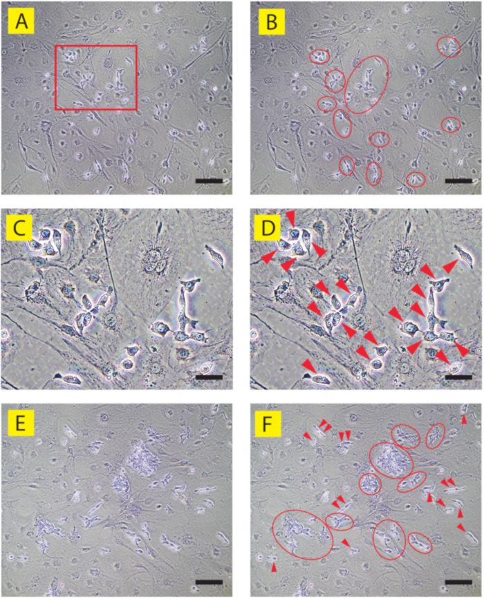
Figure 1: Phase contrast images of correct human PGCLCs on STO feeder layer (human iPSC clone 9A13). (A, C, E) are annotated in (B, D, F), respectively. (A-D) Day 6 culture. A part of (A) shown in a red rectangle is enlarged in (C, D). Most PGCLCs are observed as dispersed cells that often form loose clusters (red ovals). Individual PGCLCs (red arrowheads) do not form close contact with each other and are readily identified under phase contrast microscopy as cells protruding from the deteriorating, flat, and widely spreading STO feeder cells. (E, F) Day 15 culture. Multiple clusters of PGCLCs are readily identified (red ovals), whereas intercellular contact is still loose. Some PGCLCs are found outside the clusters, often as duplets (red arrowheads), reflecting their active proliferation while randomly migrating on and between the highly deteriorated feeder layer. Scale bars = 50 µm (A, B, E, F) or 15 µm (C, D). Please click here to view a larger version of this figure.
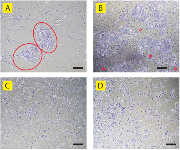
Figure 2: Phase contrast images of human PGCLCs that are deviated from the PGC-like characteristics. (A) Two anomalous clusters of PGCLCs (red ovals) appeared in the Day 9 culture of 9A13 PGCLCs on the STO feeder layer. Different from the correct PGCLC clusters (See Figure 1A-D), cells show strong intercellular contact that obscures the boundaries of each individual cell. (B) Day 18 culture of human PGCLC culture shown in (A). Multiple anomalous PGCLC clusters are observed, some of which show dense colonies resembling the precursor human iPSCs (red asterisks). Note that most of the fibroblast-like elongated cells found between the clusters are also PGCLC-derived anomalous cells, while most STO feeder cells are deteriorated and dislodged. (C) LTC-hPGCLCs (human iPSC clone B6) showing massive cell death and anomalous morphologies on BME-coated, feeder-free dishes. (D) LTC-hPGCLCs that are largely deviated to cells with anomalous morphologies on BME-coated, feeder-free dishes (human iPSC clone T548). Compare these anomalous LTC-hPGCLCs with the correct LTC-hPGCLCs shown in Figure 3B. Scale bars = 50 µm (A, B) or 100 µm (C, D). Please click here to view a larger version of this figure.
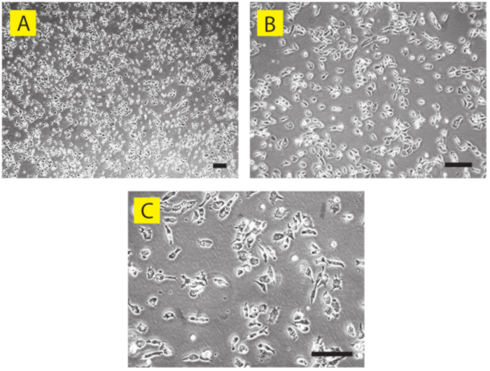
Figure 3: Phase contrast images of correct human PGCLCs as feeder-free culture (human iPSC clone A4). Day 56 feeder-free culture of LTC-PGCLCs on BME-coated dishes is shown in (A) low, (B) middle, and (C) high magnifications. Even though the cell density is relatively high, each individual PGCLC does not form close intercellular contacts except for cells that have recently completed cell division. Due to the active cytokinesis, some PGCLCs show elongated morphology while others are round-shaped. Scale bars = 50 µm. Please click here to view a larger version of this figure.
| PGCLC Basal Medium | |
| Ingredients | Volume |
| Glasgow’s MEM | 500 mL |
| Knockout Serum Replacement (KSR) | 75 mL |
| MEM NEAA (100X) | 6 mL |
| Sodium Pyruvate (100X) | 6 mL |
| Penicillin-Streptomycin (100X) | 6 mL |
Table 1: PGCLC basal medium. Composition of the PGCLC basal medium (593 mL).
| STO medium 1 | |
| Ingredients | Volume |
| Dulbecco’s MEM | 445 mL |
| Fetal Calf Serum | 50 mL |
| Penicillin-Streptomycin (100X) | 5 mL |
Table 2: STO medium 1. Composition of the STO medium 1 (500 mL).
| STO medium 2 | |
| Ingredients | Volume |
| Dulbecco’s MEM | 47 mL |
| Fetal Calf Serum | 2.5 mL |
| Penicillin-Streptomycin (100X) | 0.5 mL |
| FGF2 (200 µg/mL) | 2.5 µL |
Table 3: STO medium 2. Composition of the STO medium 2, which is supplemented with FGF2 for augmented growth of STO cells (50 mL).
| MMC-STO Storage Medium | |
| Ingredients | Volume |
| Dulbecco’s MEM | 42.5 mL |
| Fetal Calf Serum | 5 mL |
| Dimethyl sulfoxide | 2.5 mL |
Table 4: MMC-STO Storage Medium. Composition of the MMC-STO Storage Medium, which is used to prepare frozen stocks of MMC-treated STO feeder cells (50 mL).
Discussion
The method of human PGCLC maintenance described in this article is relatively simple without requiring any specific devices or non-standard materials. Previously published methods of human PGCLC maintenance require frequent FACS sorting due to significant deviation of cells from PGCLC-like status38. These methods also require a non-standard feeder layer cell line (m220-5), which is not readily available through publicly and internationally accessible repositories39,40. The inclusion of FCS in their media introduces uncertainty due to dependency on the performance of a specific lot of the serum. In contrast, the currently presented method readily obtains many millions of human PGCLCs, which can be stored frozen while maintaining high (> 90%) viability, without requiring serum or other animal-based reagents. The protocol requires the standard STO feeder layer cell line, which is commonly available from many public repositories. The method provides an unprecedented level of convenience to handle human PGCLCs as if they were conventional cell lines. The unlimited supply of human PGCLCs achieved using this method will widen the scope of human PGCLC applications in the areas such as experimental productions of human germ cells and/or gametes, assessments of human germ cell disorders such as infertility (including attempts of in vitro gametogenesis) or germ cell tumors, and toxicological assessments of prospective germ cell toxicants.
The method excludes FCS in the S-CM to minimize the emergence of unwanted types of cells from human PGCLCs41. Several iPSC clones tended to produce unwanted types of cells during the feeder-dependent stage or thereafter, and some of them had to go through two or three rounds of FACS purification of PGCLCs. However, once the LTC-PGCLC culture is established with more than 50% confluency, the necessity of FACS purification is typically diminished. Growth of human PGCLCs in vitro is strictly dependent on the presence of SCF41. If the growth rate of LTC-PGCLCs diminishes, it is recommended to test different lots or manufacturers' SCF reagents. Because human PGCLCs express telomerase, they do not experience senescence during long-term expansion in vitro41. It has been used successfully to establish LTC-PGCLCs derived from male and female human iPSCs with various ethnic backgrounds41. Examples of successful application of the currently presented method include male human iPSC clones A4 and A5 (Caucasian newborn), PGP1 (Caucasian adult), and 9A13 (Japanese adult), and female iPSC clones ATCC-BXS0116 (Caucasian) and ATCC-BXS0115/ACS-1029 (Hispanic). The general applicability of this method and prediction markers needs to be established in future studies.
The necessity of occasional FACS purification of human PGCLCs depends on the precursor iPSC clone. Whereas a few iPSC clones never required FACS enrichment after the initial isolation of PGCLCs as CD38-positive cells, some PGCLCs had to be enriched as CD38-positive cells up to three times by FACS during the feeder-dependent phase. FACS-enriched PGCLCs were inoculated into freshly prepared MMC-STO feeders. FACS enrichment is not typically required at every passage once PGCLCs start stable amplification on the BME without the feeder.
Detailed data on the proportion of cells maintaining the PGC-like characteristics versus cells deviating to unwanted cell types for the individual iPSC clone at each passaging are not currently available. In general, LTC-hPGCLC culture is stabilized after 2-4 weeks of culture with repeated passaging. When human PGCLCs tend to generate unwanted types of cells, the differential adhesion method may effectively enrich PGCLCs, as most of the unwanted cells tend to adhere to the culture dish more strongly than PGCLCs. If repeated attempts of differential adhesion do not help PGCLC enrichment, FACS enrichment of human PGCLCs as CD38-positive cells often eliminates the unwanted cells, and thereafter, the pure LTC-hPGCLC can be maintained using the differential adhesion method only. Establishing a long-term culture of human PGCLCs generated using the 2-D method described by Overeem et al. has sometimes been challenging. However, the same human PGCLCs that gained apoptosis resistance through CRISPR knockout of pro-apoptotic genes were readily expanded to LTC-hPGCLCs. Details of the compatibility of this method with each individual human iPSCs and PGCLCs need to be examined in future studies.
Whereas the MMC-treated STO feeder layer cells initially support survival and proliferation of freshly isolated human PGCLCs, rapidly proliferating PGCLCs expand in vitro without a feeder layer as long as they are supported by the STO conditioned medium and BME coating of the dishes. The unique benefit of the currently presented method over other approaches is the capability of establishing LTC-hPGCLCs as a feeder-free culture. The MMC-STO feeder layer cells show initial signs of deterioration in the S-CM medium after 5-6 days of culture. By day 6-7 (Figure 1A-D), the feeder layer shows modest damage, which becomes more evident by day 14-15 (Figure 1E,F). In the absence of the feeder cells, the survival rate of freshly isolated human PGCLCs is typically low, apparently due to their poor efficiency in adhering to the bottom of cell culture dishes. It is speculated that the extracellular matrix proteins deposited by the MMC-STO cells facilitate the adhesion and survival of PGCLCs even after the MMC-STO cells deteriorate. Once the number of human PGCLCs becomes sufficiently large to form visible clusters by day 14-15, extracellular matrix deposited by the PGCLCs themselves may be sufficient to support their further survival and proliferation. Supporting this assumption, inoculation of freshly isolated human PGCLCs at a high density (e.g., 100,000 cells in a well of a 12-well plate) to feeder-free, BME-coated wells often initiates successful expansion of the LTC-hPGCLC culture. On the other hand, the use of the MMC-STO conditioned medium is absolutely required throughout the human PGCLC culture, emphasizing the importance of continued conditioning by the MMC-STO cells.
It is important to maintain a relatively high density of LTC-PGCLC culture on BME to suppress the emergence of unwanted types of cells. It is recommended to passage PGCLCs at more than 25% confluency. Inoculating PGCLCs at a lower density increases the risk of producing unwanted cells. Since the population doubling time of human PGCLCs is approximately 4.5 days41, routine weekly passaging is typically adequate to maintain the long-term culture. If a large portion of PGCLCs deviate from their PGC-like characteristics, and if the differential adhesion technique or FACS is insufficient to remove the unwanted cells, consider inoculation of the MMC-STO feeder layer to BME-coated wells instead of gelatin coating, and/or elongate culture of MMC-STO cells to 3 or 4 days before inoculation of PGCLCs.
The morphological and marker expression characteristics among the unwanted cell types vary significantly, although they can be readily identified under the standard phase contrast microscopy (Figure 2). Cells showing aggregated growth characteristics or flattened appearance deviate from PGC-like characteristics. Expression of the standard PGC markers such as PRDM1 or TFAP2C is typically diminished in such unwanted cells, although clear criteria are not currently available to distinguish the unwanted cell population from the convincingly PGC-like cell population, which can be confirmed by immunofluorescence staining, FACS, or single-cell RNA-seq. As of today, the identities or relevance of the non-PGC cell types that emerge during the expansion of LTC-hPGCLCs are unknown.
Although the method presented in this article uses the STO feeder layer cells, the SNL 76/7 cells can also be used for the production of the conditioned medium and feeder layer. Some PGCLC cultures were able to adapt to the long-term culture without going through the feeder-dependent culture, especially when initiating the culture with a large inoculum size (>100,000 cells). On the other hand, establishing an LTC-PGCLC culture may be challenging when the initiating size is smaller than 10,000 cells. Although the key components of the STO conditioned medium have been previously described, components that support long-term PGCLC expansion have not yet been identified44.
Disclosures
The authors have no conflicts of interest to disclose.
Acknowledgements
This work was supported by NIH grants R01ES035401, R21ES034468, R21HD110839, R21DA059789, and R21CA290650 to TS. TS is also a subaward recipient of a John Templeton Foundation grant 63299 (PI, John McCarrey, University of Texas, San Antonio). A part of the presented study was also supported by the James Patrick Stowe Fund (PI, Philip Saylor, Massachusetts General Hospital).
Materials
| Name | Company | Catalog Number | Comments |
|---|---|---|---|
| 6-well cell culture plates | Corning | 3506 | |
| Accutase | Innovative Cell Technologies | AT104 | Store at -20 °C for 6 months. |
| Cell culture-treated 10 cm plastic dishes (P10 dishes) | Corning | 353003 | |
| Cell culture-treated 15 cm plastic dishes (P15 dishes) | Corning | 353025 | |
| Cryogenic vial | Millipore-Sigma | CLS431417 | |
| CryoStor CS10 | STEMCELL Technologies | 7930 | Store at 4 °C. |
| Dimethyl Sulfoxide | Fisher Scientific | BP231-1 | Store at room temperature. Cell culture grade. |
| Dulbecco;s Phosphate-Buffered Saline (Ca/Mg free) (DPBS) | Gibco | 14190144 | Store at room temperature. Cell culture grade. |
| Dulbecco's Minimum Essential Medium (Dulbecco's MEM) | Corning | 10-013-CM | Store at 4 °C. nUse medium containing 4.5 g/L glucose, 1X L-glutamine, and 1X sodium pyruvate. |
| Fetal Calf Serum | CPS Serum | FBS-500 | Store at -20 °C for 12 months. |
| Gelatin | STEMCELL Technologies | 7903 | 0.1% in water. Sterile. |
| Glasgow's Miminum Essential Medium (Glasgow's MEM) | Gibco | 11710-035 | Store at 4 °C. |
| Human recombinant fibroblast growth factor 2 (FGF2) | PeproTech | 100-18B | Store at -80 °C for 3 months. |
| Human recombinant stem cell factor (SCF) | PeproTech | 300-07 | Reconstitute in water following the manufacturer's instruction. Store at -80 °C for 3 months. |
| Knockout Serum Replacementq | Gibco | 10828-028 | Store at -20 °C for 6 months. |
| Matrigel (human embryonic stem cell qualified) | Corning | 354277 | |
| Minimum Essential Medium Non-essential amino acids (MEM NEAA) | Gibco | 11140-050 | Store at 4 °C. |
| Mitomycin-C (10 mM in water) | Millipore-Sigma | M5353 | Dissolve Mitomycin-C (MMC) in water and filter-sterile (0.22 mM pore size). Store 100 mL aliquots at -20 °C for 6 months. To thaw frozen MMC, thaw purple crystals in hot water bath (>60 °C) to dark-purple solution. |
| Penicillin-Streptomycin (100X) | Gibco | 15140-122 | Store at 4 °C. |
| Plastic centrifuge tubes (15 mL) | Corning | 352196 | |
| Plastic centrifuge tubes (50 mL) | Corning | 352070 | |
| Sodium Pyruvate (100X) | Gibco | 11360-070 | Store at 4 °C. |
| STO mouse embryonic fibroblasts | ATCC | CRL-1503 | |
| Trypan blue (0.4%) | Fisher Scientific | T10282 | Store at room temperature. |
| TrypLE Express | Gibco | 12604-013 | Store at room temperature. |
| Y-27632 (10 mM in water) | Tocris | 1254 | Dissolve Y-27362, which is a ROCK inhibitor, in water to 10 mM and filter-sterile (0.22 mM pore size). Store 100 mL aliquots at -20 °C for 6 months. |
References
- Lee, S. M., Smela, M. P., Surani, M. A. The role of KLF4 in human primordial germ cell development. Open Biol. 15 (1), 240214(2025).
- Castillo-Venzor, A., et al. Origin and segregation of the human germline. Life Sci Alliance. 6 (8), e202201706(2023).
- Saitou, M. Mammalian germ cell development: From mechanism to in vitro reconstitution. Stem Cell Rep. 16 (4), 669-680 (2021).
- Boroviak, T., et al. Single cell transcriptome analysis of human, marmoset and mouse embryos reveals common and divergent features of preimplantation development. Development. 145 (21), dec167833(2018).
- Kobayashi, T., et al. Principles of early human development and germ cell program from conserved model systems. Nature. 546 (7658), 416-420 (2017).
- Tang, W. W., Kobayashi, T., Irie, N., Dietmann, S., Surani, M. A. Specification and epigenetic programming of the human germ line. Nat Rev Genet. 17 (10), 585-600 (2016).
- Irie, N., et al. Sox17 is a critical specifier of human primordial germ cell fate. Cell. 160 (1-2), 253-268 (2015).
- Kojima, Y., et al. Evolutionarily distinctive transcriptional and signaling programs drive human germ cell lineage specification from pluripotent stem cells. Cell Stem Cell. 21 (4), 517-532.e5 (2017).
- Alves-Lopes, J. P., et al. Specification of human germ cell fate with enhanced progression capability supported by hindgut organoids. Cell Rep. 42 (1), 111907(2023).
- Chitnis, M. S., Gao, X., Marlena, J., Holle, A. W. The mechanical journey of primordial germ cells. Am J Physiol Cell Physiol. 327 (6), C1532-C1545 (2024).
- Barton, L. J., Roa-De La Cruz, L., Lehmann, R., Lin, B. The journey of a generation: Advances and promises in the study of primordial germ cell migration. Development. 151 (7), dev201102(2024).
- Grimaldi, C., Raz, E. Germ cell migration-evolutionary issues and current understanding. Semin Cell Dev Biol. 100, 152-159 (2020).
- Hen, G., Sela-Donenfeld, D. "A narrow bridge home": The dorsal mesentery in primordial germ cell migration. Semin Cell Dev Biol. 92, 97-104 (2019).
- Cantu, A. V., Laird, D. J. A pilgrim's progress: Seeking meaning in primordial germ cell migration. Stem Cell Res. 24, 181-187 (2017).
- Molyneaux, K. A., et al. The chemokine SDF1/CXCL12 and its receptor CXCR4 regulate mouse germ cell migration and survival. Development. 130 (18), 4279-4286 (2003).
- Sasaki, K., et al. The germ cell fate of cynomolgus monkeys is specified in the nascent amnion. Dev Cell. 39 (2), 169-185 (2016).
- Li, L., et al. Single-cell RNA-seq analysis maps development of human germline cells and gonadal niche interactions. Cell Stem Cell. 20 (6), 891-892 (2017).
- Gomes Fernandes, M., Bialecka, M., Salvatori, D. C. F., Chuva De Sousa Lopes, S. M. Characterization of migratory primordial germ cells in the aorta-gonad-mesonephros of a 4.5-week-old human embryo: A toolbox to evaluate in vitro early gametogenesis. Mol Hum Reprod. 24 (5), 233-243 (2018).
- Hayashi, K., et al. Offspring from oocytes derived from in vitro primordial germ cell-like cells in mice. Science. 338 (6109), 971-975 (2012).
- Ishikura, Y., et al. In vitro reconstitution of the whole male germ-cell development from mouse pluripotent stem cells. Cell Stem Cell. 28 (12), 2167-2179.e9 (2021).
- Miyoshi, N., et al. Erasure of DNA methylation, genomic imprints, and epimutations in a primordial germ-cell model derived from mouse pluripotent stem cells. Proc Natl Acad Sci U S A. 113 (34), 9545-9550 (2016).
- Sasaki, K., et al. Robust in vitro induction of human germ cell fate from pluripotent stem cells. Cell Stem Cell. 17 (2), 178-194 (2015).
- Tang, W. W., et al. A unique gene regulatory network resets the human germline epigenome for development. Cell. 161 (6), 1453-1467 (2015).
- Mitsunaga, S., et al. Relevance of ipsc-derived human pgc-like cells at the surface of embryoid bodies to prechemotaxis migrating pgcs. Proc Natl Acad Sci U S A. 114 (46), E9913-E9922 (2017).
- Yamashiro, C., et al. Generation of human oogonia from induced pluripotent stem cells in vitro. Science. 362 (6412), 356-360 (2018).
- Pierson Smela, M., et al. Rapid human oogonia-like cell specification via transcription factor-directed differentiation. EMBO Rep. 26 (4), 1114-1143 (2025).
- Hwang, Y. S., et al. Reconstitution of prospermatogonial specification in vitro from human induced pluripotent stem cells. Nat Commun. 11 (1), 5656(2020).
- Von Meyenn, F., et al. Comparative principles of DNA methylation reprogramming during human and mouse in vitro primordial germ cell specification. Dev Cell. 39 (1), 104-115 (2016).
- Alves-Lopes, J. P., Wong, F. C. K., Surani, M. A. Human primordial germ cell-like cells specified from resetting precursors develop in human hindgut organoids. Nat Protoc. 19 (4), 1149-1182 (2024).
- Overeem, A. W., et al. Efficient and scalable generation of primordial germ cells in 2d culture using basement membrane extract overlay. Cell Rep Methods. 3 (6), 100488(2023).
- Esfahani, S. N., et al. Derivation of human primordial germ cell-like cells in an embryonic-like culture. Nat Commun. 15 (1), 167(2024).
- Neupane, J., et al. The emergence of human primordial germ cell-like cells in stem cell-derived gastruloids. Sci Adv. 11 (13), eado1350(2025).
- Rodriguez-Polo, I., Moris, N. Using embryo models to understand the development and progression of embryonic lineages: A focus on primordial germ cell development. Cells Tissues Organs. 213 (6), 503-522 (2024).
- Chen, D., et al. Human primordial germ cells are specified from lineage-primed progenitors. Cell Rep. 29 (13), 4568-4582.e5 (2019).
- Zheng, Y., et al. Controlled modelling of human epiblast and amnion development using stem cells. Nature. 573 (7774), 421-425 (2019).
- Zheng, Y., et al. Single-cell analysis of embryoids reveals lineage diversification roadmaps of early human development. Cell Stem Cell. 29 (9), 1402-1419.e8 (2022).
- Gell, J. J., et al. An extended culture system that supports human primordial germ cell-like cell survival and initiation of DNA methylation erasure. Stem Cell Reports. 14 (3), 433-446 (2020).
- Murase, Y., et al. Long-term expansion with germline potential of human primordial germ cell-like cells in vitro. EMBO J. 39 (21), e104929(2020).
- Murase, Y., et al. In vitro reconstitution of epigenetic reprogramming in the human germ line. Nature. 631 (8019), 170-178 (2024).
- Ishikura, Y., Ohta, H., Nagano, M., Saitou, M. Optimized protocol to derive germline stem-cell-like cells from mouse pluripotent stem cells. STAR Protoc. 3 (3), 101544(2022).
- Kobayashi, M., et al. Expanding homogeneous culture of human primordial germ cell-like cells maintaining germline features without serum or feeder layers. Stem Cell Rep. 17 (3), 507-521 (2022).
- Mitsunaga, S., Shioda, K., Isselbacher, K. J., Hanna, J. H., Shioda, T. Generation of human primordial germ cell-like cells at the surface of embryoid bodies from primed-pluripotency induced pluripotent stem cells. J Vis Exp. (143), e58297(2019).
- Mitsunaga, S., Shioda, K., Hanna, J. H., Isselbacher, K. J., Shioda, T. Production and analysis of human primordial germ cell-like cells. Methods Mol Biol. 2195, 125-145 (2021).
- Talbot, N. C., Sparks, W. O., Powell, A. M., Kahl, S., Caperna, T. J. Quantitative and semiquantitative immunoassay of growth factors and cytokines in the conditioned medium of STO and CF-1 mouse feeder cells. In Vitro Cell Dev Biol Anim. 48 (1), 1-11 (2012).
Reprints and Permissions
This article has been published
Video Coming Soon