A 57-year-old male with diffuse large B-cell lymphoma and cardiac involvement achieved 30-month remission through dose-modified immunochemotherapy, which included rituximab, cyclophosphamide, doxorubicin hydrochloride liposomes, vincristine, and prednisone.
Case Report
Case Report of Long-Term Complete Remission with Liposomal Doxorubicin for Treatment of Cardiac Involvement in Diffuse Large B-Cell Lymphoma
In This Article
Summary
Abstract
Cardiac involvement in diffuse large B-cell lymphoma (DLBCL) is rare and poses diagnostic and therapeutic challenges. We report a 57-year-old male presenting with dysphagia, right-sided tonsillar enlargement, and nocturnal cough. Imaging showed bilateral cervical lymphadenopathy, a right tonsillar mass, and a cardiac mass on echocardiography. Biopsy confirmed DLBCL (Lugano IV, IPI 4). Positron emission tomography-computed tomography (PET-CT) showed hypermetabolic lesions in the heart, testes, and multiple lymph nodes. Immunochemotherapy with rituximab, cyclophosphamide, doxorubicin hydrochloride liposomes, vincristine, and prednisone (R-CDOP) was initiated, with dose adjustments for cardiac toxicity. After three cycles, partial remission was achieved. Subsequent cycles using the Rituximab, Vincristine, Cyclophosphamide, Prednisone (R-CVP) regimen led to a complete metabolic response on PET-CT. However, persistent atrial flutter required beta-blockade. The patient remains in remission at 24-month follow-up. This case highlights that DLBCL, with cardiac involvement responds to aggressive but tailored chemotherapy. Close monitoring and management of cardiac complications are critical for favorable outcomes. The individualized treatment approach, including dose modification and specific management of cardiac complications, played a key role in the patient's successful treatment.
Introduction
Lymphoma is one of the most common hematological malignancies. According to the 2017 World Health Organization (WHO) classification of hematolymphoid tumors, it is divided into Hodgkin lymphoma (HL) and non-Hodgkin lymphoma (NHL), with NHL accounting for approximately 70%1. Most patients present with lymphadenopathy, and about 40% have extranodal involvement at onset. The gastrointestinal tract is the most common extranodal site, while cardiac involvement is relatively rare2. DLBCL involving the heart is characterized by nonspecific clinical manifestations, making it highly susceptible to misdiagnosis or missed diagnosis, and it is associated with a poor prognosis3. Conventional doxorubicin serves as a cornerstone agent for the treatment of DLBCL yet it is associated with significant cardiotoxicity, particularly in elderly patients or those with pre-existing cardiac conditions. Liposomal doxorubicin is designed to reduce drug exposure in normal tissues, specifically aiding in lowering the risk of doxorubicin-associated cardiotoxicity. Through targeted delivery, liposomal encapsulation systems reduce exposure to cardiac tissue, which may lower the risk of cardiac events (such as myocardial injury or heart failure)4. This case report aims to contribute to the limited existing knowledge by detailing a unique case managed with dose-modified chemotherapy and heart rate control.
Case Presentation:
A 57-year-old male presented on 21 November 2022 with a 1 month history of sore throat, right-sided III-degree tonsillar enlargement, dysphagia, and nocturnal cough without expectoration, nausea, vomiting, low-grade fever, night sweats, dizziness, headache, fatigue, and weight loss. The patient had been in good health in the past and had not been treated in any other hospital before. Physical examination revealed right pharyngeal wall congestion and swelling, diffuse III-degree enlargement of the right tonsil with a lobulated surface and local erosion, and bilateral cervical lymphadenopathy. The remaining physical examination was unremarkable. A smooth-surfaced mass was noted at the base of the tongue. Lung auscultation showed clear breath sounds without significant dry or wet rales or wheezing. The heart rate was 80 beats per min, regular, with no pathological murmurs heard in any valve area. The abdomen was soft, non-tender, and non-rebound, with no palpable spleen.
Diagnosis, Assessment, and Plan:
Pathological examination of the right tonsil confirmed an invasive B-cell lymphoma expressing Bcl2 but not c-myc protein. Immunohistochemistry showed Bcl-2 (approximately 100%+), Bcl-6 (approximately 95%+), CD10 (approximately 100%+), CD20 (+), CD3 (scattered cells +), CD30 (approximately 4% scattered +), CD5 (-), Cyclin-D1 (-), Ki67 (hotspot area >95%+), Mum-1 (approximately 40%+), C-MYC (approximately 20%+). MYC, Bcl-2, and Bcl-6 FISH tests were negative. EBV antibody quantification was negative. PET/CT on November 24, 2022, showed multiple hypermetabolic enlarged lymph nodes above and below the diaphragm, significant enlargement and hypermetabolism of the right tonsil and testis, suggestive of lymphoma involving the pericardium and heart (Figure 1A). Echocardiography revealed a right atrial mass, likely a metastatic tumor, and pericardial and myocardial metastases (Figure 2A). Bone marrow biopsy did not show lymphoma cells. Lymphoma gene testing identified KDM6A, PRDM1, and SETD1B mutations (Table 1).
Diagnosis: DLBCL, Lugano stage IV, IPI score 4 (high risk).
Due to cardiac involvement, the patient was treated with a modified R-CDOP regimen: Rituximab 375 mg/m² day 0, Vincristine 2 mg day 1, Cyclophosphamide 375 mg/m² days 2-3, Doxorubicin 20 mg day 4, Prednisone 100 mg days 1-5, q3w, for two cycles. After two cycles, echocardiography showed a reduction in the size of the right atrial lesion, with partial remission. The regimen was then modified to R-CDOP: Rituximab 375 mg/m² day 0, Vincristine 2 mg day 1, Cyclophosphamide 750 mg/m² day 1, Doxorubicin 20 mg/m² day 1, Prednisone 100 mg days 1-5, q3w, for three cycles. PET-CT on 21 April 2023 showed no significant lymphadenopathy or increased metabolic activity, with normalization of metabolism in the tonsils, testis, and mediastinal lymph nodes, indicating complete remission. Electrocardiograms showed junctional rhythm on 21 November 2022, sinus tachycardia from 9 January 2023 to 21 March 2023, and atrial flutter on 20 April 2023. The treatment regimen was changed to R-CVP: Rituximab 375 mg/m² day 0, Vincristine 2 mg day 1, Cyclophosphamide 750 mg/m² day 1, Prednisone 100 mg days 1-5, for three cycles, with intervals of about 4 weeks. Echocardiography showed a slight reduction in the right atrial lesion, but atrial flutter persisted. After consultation with a cardiologist, Bisoprolol fumarate 2.5 mg daily was prescribed, and the patient reported no symptoms of palpitations or chest tightness. Standard R-CVP chemotherapy was administered for four cycles with intervals of about 5.5 months, and the disease remained stable. Now the patient remains in remission with no discomfort.
Protocol
This study was approved by the Ethics Committee of The Second Affiliated Hospital of Medical College of Shantou University (Approval No. 2022-086). Written informed consent was obtained from the patient before all procedures.
1. Induction therapy
- Administer a modified R-CDOP regimen (1 cycle) to the patient (due to cardiac involvement) as follows:
Rituximab: 375 mg/m2 intravenously (IV) on Day 0 for 2 h
Vincristine: 2 mg IV push on Day 1 dilute with 0.9% normal saline (NS) to a concentration of 20 mg/mL, and infuse over 30 min
Doxorubicin hydrochloride liposomes: 20 mg IV infusion on Day 4; dilute with 5% dextrose injection to a concentration of 1 mg/mL, infuse over 60 min
Prednisone: 100 mg orally 1x daily on Days 1-5. - Repeat the regimen every 3 weeks for two cycles.
- Before each cycle, check complete blood count (CBC), liver function (ALT, AST), renal function (creatinine, urea), and LVEF (via echocardiography). During drug infusion, monitor heart rate (HR) and blood pressure (BP) every 15 min.
NOTE: Adjust dose if grade ≥3 hematological toxicity (neutrophil count <1.0 x 10⁹/L) or LVEF <50% occurs. - After 2 cycles of the above regimen, perform echocardiography to assess cardiac mass size and LVEF.
- If partial remission is achieved (defined as ≥50% reduction in cardiac mass size), switch to a standard R-CDOP regimen (1 cycle) as follows:
Rituximab: 375 mg/m2 IV on Day 0 for 2 h
Vincristine: 2 mg IV push on Day 1
Cyclophosphamide: 750 mg/m2 IV infusion on Day 1, diluted with 0.9% normal saline to 20 mg/mL, infused over 30 min
Doxorubicin hydrochloride liposomes: 20 mg/m2 IV infusion on Day 1, diluted with 5% dextrose to 1 mg/mL, infused over 60 min.
Prednisone: 100 mg orally 1x daily on Days 1-5. - Repeat every 3 weeks for 3 cycles. Monitor as described in steps 1.3-1.4.
2. Consolidation therapy
- After 5 cycles of induction therapy (2 cycles of modified R-CDOP and 3 cycles of standard R-CDOP), switch to the R-CVP regimen if a complete metabolic response (CMR) is confirmed by PET-CT (no hypermetabolic lesions) and cardiac arrhythmia (e.g., atrial flutter) develops:
Rituximab: 375 mg/m2 IV on Day 0 for 2 h
Vincristine: 2 mg IV push on Day 1
Cyclophosphamide: 750 mg/m2 IV infusion on Day 1, diluted with NS to 20 mg/mL, infused over 30 min
Prednisone: 100 mg orally 1x daily on Days 1-5 - Repeat every 4 weeks for 3 cycles. Before each cycle, perform ECG and echocardiography; during infusion, monitor HR/BP every 15 min.
3. Maintenance therapy
- After consolidation therapy, administer standard R-CVP chemotherapy (same as step 2.1) every 5.5 months for 4 cycles.
- Perform PET-CT every 6 months and ECG/echocardiography every 3 months to assess disease status and cardiac function. Discontinue therapy if disease progression or grade ≥3 cardiac toxicity (e.g., LVEF <45%) occurs.
4. Cardiac management
- ECG monitoring: Perform ECG before each cycle of chemotherapy (baseline and during treatment). For abnormal rhythms (e.g., junctional rhythm, sinus tachycardia, atrial flutter), document rhythm type and HR. Consult a cardiologist for further management.
- Arrhythmia management: If atrial flutter persists (HR >100 beats/min) despite rhythm monitoring, prescribe bisoprolol fumarate 2.5 mg orally 1x daily. Instruct the patient to record daily HR and report symptoms (palpitations, chest tightness) immediately.
- Recheck ECG every 2 weeks to adjust the dose (maximum 10 mg/day if HR remains >80 beats/min). Check HR and BP weekly during the 1st month of bisoprolol use.
- In case of drug extravasation (e.g., vincristine), stop infusion immediately, aspirate residual drug, and administer local antidote (hyaluronidase for vincristine).
CAUTION: All chemotherapy drugs (rituximab, cyclophosphamide, doxorubicin hydrochloride liposomes, vincristine) are cytotoxic. Handle them in a Class II biological safety cabinet. Wear personal protective equipment (PPE): nitrile gloves, goggles, a disposable gown, and a mask during drug preparation and administration. Dispose of unused drugs, syringes, and infusion sets as hazardous medical waste (per local regulations).
Results
Treatment response
Lymphoma is characterized by multifocality and a high tendency to exhibit either lesion shrinkage with persistent viability or unchanged lesion size accompanied by necrosis following treatment. Conventional CT can only evaluate treatment response based on the maximum diameter of lesions, which often results in misclassification of residual disease or recurrence. In contrast, 18F-FDG PET-CT integrates metabolic activity-an indicator of tumor cell proliferative capacity-with CT structural characteristics, enabling accurate differentiation between post-treatment fibrosis (lacking metabolic activity) and viable residual tumor (exhibiting increased metabolic activity). This capability establishes it as the current first-line imaging modality for post-treatment response assessment in lymphoma.
After two cycles of modified R-CDOP and three cycles of standard R-CDOP, PET-CT on April 21, 2023, showed no significant lymphadenopathy or increased metabolic activity; metabolism in the tonsils, testis, and mediastinal lymph nodes normalized, indicating CMR (Figure 1B). Echocardiography showed a reduction in the right atrial mass (Figure 2B).
After seven cycles of R-CVP consolidation (step 2.1), follow-up PET-CT on June 30, 2025, confirmed persistent CMR (Figure 1C).
Cardiac outcomes
Rhythm abnormalities: On November 21, 2022, the ECG showed junctional rhythm (Figure 3A), then sinus tachycardia (Figure 3B) from January 9 to March 21, 2023. On April 20, 2023, to November 25, 2024, the ECG showed persistent atrial flutter, which became asymptomatic after initiating bisoprolol fumarate 2.5 mg/day. On June 30, 2025, ECG showed bradycardia (Figure 3C); echocardiography revealed a further reduction in the right atrial mass compared to April 2023 (Figure 2C). Cardiac Function: LVEF remained stable throughout treatment.
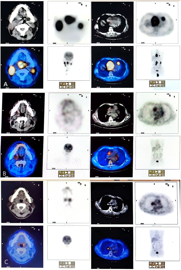
Figure 1: PET-CT. (A) Transaxial images (NM Transaxials) of pre-treatment PET-CT on November 24, 2022, showing hypermetabolic lesions in the heart, tonsils, testes, and multiple groups of lymph nodes, indicating extensive invasion of diffuse large B-cell lymphoma (DLBCL). (B) After two cycles of modified R-CDOP and three cycles of standard R-CDOP, PET-CT on April 20, 2023, showed the complete disappearance of hypermetabolic lesions in the original heart, tonsils, testes, and multiple groups of lymph nodes, with metabolic levels returning to normal, indicating complete remission of the disease. (C) After 7 cycles of R-CVP consolidation, PET-CT on June 30, 2025, confirmed persistent complete remission. Please click here to view a larger version of this figure.
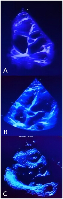
Figure 2: Echocardiography. (A) Pre-treatment on November 24, 2022, echocardiography revealed the right atrial mass and pericardial and myocardial metastases. (B) On April 20, 2023, echocardiography showed a reduction in the right atrial mass. (C) On June 30, 2025, echocardiography revealed a further reduction in the right atrial mass compared to April 2023. Please click here to view a larger version of this figure.
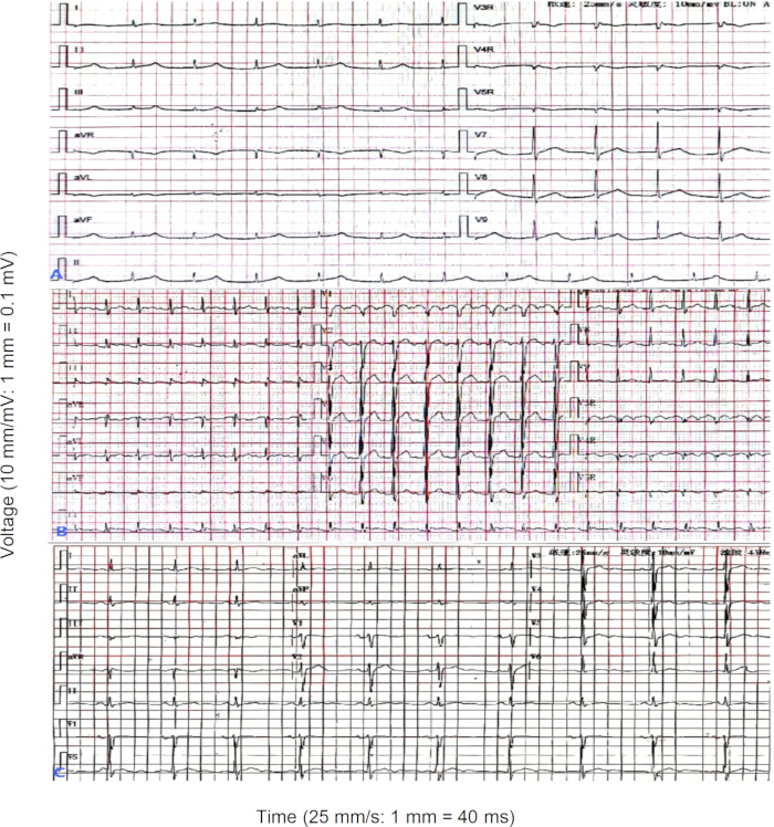
Figure 3: ECG. (A) November 21, 2022, ECG showed a junctional rhythm. (B) On April 20, 2023, ECG showed Sinus tachycardia. (C) On June 30, 2025, ECG showed bradycardia. Please click here to view a larger version of this figure.
| Gene | Variation content | Variation ratio |
| KDM6A | Exon 17 NM_02114:c.2578G>T (p.E860) | 69.7% (1750X) |
| PRDM1 | Exon 17NM_001198:c.2173C>T (p.R725*) | 56.3% (1904X) |
| SETD1B | Exon 2 NM_001353345:c.22dupC(p.H8fs) | 53.3% (1989X) |
Table 1: Lymphoma gene testing identified KDM6A, PRDM1, and SETD1B mutations.
Discussion
Critical protocol steps for success
Successful management of this case relied on three key steps:
Dose-Modified Liposomal Doxorubicin Regimen: Replacing conventional doxorubicin with liposomal doxorubicin (20 mg/m²) reduced cardiac toxicity while maintaining efficacy, critical for a patient with pre-existing cardiac involvement. Dose adjustments (e.g., cyclophosphamide reduced from 750 mg/m² to 375 mg/m² initially) minimized early cardiac stress, and escalation after partial remission balanced efficacy and safety. The patient's presentation with cardiac metastases necessitated a tailored treatment approach to balance the risk of cardiac toxicity with the need for effective lymphoma control5,6.
Close Cardiac Monitoring: Serial echocardiography and ECG allowed timely detection of cardiac changes. For example, atrial flutter was identified early and managed with bisoprolol, preventing progression to more severe arrhythmias. Close monitoring of cardiac function allowed for disease control while minimizing adverse effects7,8.
Multidisciplinary Collaboration: Hematologists, cardiologists, and radiologists collaborated to tailor therapy: cardiologists guided beta-blocker use, radiologists interpreted multimodal imaging (PET-CT/echocardiography) for response assessment, and hematologists adjusted chemotherapy based on both disease and cardiac status. Multidisciplinary diagnosis and treatment can improve the survival rate and quality of life of patients9.
Limitations of the approach
Diagnostic Limitations: Cardiac involvement in DLBCL is rare but increasingly recognized, often presenting as arrhythmias or cardiomegaly. Due to the difficulty and high risk of cardiac biopsy, the diagnosis of cardiac tumors is a clinical challenge10,11,12. Cardiac biopsy was not performed due to high procedural risk, so the cardiac mass was diagnosed by imaging and clinical correlation-this may lead to underdiagnosis of subtle cardiac involvement. Multimodal imaging (PET-CT + echocardiography) partially mitigated this, but tissue confirmation remains the gold standard.
Therapeutic Limitations: This is a single-case report, so the efficacy of the dose-modified regimen cannot be generalized to all DLBCL patients with cardiac involvement.
Data gaps: The lack of serial troponin I and brain natriuretic peptide (BNP) measurements (biomarkers for cardiac injury) limited the assessment of subclinical cardiac toxicity. Future studies should include these biomarkers to refine dose-modification strategies.
Future research directions
Clinical Trials: Future studies should conduct a multicenter, retrospective study comparing liposomal doxorubicin-based regimens (R-CDOP) versus conventional R-CHOP in DLBCL patients with cardiac involvement, focusing on cardiac toxicity and remission duration.
Biomarker Research: Research should also explore the role of KDM6A/PRDM1/SETD1B mutations (identified in this case) in treatment response-preliminary data suggest KDM6A mutations correlate with anthracycline sensitivity, so these mutations may serve as predictive biomarkers for liposomal doxorubicin efficacy.
Guideline Development: In future, development of consensus guidelines for dose modification of liposomal doxorubicin based on LVEF and cardiac biomarkers (troponin I/BNP), to standardize therapy for high-risk patients should be carried out.
Role of liposomal doxorubicin
Liposomal doxorubicin was the cornerstone of successful treatment in this case, with two key rationales.
Targeted Delivery: Its liposomal matrix (composed of cholesterol and sucrose, Table of Materials) enables passive targeting to tumor tissues by enhanced permeability and retention (EPR) effect, reducing drug accumulation in the heart4. This is critical for patients with cardiac involvement, as conventional doxorubicin accumulates in cardiac myocytes, causing oxidative stress and cell death3.
Efficacy-Safety Balance: Despite dose reduction (20 mg/m2 versus 50 mg/m2 conventional doxorubicin), liposomal doxorubicin maintained anti-lymphoma efficacy (achieved CMR) because its liposomal formulation increases intratumoral drug concentration4. This balance is unavailable with conventional doxorubicin, which requires dose reduction (to <30 mg/m2) for cardiac-involved patients, often compromising efficacy.
In summary, liposomal doxorubicin-based, dose-modified chemotherapy combined with close cardiac monitoring and multidisciplinary care is a promising approach for DLBCL patients with cardiac involvement, addressing the unmet need for effective, cardiac-sparing therapy.
Disclosures
The authors declare no conflicts of interest.
Acknowledgements
The authors thank the patient and family for their cooperation.
This work was supported by Shantou Science and Technology Planning Project of Guangdong Province (grant number 221122176494996).
Materials
| Name | Company | Catalog Number | Comments |
|---|---|---|---|
| Bisoprolol fumarate | Merk KGaA | B202202A02 | Bisoprolol fumarate |
| Cyclophosphamide | Tonghua Maoxiang Pharmaceutical Co., Ltd | MH20220203 | Cyclophosphamide |
| Doxorubicin Hydrochloride Liposomes | Shiyao Group Ouyi Pharmaceutical Co., Ltd. | SHPL202202A | Doxorubicin Hydrochloride Liposomal matrix, cholesterol, sucrose, histidine |
| Prednisone | Henan Lihua Pharmaceutical Co., Ltd. | LH202201A21 | Prednisone Acetate |
| Rituximab | Shanghai Fuhong Hanlin Biopharmaceutical Co., Ltd. | HLX202201A04 | Rituximab |
| Vincristine | Northeast Pharmaceutical Group Co., Ltd | DB202202A03 | Vincristine Sulfate |
References
- Swerdlow, S. H., et al. WHO Classification of Tumours of Haematopoietic and Lymphoid Tissues. , 4th ed, International Agency for Research on Cancer (IARC). Lyon, France. (2017).
- Ido, T., et al. Cardiac involvement of diffuse large B-cell lymphoma presenting as various arrythmias. Clin Case Rep. 10 (11), e6504(2022).
- Zhao, Y., et al. Clinical features of cardiac lymphoma: an analysis of 37 cases. J Int Med Res. 49 (3), 300060521999558(2021).
- Visco, C., et al. Efficacy of R-COMP in comparison to R-CHOP in patients with DLBCL: A systematic review and single-arm metanalysis. Crit Rev Oncol Hematol. 163, 103377(2021).
- Tekinalp, A., et al. Treatment with doxorubicin-based protocol in cardiac involvement diffuse large B-cell lymphoma: A case report. J Oncol Pharm Pract. 28 (2), 449-452 (2022).
- Pennacchioni, A., et al. Diffuse large B-cell lymphoma with cardiac involvement: the importance of a multidisciplinary and multiparametric approach. G Ital Cardiol. 25 (7), 526-529 (2024).
- Shaw, B., et al. Acute Heart Failure and Complete Heart Block in a Patient with Recurrent Diffuse Large B-Cell Lymphoma: A Case Report. Am J Case Rep. 25, e945085(2024).
- Rezvani, A., Shah, S. Treatment of primary cardiac diffuse large B-cell lymphoma involving the coronary sinus with R-EPOCH: a case report and literature review. Ann Hematol. 103 (7), 2557-2560 (2024).
- Xia, J., et al. Surgical treatment of a primary cardiac lymphoma presenting with cardiac tamponade. Gen Thorac Cardiovasc Surg. 69 (2), 356-359 (2021).
- Osmani, A. H., et al. A Case of Diffuse Large B-Cell Lymphoma with Cardiac Involvement Incidently Diagnosed on Cardiac Imaging. J Coll Physicians Surg Pak. 31 (9), 1102-1104 (2021).
- Yang, Y., et al. The value of 18F-FDG PET/CT in evaluating the efficacy of chemotherapy for diffuse large B-cell lymphoma with cardiac involvement. J Nucl Cardiol. 29 (6), 3548-3553 (2022).
- Drewniowska, J., et al. The role of multimodal imaging in diffuse large B-cell lymphoma with primary cardiac involvement. Pol Arch Intern Med. 134 (9), 16796(2024).