Drug-resistant cell lines enable understanding of treatment resistance mechanisms and nonclinical evaluation of new compounds, repurposed drugs, and combination therapies. This paper introduces a protocol for creating resistant cell lines by exposing parent cell lines to stepwise increases in the concentration of the target drug.
Method Article
Development of Drug-resistant Cell Lines for Experimental Procedures
Opens in a new tab
In This Article
Summary
Loading...
$$\rightleftharpoonup{xx}$$
$$\longleftharp{xx}$$,
$$\longrightharp{xx}$$,
Abstract
Loading...
$$\rightleftharpoonup{xx}$$
$$\longleftharp{xx}$$,
$$\longrightharp{xx}$$,
The development of drug-resistant cell lines is essential for understanding the mechanisms of drug resistance and identifying strategies to overcome treatment failure in cancer therapy. Resistance models enable preclinical evaluation of novel compounds, repurposed drugs, and combination therapies. To generate resistant cells, parental cancer cell lines are repeatedly exposed to incrementally increasing concentrations of the target drug over several weeks. Cells that survive and proliferate at each stage are selected, expanded, and exposed to higher drug doses. The development of resistance is confirmed by quantifying and comparing the half-maximal inhibitory concentration (IC50) values between parental and resistant cells using cell viability assays and nonlinear regression analysis. Significantly increased IC50 values indicate successful adaptation to drug pressure and the development of resistance. These drug-resistant cell lines are available for comprehensive analysis, such as microarray and single-cell sequencing, as well as various in vitro or in vivo experiments. These models provide valuable tools for investigating potential therapeutic strategies to overcome drug resistance.
Introduction
Loading...
$$\rightleftharpoonup{xx}$$
$$\longleftharp{xx}$$,
$$\longrightharp{xx}$$,
Chemotherapy is the most frequently used treatment option for local cancers not amenable to surgery or radiation therapy or metastatic cancers. Molecularly targeted drugs and chemotherapy are used as first-line treatments for many malignancies, and their efficacy is widely recognized. Some cancers show resistance to certain drugs from the start of treatment, making treatment ineffective. However, in more common cases, treatment that was initially effective becomes ineffective due to the development of drug resistance, leading to the progression of cancer.
Drug resistance is caused by both genetic mutations or epigenetic response in cancer cells that induce the expression of gene products that evade drugs or aid cell growth1,2. Furthermore, in many cases, this resistance evolves into multidrug resistance, where the cancer cell becomes resistant to multiple drugs3,4. Therefore, identifying the mechanisms of drug resistance and developing strategies to overcome it are extremely important for the advancement of cancer treatment.
Drug-resistant cell lines are frequently used models for studying the mechanisms of therapeutic resistance and play an important role in both in vitro and in vivo experiments5. These models are essential tools for gaining a deeper understanding of drug action pathways, identifying biomarkers that influence therapeutic efficacy, and finding therapeutic strategies to inhibit drug resistance formation. The basic process of creating a drug-resistant cell line is to induce resistance by exposing a cell line to increasing levels of the target drug3,6.
The concentration used and duration of the drug exposure are critical components of this process. At each concentration level, the subset of cells that survive is amplified and then exposed to the next concentration of drug in a repetitive fashion. Ultimately, a drug-resistant cell line is derived that has an increased half-maximal inhibitory concentration (IC50) for the drug compared to the parental cell line. This report details the procedure for generating a paclitaxel-resistant cell line (DU145-TxR) from the human prostate cancer cell line DU145, employing a stepwise method with intermittent drug exposures.
Protocol
Loading...
$$\rightleftharpoonup{xx}$$
$$\longleftharp{xx}$$,
$$\longrightharp{xx}$$,
1. Cell culture and cell viability assays
NOTE: Paclitaxel is an anticancer drug and is cytotoxic. When handling it, wear a gown, goggles, and gloves, and work inside a safety cabinet to prevent inhalation into the body. DMSO is not highly cytotoxic, but when it comes into contact with skin or mucous membranes, the permeability of dissolved drugs increases, so wear a gown, goggles, and gloves when handling it.
- Culture DU-145 cells in a 10 cm cell culture dish using complete medium (RPMI-1640 + 1% penicillin-streptomycin [pen-strep] + 10% fetal bovine serum [FBS]) in a 37 °C incubator at 5% CO2 until 80% confluent.
- Aspirate the culture medium, wash with DPBS, add 0.05% trypsin, and leave in the incubator for 5 min.
- After tapping and confirming by microscopy that the cells are detached from the plate and floating, add 1 mL of complete medium to stop the action of trypsin.
- Count the number of cells and seed them at a density of 1.0 × 10⁴/99 µL per well in complete medium in each well of a 96-well plate. Incubate for 2 h to allow the cells to adhere to the bottom of the plate. Cells should be plated in triplicate or quadruplicate for each test group.
- Adjust the paclitaxel and DMSO to be added to each well. Ensure that the amount of DMSO added to each well is the same, and that the final concentration of DMSO is no more than 1%.
- To perform this and following our protocol, start with 10 mM paclitaxel in DMSO and add 87.2 µL of DMSO to 12.8 µL of this drug in a tube to obtain 100 µL of paclitaxel at 1,280 µM.
- Transfer 50 µL of this solution to another tube and dilute it to half strength with 50 µL of DMSO (640 µM).
- Repeat this procedure to perform serial dilution and prepare solutions of paclitaxel at concentrations of 0 (DMSO only), 0.1, 0.5, 1, 2, 4, 10, 20, 40, 80, 160, 320, 620, and 1,280 µM.
NOTE: Paclitaxel diluted with DMSO should be stored frozen for reuse in subsequent experiments (to minimize freeze-thaw cycles, store in aliquots).
- Dilute 1 µL of the paclitaxel solution prepared earlier with 9 µL of complete medium in a separate tube, and add 1 µL of this to each well 2 h after seeding the cells. The paclitaxel concentrations in each well will be 0, 0.1, 0.5, 1, 2, 4, 10, 20, 40, 80, 160, 320, 620, and 1,280 nM, with each well containing 100 µL of medium.
NOTE: When setting the concentration of drugs used in cell viability assays, check the IC50 values published in previous papers and ensure that the concentration falls within the range3. The DMSO concentration in each well was 0.1% throughout (less than 1% is desirable). - Incubate for 48 h. Add 10 µL of Cell Proliferation Reagent WST-1 to each well (100 µL of culture medium) and incubate for an additional 0.5 to 4.0 h.
NOTE: Optimal incubation time depends on the cell line and cell density. In this study, we cultured cells for approximately 1.5 h. If the incubation time is too short or too long, the absorbance of each well will be too low or too high in the next step. As a result, it will not be possible to confirm the difference in cell viability. Thus, for the initial testing, one should test several incubation times to confirm the appropriate incubation time. - Measure WST-1 absorbance using a microplate reader.
NOTE: The target absorbance is 450 nm (420-480 nm is recommended), and the background absorbance is 650 nm (600 nm or higher is recommended).
2. Calculation of cell viability and half-maximal inhibitory concentration (IC50) value
- Calculate cell viability from absorbance. Absorbance is (A450nm-A650nm).
Cell viability (%) = [(As-Ab)/(Ac-Ab)] × 100
As: sample absorbance (drug-treated cells)
Ab: blank absorbance (medium only, no cells)
Ac: control absorbance (drug-untreated cells) - Summarize the cell viability calculated in the previous section in a spreadsheet software.
- Calculate and record the IC50 for the parental cells by nonlinear regression analysis.
NOTE: The four-parameter logistic model (4PL) is often a suitable calculation method. Online IC50 calculation tools are available7. However, the 4PL model should not be used when the efficacy of the compound is outside the concentration range, and the data cannot fully represent the lower and upper asymptotes of the sigmoid curve.- To avoid such an increase, measure cell viability at as many concentration points as possible. If a sigmoid curve cannot be obtained, use a two- or three-parameter mode with fixed minimum and maximum values7.
- If not using calculation software or statistical software, including those available on the web, use the following formula:
IC50=((50-C)(B-A) + A(D-C))/(D-C)
A: The lowest value among the drug concentrations measured with a cell viability of ≤50%
B: The highest value among the drug concentrations measured with a cell viability of >50%
C: Cell viability at B
D: Cell viability at A
NOTE: This formula assumes that the graph between the two points sandwiching the IC50 value is a straight line.
3. Drug exposure
NOTE: An overview of the procedures described in this section is shown in Figure 1.
- Seed parental cells on cell culture plates (2.0 × 10⁶ cells/dish for 100 mm dishes).
- Add paclitaxel at a cell viability inhibitory concentration of ~10-20% (IC10-20, ~0.5 nM in this case) to the medium and culture in an incubator for 2 days.
- Replace with paclitaxel-free medium and incubate for several days.
- Once cells have grown and become 80% confluent, passage them to a new cell culture plate. Cryopreserve the remaining cells.
- Add paclitaxel to the passed cells at a concentration of ~1.5-2.0-fold the starting concentration (0.75-1 nM). Incubate in an incubator for 2 days.
- Replace with paclitaxel-free medium and incubate for several days.
- If the cells grow similarly, repeat steps 3.4-3.6, increasing the concentration of paclitaxel exposure step by step.
NOTE: The increase in drug concentration (1.5-2.0-fold) can be freely set by the experimenter, and the experiment should be repeated at that concentration. We recommend freezing and storing the cells each time the drug concentration is increased. If the paclitaxel concentration continues to increase further, cell proliferation may not recover, and the cells may die. In such cases, thaw the cells that proliferated at the concentration before death, reduce the paclitaxel increase to 1.1-1.5-fold (1.2-fold is recommended), and repeat the culture. - Gradually increase the resistance of the cell line to the drug and then, calculate and record the IC50 using WST-1 and software as described in Section 2.
NOTE: If an increase in IC50 of at least 3-5-fold is observed, it can be determined that the strain is a drug-resistant cell line.
4. Maintenance of drug resistance
- To maintain the resistant phenotype of the drug-resistant cell line, incubate with a drug concentration of IC10-20.
NOTE: It is recommended that the IC50 in a cell survival assay be measured periodically, as drug resistance may change.
Results
Loading...
$$\rightleftharpoonup{xx}$$
$$\longleftharp{xx}$$,
$$\longrightharp{xx}$$,
Using this method, we previously spent 9 months successfully establishing paclitaxel-resistant DU145 cells (DU145-TxR). The results of the cell viability assay showed that the IC50 value of paclitaxel for the DU145 parent cell line was 1.1 nM, whereas the IC50 value for DU145-TxR was 149.6x higher than that of the parent cell line (IC50: 164.6 nM) (Table 1 and Figure 2). This result indicates that DU145-TxR has acquired very high drug resistance.
Cell morphology was observed using digital inverted microscopy immediately prior to the cell viability assay at 48 h after cell seeding and paclitaxel addition. As a result, as the paclitaxel concentration increased from 1 nM to 10 nM, the parental cell line DU145 showed clear cell atrophy and morphological changes suggestive of cell death. However, no obvious morphological changes were observed in the DU145-TxR cell line (Figure 3).

Figure 1: Overview of drug-resistant cell line creation. On day 1, cells are seeded into plates containing growth medium and incubated with paclitaxel for 48 h at which time the medium is replaced with medium that does not contain paclitaxel. If the cells regrow after several days of culture, they are harvested, some of the cells are replated and maintained with the original paclitaxel concentration and some of the cells are re-plated with paclitaxel at a concentration of 1.5-2.0-fold the previous concentration. If the cells grown in the increased paclitaxel concentration do not survive the backup cells from the previous concentration should be harvested and some of them grown at a concentration of 1.1-1.5-fold the previous paclitaxel concentration. Please click here to view a larger version of this figure.
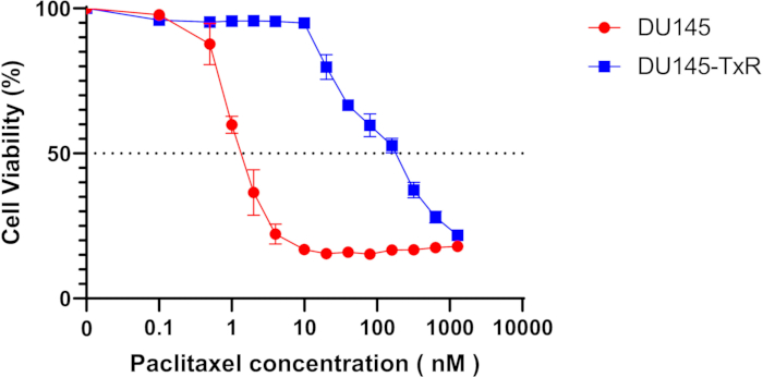
Figure 2: Establishment of paclitaxel-resistant DU145 (DU145-TxR). DU145 and DU145-TxR cells were exposed to paclitaxel at concentrations of 0, 0.1, 0.5, 1, 2, 4, 10, 20, 40, 80, 160, 320, 620, 1,280 nM for 48 h. Cell viability assay (WST-1) was performed. The IC50 of DU145 was 1.1 nM, and the IC50 of DU145-TxR was 164.6 nM. Data are shown as mean ± SEM. Please click here to view a larger version of this figure.
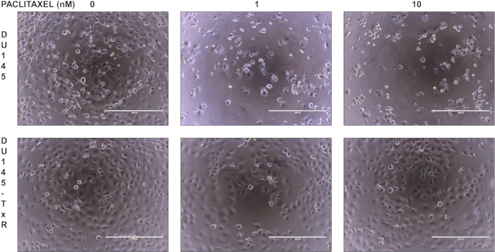
Figure 3: Cell morphology of DU145 and DU145-TxR using digital inverted microscopy. DU145 or DU145-TxR ells were seeded in 96-well plates at 1.0 × 10⁴ cells/well and grown in the indicated concentration of paclitaxel. Images were taken 48 h later at magnification of 100x (scale bar = 400 µm). Please click here to view a larger version of this figure.
| Paclitaxel concentration (nM) | Cell viability (%) | |
| DU145 | DU145-TxR | |
| 0 | 100 | 100 |
| 0.1 | 97.8 | 96 |
| 0.5 | 87.7 | 95.3 |
| 1 | 59.8 | 95.6 |
| 2 | 36.5 | 95.7 |
| 4 | 22.2 | 95.5 |
| 10 | 16.8 | 95 |
| 20 | 15.5 | 79.8 |
| 40 | 15.9 | 66.6 |
| 80 | 15.3 | 59.7 |
| 160 | 16.7 | 52.7 |
| 320 | 16.7 | 37.4 |
| 640 | 17.5 | 28 |
| 1280 | 18 | 21.8 |
| IC50 (nM) | 1.1 | 164.6 |
Table 1: Paclitaxel concentration, cell viability, and IC50 calculation. The IC50 for DU145 was calculated using an online calculation tool (1.1 nM) using cell viability values ranging from 0 to 80 nM as listed in the table. Since the cell viability curve for DU145-TxR is not a complete sigmoid curve, the IC50 for DU145-TxR was calculated using an online calculation tool (164.6 nM) with the minimum value fixed at 0. (See NOTE in Protocol Procedure 2.3.)
Discussion
Loading...
$$\rightleftharpoonup{xx}$$
$$\longleftharp{xx}$$,
$$\longrightharp{xx}$$,
The development of drug-resistant cancer cell lines is an established method for investigating drug toxicity and resistance mechanisms, but the first challenge researchers face is selecting an appropriate model for expressing resistance. A common method for creating drug-resistant cell lines in vitro is to expose the cell line to the target drug, but it is necessary to consider how to set the exposure concentration (stepwise or all at once) and whether to set a drug-free period (pulsed or continuous).
The pulse method, with its drug-free period, is similar to a cancer patient undergoing several cycles of chemotherapy, as there is a short period of drug exposure followed by a recovery period. Therefore, this method is close to the clinical model and is widely used. Conversely, the method of continuous drug exposure mimics a model in which drug levels in the blood are stable, such as some chemotherapies and molecular targeted therapies that are administered daily.
The maximum drug concentration to which a cell line is exposed is determined by the blood concentration of the drug at the time the patient is treated with the drug. That concentration of drug is considered to be the exposure of the cancer cells in the patient's body. Comparing the stepwise method with the one-step method, the one-step method is easier. However, the stepwise method has the advantage of establishing a series of highly drug-resistant cell lines by gradually increasing the drug concentration.
A 3- to 10-fold increase of IC50 compared to the parent cell would represent drug resistance based on what is observed in cell lines established from cancer patients before and after chemotherapy8,9,10. This is very dependent on the drug, the cancer type, and the timeline of drug exposure. It is possible, in some cases, that the IC50 could be much higher. A small increase in IC50 (3-10-fold) may make it difficult to study the mechanism of resistance because of its low stability in vitro and small changes in gene expression involved in resistance. Conversely, high-level resistant cell lines are highly stable, making the cells easy to manage. In addition, the genes and molecular changes involved in the resistance mechanism are large and can be easily detected by comparison with the parent cells. However, these may not reflect the true clinical scenario accurately, as these drug levels would not be typically achieved in patients. Accordingly, a balance between IC50 levels attainable in vivo and modulating the IC50 to highlight possible mechanisms needs to be considered.
The most important step in the protocol for creating drug-resistant cell lines that we have introduced here is drug exposure. To establish a resistant cell line with an IC50 more than 10 times that of the parent cell, it is necessary to expose the cells to high concentrations of the drug. However, as the drug concentration increases, the likelihood of cell death also increases. Therefore, cell lines grown at new drug concentrations should be frozen separately from those that are to be passed on. This allows us to restart from the previous stage even if the cells die. Although there is no consistent opinion on the amount of drug concentration increase at each stage in the creation of resistant cell lines, we have successfully created multiple drug-resistant cell lines using the protocol introduced here. We recommend starting with an increase of 1.5-2.0-fold, and if cell death occurs, changing to a lower increase (1.1-1.5-fold).
The complete reproducibility of the drug-resistant cell lines created using this procedure has not been sufficiently evaluated. This is because the method we have presented requires a long period of time, ranging from 6 months to 1 year, making it difficult to repeat the process of creating resistant cell lines from different initial parent cells multiple times. Even if drug-resistant cell lines are created following the same protocol from the beginning, it is unclear whether it is possible to create exactly the same resistant cell lines. To the best of our knowledge, there are no reports that sufficiently prove this issue. It is also possible to create paclitaxel-resistant cell lines in other cancers using this method. We have also succeeded in creating other drug-resistant prostate cancer cell lines. This method is thought to be widely applicable to many drugs and cell lines.
The mechanism of the drug-resistant cell lines created is evaluated by a comprehensive search. In the past, we performed a cDNA microarray using mRNA and reported that the expression of multidrug resistance protein 1 (MDR1), a gene involved in drug efflux, was increased in paclitaxel-resistant cell lines3. Additionally, through single-cell RNA sequencing, we identified nuclear protein 1 as a mediator of drug resistance11.
However, the mechanisms of drug-resistant cell lines created by drug exposure are not necessarily identical. Using a similar procedure, Grigoreva et al. created the paclitaxel-resistant human colorectal cancer cell line HCT116tax12. Similar to our DU145-TxR, their HCT116tax showed increased expression of P-glycoprotein and MDR13,12. Interestingly, unlike our DU145-TxR, their HCT116tax exhibited enhanced cell proliferation in the presence of low concentrations of paclitaxel12. This suggests that while some major mechanisms of resistance acquisition may be common, others may vary depending on the tumor or method.
In summary, we described detailed protocols for the development and maintenance of drug-resistant cancer cell lines and the characteristics of the cell lines. Although there are some issues with the resistant cell lines created using our protocol, we believe that they play an important role in elucidating the mechanisms involved.
Disclosures
Loading...
$$\rightleftharpoonup{xx}$$
$$\longleftharp{xx}$$,
$$\longrightharp{xx}$$,
The authors declare that they have no potential conflicts of interest.
Acknowledgements
Loading...
$$\rightleftharpoonup{xx}$$
$$\longleftharp{xx}$$,
$$\longrightharp{xx}$$,
This work was supported in part by the National Institutes of Health grant P01 CA093900.
Materials
| Name | Company | Catalog Number | Comments |
|---|---|---|---|
| 0.05% Trypsin-EDTA | Gibco | 25300-054 | |
| 10 mL Disposable serological pipette | Fisher scientific | 13-678-11E | |
| 100 mm dish | Corning | 353003 | |
| 15 mL conical tube | Corning | 352097 | |
| 2.0 mL Cryogenic Vials | Corning | 431386 | |
| 96 well cell culture plate | Corning | 353072 | |
| Cell counting chamber slide | Invitrogen | C10228 | |
| Cell Proliferation Reagent WST-1 | Sigma | 5015944001 | |
| Centrifuge 5702 R | eppendorf | ||
| countless II | Invitrogen | Cell counter | |
| Dimethyl sulfoxide | Sigma | D2650 | |
| DPBS | Gibco | D4W2J | |
| DU145 | ATCC | HTB-81 | Prostate cancer cell line |
| EVOS FLc | Thermo Fisher Scientific | Digital Inverted Microscope | |
| FBS | Gibco | A5670701 | |
| Paclitaxel | Selleck | S1150 | |
| Penicillin–streptomycin | Invitrogen | 15140-122 | |
| RPMI 1640 | Gibco | 11875-093 | |
| Synergy H1 | BioTek | Microplate Reader | |
| Trypan Blue solution 0.4% | Invitrogen | 15250061 |
References
Loading...
$$\rightleftharpoonup{xx}$$
$$\longleftharp{xx}$$,
$$\longrightharp{xx}$$,
- Mechanisms and insights into drug resistance in cancer. Fron Pharmacol. 4, 28(2013).">Zahreddine, H., Borden, K. L. Mechanisms and insights into drug resistance in cancer. Fron Pharmacol. 4, 28(2013).
- Multidrug resistance in cancer: understanding molecular mechanisms, immunoprevention and therapeutic approaches. Fron Oncol. 12, 891652(2022).">Emran, T. B., et al. Multidrug resistance in cancer: understanding molecular mechanisms, immunoprevention and therapeutic approaches. Fron Oncol. 12, 891652(2022).
- The establishment of two paclitaxel-resistant prostate cancer cell lines and the mechanisms of paclitaxel resistance with two cell lines. Prostate. 67 (9), 955-967 (2007).">Takeda, M. The establishment of two paclitaxel-resistant prostate cancer cell lines and the mechanisms of paclitaxel resistance with two cell lines. Prostate. 67 (9), 955-967 (2007).
- TOP1 mutations and cross-resistance to antibody-drug conjugates in patients with metastatic breast cancer. Cli Cancer Res. 31 (10), 1966-1974 (2025).">Abelman, R. O., et al. TOP1 mutations and cross-resistance to antibody-drug conjugates in patients with metastatic breast cancer. Cli Cancer Res. 31 (10), 1966-1974 (2025).
- In vitro development of chemotherapy and targeted therapy drug-resistant cancer cell lines: a practical guide with case studies. Fron Oncol. 4, 40(2014).">McDermott, M. In vitro development of chemotherapy and targeted therapy drug-resistant cancer cell lines: a practical guide with case studies. Fron Oncol. 4, 40(2014).
- Development of taxane resistance in a panel of human lung cancer cell lines. Toxico In Vitro. 22 (5), 1234-1241 (2008).">Breen, L., Murphy, L., Keenan, J., Clynes, M. Development of taxane resistance in a panel of human lung cancer cell lines. Toxico In Vitro. 22 (5), 1234-1241 (2008).
- IC50 calculator. , AA Bioquest. https://www.aatbio.com/tools/ic50-calculator (2025).">Bioquest, A. A. IC50 calculator. , AA Bioquest. https://www.aatbio.com/tools/ic50-calculator (2025).
- Characterization of non small-cell lung cancer cell lines established before and after chemotherapy. Lun Cancer. 35 (3), 305-314 (2002).">Kawai, H. Characterization of non small-cell lung cancer cell lines established before and after chemotherapy. Lun Cancer. 35 (3), 305-314 (2002).
- Characterization of three small cell lung cancer cell lines established from one patient during longitudinal follow-up. Cance Res. 48 (23), 6891-6899 (1988).">Berendsen, H. H., et al. Characterization of three small cell lung cancer cell lines established from one patient during longitudinal follow-up. Cance Res. 48 (23), 6891-6899 (1988).
- Functional restoration of BRCA2 protein by secondary BRCA2 mutations in BRCA2-mutated ovarian carcinoma. Cance Res. 69 (16), 6381-6386 (2009).">Sakai, W. Functional restoration of BRCA2 protein by secondary BRCA2 mutations in BRCA2-mutated ovarian carcinoma. Cance Res. 69 (16), 6381-6386 (2009).
- Single-cell transcriptomics analysis identifies nuclear protein 1 as a regulator of docetaxel resistance in prostate cancer cells. Mo Cancer Res. 18 (9), 1290-1301 (2020).">Schnepp, P. M., et al. Single-cell transcriptomics analysis identifies nuclear protein 1 as a regulator of docetaxel resistance in prostate cancer cells. Mo Cancer Res. 18 (9), 1290-1301 (2020).
- New insights into chemoresistance mediated by Mdm2 inhibitors: the benefits of targeted therapy over common cytostatics. Biomedicines. 12 (3), 547(2024).">Grigoreva, T., Sagaidak, A., Novikova, D., Tribulovich, V. New insights into chemoresistance mediated by Mdm2 inhibitors: the benefits of targeted therapy over common cytostatics. Biomedicines. 12 (3), 547(2024).
Reprints and Permissions
Request permission to reuse the text or figures of this JoVE article
Request Permission