This protocol describes a method for combining telomere FISH (native or denaturing) with immunofluorescence and/or visualization of a fluorescent protein.
Method Article
A Flexible Procedure for Performing Telomere FISH and Immunofluorescence in Cells Expressing Fluorescent Proteins
In This Article
Summary
Abstract
Visualizing telomeres using fluorescence in situ hybridization (TelFISH) has long been an essential tool in telomere biology experiments. Combining TelFISH with immunofluorescence (IF) is also a well-established method (IF-TelFISH), for example, in identifying telomere dysfunction-induced foci, where telomeres co-localize with 53BP1. More recently, native telomere FISH (nTelFISH) has become an important tool for assaying Alternative Lengthening of Telomeres, a telomere maintenance mechanism used in some tumor cells. Expressing fluorescent proteins is also an essential tool in cell biology, allowing visualization of proteins and structures that may remain undetectable by other methods. Because FISH buffers denature proteins, performing the assay in cells with an expressed fluorescent protein (FP) results in loss of FP signal. A method for integrating IF and telomere FISH in cells with an expressed fluorescent protein that preserves the FP signal even after FISH is described here. Alternatives are provided for denaturing and native telomere FISH in combination with several IF and FP scenarios. Taken together, this protocol offers a flexible framework for exploring telomere biology in both normal and tumor cells.
Introduction
Organisms with linear chromosomes must contend with two problems. First, due to the end-replication problem, the ends of chromosomes erode slightly each time DNA is replicated. Second, cells must be able to distinguish natural DNA ends from DNA breaks. Telomeres evolved as a solution to these problems. Through the action of the ribonucleoprotein telomerase, chromosomes are lengthened by the addition of short tandem repeats-TTAGGG in humans. These repeats are bound by the shelterin complex, which conceals the chromosome end from cellular DNA damage repair machinery1. Short or dysfunctional telomeres lead to cellular senescence and aging phenotypes2, while escape from telomere crisis is a major milestone in tumorigenesis3. For these reasons, telomere biology is of special interest in understanding human health and disease.
Telomere fluorescent in situ hybridization (FISH) has long been an important method for visualizing telomeres in interphase cells or metaphase spreads4. For decades, telomere FISH has been coupled with 53BP1 immunofluorescence staining (IF) to identify telomere dysfunction-induced foci (TIFs), a marker of dysfunctional, uncapped telomeres5,6. Similarly, a hallmark of tumors that use Alternative Lengthening of Telomeres (ALT) for telomere maintenance is the association of telomeres with nuclear PML bodies to form ALT-associated PML bodies or APBs7. APBs can be assayed by pairing telomere FISH with PML IF staining. Thus, IF-FISH is a well-established tool in telomere biology.
Here, the use of IF-FISH has been extended in two ways. First, IF-FISH is performed on cells with expressed fluorescent proteins (FP). The expression of fluorescently tagged proteins has become an ubiquitous tool in cell biology. Expressing a tagged protein may be the only option for visualizing a protein for which good antibodies do not exist. For example, the expressed GFP-tagged nuclear actin chromobody (nAC-GFP) reveals nuclear actin dynamics8. This reagent has opened new opportunities in the exploration of nuclear f-actin, which was once considered a controversial topic. While FPs like GFP and mCherry typically maintain fluorescence activity after fixation, the formamide in FISH hybridization buffers will render them dark9. To circumvent this issue, this protocol offers the innovation of a fluorophore-tagged anti-FP "booster" in the secondary step of IF staining. The brightness of the fluorophore is unaffected by formamide, allowing IF-FP-FISH.
Second, IF is performed in conjunction with Native Telomere FISH (nTelFISH), an important diagnostic for ALT status. In denaturing FISH, one must use some method to melt the DNA strands10 (e.g., heat or alkali), allowing probe access to DNA that is normally double-stranded. In contrast, native FISH does not include a denaturing step, so the probe can only access DNA that is already single-stranded. Native telomere FISH specifically detects single-stranded C-strand telomere repeats, another hallmark of ALT in tumor cells11,12. Coupling this assay with IF-FP staining allows us to address new questions in understanding the mechanism of ALT, which is now considered a strong candidate for targeted tumor therapies.
The protocol described here can be used in a "choose your own adventure" style to stain any combination of IF, FP, and either native or denaturing telomere FISH. Because the protocol is highly configurable, the timing can vary substantially. Generally speaking, hands-on time is required on four days, but these days might not be sequential. Estimates of time required to complete a typical denaturing FISH workflow are given in Figure 1. Native FISH need not be hybridized overnight, so one can potentially eliminate an overnight step (combining Day 2 and Day 3) if a long day at the bench is feasible.
A typical confocal microscope will accommodate imaging four color channels: roughly speaking, blue, green, red, and far-red. Four examples of potential four-color combinations are demonstrated, but the potential permutations are endless. It is possible to use two IF antibodies or two expressed FPs with equal success. The protocol is straightforward and robust. Any antibody that produces good IF results on its own is suitable. While only telomere probes for FISH are included here, denaturing FISH with a centromere probe also produces good results. Thus, this protocol offers a wide degree of flexibility for answering important questions in nuclear biology.
Protocol
Before beginning, consider the microscope, the FP-expressing cell line, and the available probes and antibodies. Blue is typically used for a DNA stain such as DAPI, as blue fluorophores are generally not bright. Blue secondary antibodies can be used if necessary. If the cells express GFP, a FISH probe tagged with a red fluorophore may be used along with a secondary antibody for IF tagged with AF-647. Alternatively, for imaging cells expressing mCherry, an IF secondary antibody tagged with AF-488 and a FISH probe labeled with AF-647 may be selected. Examples of four possible IF-FP-FISH combinations are shown in Figure 2, and are intended as a guide to encourage experimental design flexibility. It is important to choose either native or denaturing FISH, as these methods cannot be combined. A reliable method for acid-washing coverslips to allow for cell adherence is included here. This can be completed at any time prior to use, and sterile coverslips can be stored indefinitely. The reagents and the equipment used are listed in the Table of Materials.
1. Acid-washing of coverslips
NOTE: Glass coverslips must be cleaned and acid-washed to permit cultured cells to adhere. For growth in 12-well plates, 18 mm round coverslips are ideal. Consult with the microscopy core facility to determine whether #1 or #1.5 coverslips are preferred. The stated times for cleaning coverslips are not critical, but do not stint on the acid wash.
- Use coverslip forceps (Figure 3A) to place coverslips in the racks so that their surfaces do not touch. Place additional coverslips diagonally to increase capacity if needed (Figure 3B). Fit several racks comfortably into the staining dish (Figure 3C).
- Fill the staining dish with water and a small amount of detergent. Gently lower the racks with the coverslips into the dish.
- Agitate the coverslips in the soapy water for 30 min using a rocker or nutator.
- Lift the racks of coverslips out of the staining dish. Dispose of the soapy water.
- Rinse the staining dish and fill with deionized water. Return the racks of coverslips to the clean water and place on a rocker to agitate gently for 5 min.
- Repeat the rinse with two additional changes of clean deionized water, for a total of three rinses.
- Lift the racks of coverslips out of the staining dish. Add 1 N HCl to the dish.
CAUTION: HCl is caustic. Handle in a fume hood and use personal protective equipment. Carefully lower the racked coverslips into the acid solution. Ensure that the 1 N HCl fully covers the coverslips. Place the lid on the staining dish. - Place the staining dish containing the coverslips and acid solution into the tank of a bath sonicator. Sonicate for 15 min at 55 °C.
- Leave the coverslips in the acid bath overnight.
- Carefully remove the coverslip racks from the staining dish the next day. Dispose of the 1 N HCl according to institutional safety protocols.
- Rinse the staining dish and fill it with deionized water. Return the racks of coverslips to the clean water and place on a rocker to agitate gently for 5 min.
- Repeat the rinse with two more changes of clean deionized water, for a total of three rinses.
- Lift the racks of coverslips out of the staining dish. Empty the dish and fill it with 70% ethanol. Return the racks of coverslips to the dish and agitate gently for 5 min.
- Lift the racks of coverslips out of the staining dish. Empty the dish and fill it with 100% ethanol. Return the racks of coverslips to the dish and agitate gently for 5 min.
- Take the dish with 100% ethanol and the coverslips to a tissue culture hood. In the hood, remove the racks of coverslips from the dish and allow them to air-dry.
- Empty the dish and return the racks of coverslips to it. Store with the lid on to maintain sterility. The staining dish is not airtight; if the coverslips are left in ethanol, it will eventually evaporate. The coverslips will remain usable.
2. Plating cells on coverslips
NOTE: Handling coverslips requires practice. It is easier to start with a 6-well plate until confident. When using a 6-well plate, double the wash volumes given for 12-well plates.
- Culture healthy, growing cells expressing the protein of interest tagged with GFP or mCherry (several red or green FPs are suitable; see notes in Table of Materials). U2OS osteosarcoma cells, which use ALT for telomere maintenance, are used in the examples shown.
- Use transiently transfected cells or cell lines with stable FP expression. If the experiment requires drug treatment or transfection, consider the desired confluency at fixation (typically 70%-80%), the treatment duration, and the cell growth rate to calculate the appropriate number of cells to plate per coverslip.
- Prepare coverslips in the tissue culture hood to maintain sterility. To each well of a 12-well plate, add 1 mL of sterile PBS. Using coverslip forceps, place one clean, acid-washed coverslip in each well. Agitate gently by hand for a few seconds.
- Aspirate PBS. Add 1 mL of fresh sterile PBS per well. Set the plate aside.
- (Optional) Coat coverslips with fibronectin to improve cell adhesion. Many adherent cell lines will adhere well to acid-washed coverslips when plated for at least 18 h before fixation. If cells do not adhere well or require fixation soon after plating, coat coverslips to promote adhesion.
- Prepare a 5 µg/mL solution of fibronectin in sterile PBS.
- Aspirate PBS from the coverslips. Gently apply approximately 300 µL of fibronectin solution to each coverslip, forming a droplet held by surface tension.
- Incubate for several hours at 37 °C or overnight at 4 °C.
- Rinse several times with PBS, holding the coverslips in a final change of PBS. Set the plate aside while preparing cells.
- If fibronectin is not suitable, consider poly-L-lysine, collagen, or gelatin as alternatives.
- Trypsinize cells and dilute an appropriate number in 12 mL of culture medium based on experimental design and desired cell density at fixation.
- Aspirate PBS from the coverslips and add 1 mL of the cell suspension per well.
- Place the cells in the incubator.
- Treat cells as required with drugs, siRNAs, or other reagents according to the experimental design. Grow to the desired confluency.
3. IF-FP staining
NOTE: Throughout the protocol, do not let coverslips dry out. It is advisable to pipette with one hand and aspirate solutions with the other so that coverslips are out of the solution for as little time as possible. Pipette solutions gently against the wall of the well, not directly onto the coverslip. Handle coverslips firmly but gently with forceps. It is easy to break a coverslip by grasping it with too much force. In the beginning, plan for duplicate coverslips in case of breakage. With experience, broken coverslips become a rare occurrence. Coverslips cannot be picked up out of a dry well. Coverslips must be covered in buffer to be picked up.
- Remove media and fix cells using the fixative of choice. Rinsing cells before fixation is not necessary. Use 1 mL per well in a 12-well plate or 2 mL per well in a 6-well plate.
- Option 1: Use 4% Formaldehyde in PBS.
CAUTION: Formaldehyde is toxic. Handle with gloves and dispose of properly. Prepare fresh using 16% methanol-free formaldehyde and 10x PBS stock. Fix for 15 min at room temperature. - Option 2: Use 4% Formaldehyde in cytoskeleton buffer with sucrose (CBS).
NOTE: This buffer is preferred for preserving cytoskeletal structure13. CBS has a final concentration of 10 mM MES pH 6.1, 138 mM KCl, 3 mM MgCl2, 2 mM EGTA, and 0.32 M sucrose. To prepare fixative, make a 2x CBS stock with 20 mM MES pH 6.1, 276 mM KCl, 6 mM MgCl2, 4 mM EGTA. Prepare a separate 1.28 M stock of sucrose. Store both stocks at 4 °C. For fresh working fixative, mix 1 volume of 16% methanol-free formaldehyde, 1 volume sucrose stock, and 2 volumes 2x CBS stock. Fix for 15 min at room temperature. - Option 3: Fix with -20 °C methanol. Recommended for nucleic acid antibody targets (e.g., ssDNA, G-quadruplexes, RNA-DNA hybrids). Fix for 5 min at -20 °C.
- Option 1: Use 4% Formaldehyde in PBS.
- Aspirate fixative and dispose of according to institutional policies.
- Rinse coverslips several times with PBS.
- STOPPING POINT: Fixed coverslips may be stored in PBS at 4 °C for several days with minimal signal loss. For longer storage, dehydrate to 100% ethanol (2 min in 70% ethanol, 2 min in 90% ethanol, store in 100% ethanol), seal the plate with lab film, and store at -20 °C. Coverslips can be stored this way for weeks to months. When ready to stain, rehydrate coverslips (2 min in 90% ethanol, 2 min in 70% ethanol, 15 min in PBS) and proceed to the next step.
- Permeabilize the coverslips in 0.5% Triton X-100 in PBS for 5 min at 4 °C. Perform this step in the plate.
- Rinse coverslips several times with PBS.
- Prepare the humidified chamber (Figure 3D). Place damp paper towels in the bottom of the chamber. To create a support for staining, label the top of a lid from a 12-well plate to correspond with the coverslips.
- Cover the lid with lab film so the labels remain visible. Ensure the chamber is light-tight with a close-fitting lid. Cover with foil if needed (Figure 3E).
- Pipette 75 µL drops of 100 µg/mL RNase A in PBS onto the labeled support. Gently pick up each coverslip and dab the edge on lab tissue to remove excess buffer. Invert coverslips onto the RNase A drops (cell-side down). Place the support in the humidified chamber and close the lid. Incubate for 45 min at 37 °C.
- Add fresh PBS to the 12-well plate. Return coverslips to the plate, cell-side up. Rinse several times with PBS.
- Block for 30 min or longer at room temperature. Wipe off the RNase solution from the film-covered support. Pipette 75 µL drops of 20% donkey serum in PBS onto the film. Invert coverslips onto the blocking buffer (cell-side down).
- Prepare the primary antibody.
- Use 75 µL per coverslip. Prepare 10% extra to allow for pipetting error.
- Dilute the antibody in 5% donkey serum. Determine antibody dilutions empirically. A 1:100 dilution is a reasonable starting point.
- While blocking, prepare a second labeled support for primary antibody incubation. Cover the lid with film and apply 75 µL drops of the diluted antibody at designated spots.
- At the end of the blocking step, transfer coverslips to the antibody droplets (cell-side down). Place the support in the humidified chamber, close the lid, and incubate for at least 2 h at room temperature or overnight at 4 °C.
- Add fresh PBS to the 12-well plate. Return coverslips to the plate, cell-side up. Wash in PBS three times for 5 min each at room temperature with rocking.
- During the final wash, prepare secondary antibodies.
- Use 75 µL per coverslip. Prepare 10% extra to allow for pipetting error.
- A 1:400 dilution is recommended for fluorescent secondary antibodies and GFP/RFP booster antibodies. Adjust as needed based on experience with the target.
- Dilute secondary antibodies in 5% donkey serum. Apply 75 µL drops to the appropriate spots on the labeled support.
- After washing, transfer coverslips to the secondary antibody droplets (cell-side down). Incubate for at least 30 min at room temperature in the sealed humidified chamber.
- Add fresh PBS to the 12-well plate. Return coverslips to the plate, cell-side up. Wash in PBS three times for 5 min each at room temperature with rocking.
4. Re-fixing and dehydrating
- Prepare 4% formaldehyde in PBS at room temperature for re-fixation. Use formaldehyde for this step, even if methanol was used for the initial fixation. Remove the final PBS wash and re-fix cells for 15 min with gentle rocking.
- Aspirate the fixative and dispose of it in accordance with institutional policies.
- Rinse the coverslips several times with PBS.
- Dehydrate the coverslips and air-dry to prepare for hybridization:
- Incubate in 70% ethanol for 2 min.
- Incubate in 90% ethanol for 2 min.
- Incubate in 100% ethanol for 2 min.
- After 100% ethanol, place coverslips cell-side up on labeled filter paper to maintain orientation. Allow them to air-dry completely while preparing the FISH probe.
5. FISH (OPTION 1): Denaturing FISH
- Prepare two foil supports (see Figure 3F).
- Fold a piece of aluminum foil into quarters to create four layers. Ensure the dull side is facing outward.
- Trim the folded foil to fit inside the lid of a 12-well plate. Using a lab wipe, rub the foil against the inside of the lid so that the ridges of the lid emboss a pattern onto the foil. This will create ridges that prevent the coverslips from sliding into each other during subsequent steps.
- Prepare two such supports. They may be wiped and reused in later experiments.
- Dilute the 10 µM probe 1:50 in hybridization buffer (70% deionized formamide, 10 mM Tris, pH 7.4, 0.5% 10× Roche blocking solution). Use 75 µL of this diluted probe per coverslip and prepare 10% extra to compensate for pipetting loss. The final probe concentration should be 200 nM.
- Preheat a heat block to 90 °C and a thermocycler to 85 °C.
- Denature the coverslips: Place air-dried coverslips cell-side up on one of the prepared foil supports. Gently place the foil on the thermocycler block and close the lid. Incubate for 5 min to denature.
- Simultaneously, heat the diluted probe in the heat block at 90 °C for 5 min.
- Pipette 75 µL drops of the denatured probe onto the second prepared foil support. Ensure the dull side of the foil is facing up to allow well-formed droplets. If the shiny side is up, the probe will spread and lose surface tension.
- Invert the denatured coverslips onto the probe drops, placing them cell-side down.
- Transfer the foil support with probe and coverslips to the thermocycler block set to 85 °C and incubate for 5 min. Use a spatula to assist with the transfer. Move with caution to avoid bumping or shifting the coverslips.
- Move the foil with coverslips and probe into a humidified chamber. Cover with the lid and incubate overnight at room temperature.
6. FISH (OPTION 2): Native FISH
- Dilute the 10 µM probe 1:200 in hybridization buffer (70% deionized formamide, 10 mM Tris, pH 7.4, 0.5% 10× Roche blocking solution). The final probe concentration is 50 nM. Use 75 µL per coverslip and prepare 10% extra to allow for pipetting error.
- Apply 75 µL drops of diluted probe to the designated spots on the film-covered support.
- Invert the denatured coverslips onto the 75 µL drops of probe, placing them cell-side down.
- Place the support in a humidified chamber and close the lid. Incubate for at least 45 min at room temperature. The incubation may be extended to overnight if preferred.
7. Washing and mounting
NOTE: The optimal mounting medium depends on the numerical aperture of the microscope objective. Consult the local microscopy core facility for recommendations. One suitable choice may be ProLong Glass with NucBlue, which has a refractive index of 1.52. However, an alternative mounting medium may be more appropriate depending on the microscope at hand.
- Return coverslips to the 12-well plate for washing. Invert them so they are cell-side up.
- Wash 3× 10 min in 70% formamide/10 mM Tris, pH 7.5.
CAUTION: Formamide is toxic; dispose of it according to institutional policies. - Wash 3× 5 min in TNT (0.05 M Tris/0.15 M NaCl/0.05% Tween-20, pH 7.5).
- Rinse several times in PBS. If a DNA stain is not included in the mounting medium, consider adding DAPI in the final wash. To stain with DAPI, add 300 nM DAPI to PBS, apply to coverslips, and incubate for 5 min. Dispose of the DAPI solution according to institutional guidelines.
- Mount coverslips on glass slides using a drop of mounting medium. Invert coverslips onto the drop: cell-side down.
- Allow the mounting medium to cure overnight before imaging.
Results
Typical results from this protocol are shown in Figure 4. If properly executed, cell morphology will be well maintained, and fluorescent signals will be bright. Denaturing telomere FISH should reveal a pattern of dozens of puncta in each nucleus representing each telomere (see Figure 4, Examples 1 and 2). Telomere foci should be present almost exclusively in the nucleus, though cytoplasmic telomere staining does sometimes occur, especially in ALT cell lines. The resulting telomere signals tend to be extremely bright, so much so that care must be taken to ward against telomere signal bleeding into other detection channels. In ALT cell lines, telomeres often cluster at PML bodies14, resulting in fewer foci than would normally be expected. This clustering can be observed in Figure 4, Example 2.
In cell lines that use ALT for telomere maintenance, native telomere FISH detects the telomeric ssDNA that is a hallmark of ALT11,12. It is not expected that every nucleus will have telomere ssDNA, nor that more than a small number of puncta will be present in a single nucleus (see Figure 4, Examples 3 and 4). Native telomere FISH signal will not be as bright as denaturing telomere FISH. The osteosarcoma cell line U2OS is a good positive control for native FISH. Telomere ssDNA is not expected to be observed in cell lines with telomeres maintained by telomerase.
For both denaturing and native FISH, strong nucleolar fluorescence is artifactual. If an excessive nucleolar signal is observed, try using a fresh solution of RNase A, or extending RNase A treatment15 (Protocol step 3.8). For native telomere FISH experiments using cells with unknown ALT status, a positive ALT cell line such as U2OS is used to calibrate detection settings.
Observations of FPs in IF-FP-FISH experiments may be readily compared with live-cell imaging experiments for validation. If discrepancies are observed, try varying fixation conditions. Fixing in CSB buffer (Protocol Step 3.1.2) may be beneficial for preserving cellular structures, revealing more accurate FP localization13.
Immunofluorescence staining varies widely according to the protein of interest. IF results should be compared with samples prepared without FISH to assess the impact of the FISH protocol on the localization and intensity of the protein of interest. In general, IF-FP-FISH experiments are suitable for counting of foci, but intensities may not be quantitative6.
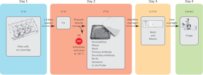
Figure 1: Example workflow with estimated durations. An additional day may be required if primary antibody incubation is performed overnight. In the case of native FISH, Days 2 and 3 are sometimes combined, although this results in an extended bench time. Please click here to view a larger version of this figure.
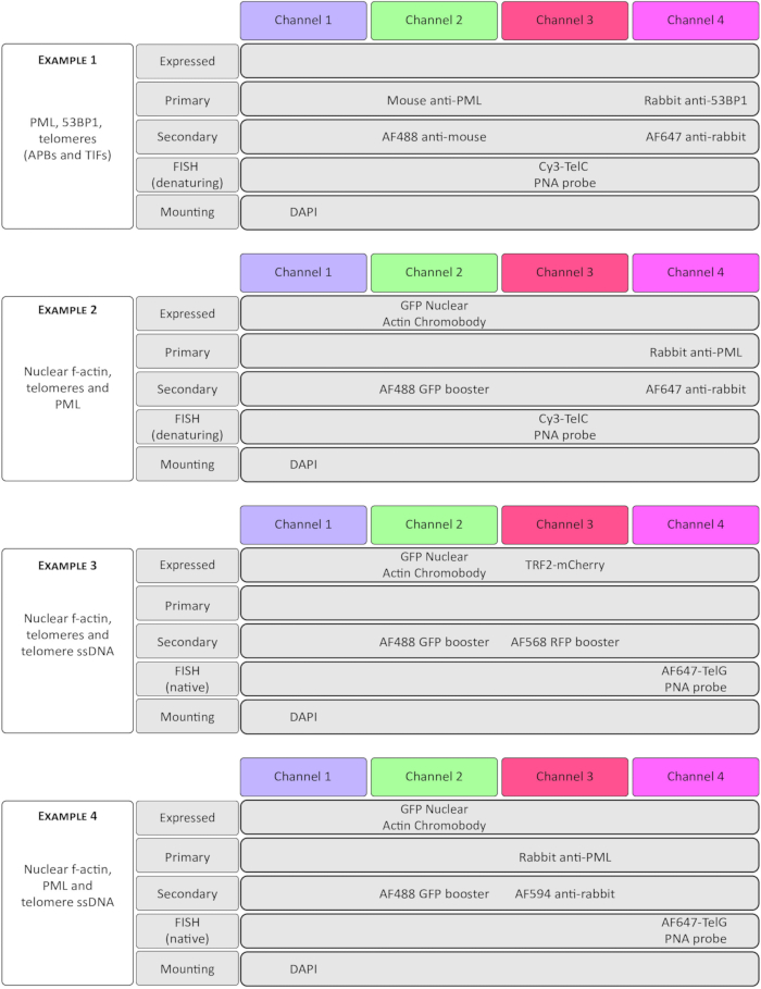
Figure 2: Examples of successful experimental combinations used with four-color imaging. Examples 1 and 2 demonstrate denaturing telomere FISH, while 3 and 4 use native FISH. Note that this protocol is useful whether immunostaining two target proteins (Example 1), combining one FP and one IF stain (Examples 2 and 4), or two FPs (Example 3). Representative results for these experiments are given in Figure 4. Please click here to view a larger version of this figure.
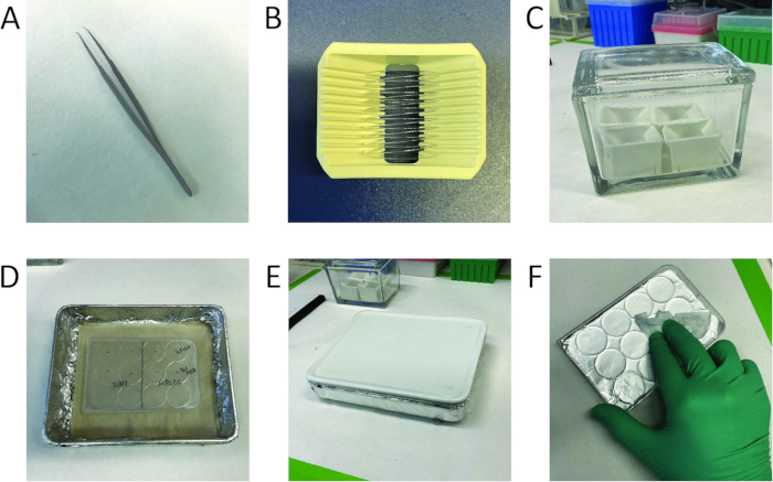
Figure 3: Equipment used in this protocol. (A) Coverslip forceps. While straight forceps can be used to handle coverslips, angled coverslip forceps make the task much easier. (B) Coverslips racked for washing. Note that in addition to racking straight across, coverslips are also angled between adjacent grooves to increase capacity. (C) Four racks of coverslips in a staining dish. A glass staining dish holds dozens of racked coverslips and is impervious to the HCl acid wash. (D) A prepared humidified chamber. The outside is covered in foil to create a dark environment, preserving the fluorophores. Damp paper towels at the bottom provide humidity. (E) The sealed humidified chamber. A tight-fitting lid is essential to prevent drying out. (F) Making the aluminum foil support. Embossing ridges onto the trimmed foil helps to prevent the coverslips from sliding into each other during the denaturing step. Please click here to view a larger version of this figure.
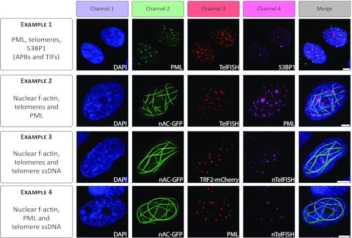
Figure 4: Representative results of four-channel imaging experiments, including IF, FISH, and expressed FPs. Panels depict actual results of the experiments described in Figure 2. DAPI stains DNA, nAC-GFP highlights nuclear f-actin, TelFISH and TRF2-mCherry mark telomeres, PML immunostaining marks nuclear PML bodies, and nTelFISH reveals telomeric ssDNA. All cells are U2OS osteosarcoma cells. Images were acquired on a confocal microscope with super-resolution capability. Example 1 images were acquired with laser-scanning confocal microscopy; all other images were acquired with Structured Illumination Microscopy. All scale bars represent 5 µM. Please click here to view a larger version of this figure.
Discussion
Experiments using IF-FISH have long been a mainstay in the telomere field as an assay for telomere stress or detection of ALT5,6,16. Here, the protocol is extended to include a fluorescent protein. Additionally, the option of native telomere FISH is offered as an orthogonal ALT assay11. This protocol is straightforward to perform and reliably produces satisfactory results.
By operating on coverslips with droplets of antibodies or PNA probes, this protocol is designed to be thrifty. Using droplets conserves valuable reagents. In contrast, alternatives such as chambered slides are expensive and offer less flexibility. While handling coverslips takes practice to develop proficiency, the cost savings make the investment of time worthwhile. It should be noted that reagent droplets larger than 75 µL are not recommended. Larger droplets simply cause the coverslips to slide around and do not improve results.
The expression of nAC-GFP is an indispensable tool for the visualization of nuclear f-actin8, as seen in Figure 4. Many other tagged reagents exist that may help to answer important biological questions. For example, the FUCCI plasmid system can be used to visualize the cell cycle, and using this protocol could be combined with telomere FISH17. A different approach to assaying the cell cycle might use the PCNA chromobody to gain cell cycle information18. Depending on the question being asked, many FP-tagged reagents are commercially available.
Here, this protocol is demonstrated with telomere FISH, but successful results have also been achieved with centromere FISH, using a PNA probe at the same concentration. It is possible to multiplex a FAM-labeled centromere probe with a Cy3-labeled telomere probe, for example. It is important to emphasize that the protocol is unlikely to work with single-locus DNA FISH. For probing a single locus, protease digestion of the sample is necessary, and this digestion is clearly incompatible with IF or FP detection19. However, success has been achieved in combining IF and mRNA FISH with a similar approach; obviously, the RNase treatment step must be omitted in this scenario.
Finally, it is apparent that native telomere FISH is rapidly becoming an essential assay for ALT status20,21 . Unlike the C-circle assay for ALT, which is population-based22, native telomere FISH provides an ALT readout at the cellular level, allowing sensitive detection of changes in ALT dynamics. By uniting native telomere FISH with IF and FP expression, important insights into the ALT mechanism can be revealed. In time, we hope these insights will lead to targeted therapeutics for tumors that rely on ALT for telomere maintenance.
Disclosures
The authors declare that they have no competing interests.
Acknowledgements
This research was supported by the Center for Cancer Research, National Cancer Institute, National Institutes of Health Intramural Research Program project number ZIA BC 011091. The contributions of the NIH authors were made as part of their official duties as NIH federal employees, are compliant with agency policy requirements, and are considered Works of the United States Government. The findings and conclusions presented in this paper are, however, those of the authors and do not necessarily reflect the views of the NIH or the U.S. Department of Health and Human Services. We thank the Lazzerini Denchi lab for their telomere native FISH protocol and for fruitful discussions. Microscopy was performed at the NCI CCR microscopy core.
Materials
| Name | Company | Catalog Number | Comments |
|---|---|---|---|
| 10 % Tween-20 | Bio-Rad | 1610781 | Discard if the solution is cloudy. |
| 10x PBS | Gibco | 70011044 | For preparing fixative and other buffers use post-fixation |
| 12-well cell culture plates | Nunc | 150628 | |
| 16% methanol-free formaldehyde | Thermo Scientifc | 28908 | |
| 5 M sodium chloride | Millipore Sigma | S6546 | |
| Adherent cells, e.g. U2OS osteosarcoma cells | ATCC | HTB-96 | |
| Airtight box for humidified chamber | Mepal | 106415032500 | Any suitable airtight box will work, but beware of boxes with uneven bottoms. |
| Alconox | Millipore Sigma | Z742914 | |
| aluminum foil | Fisher Scientific | 01-213-100 | |
| Appropriate cell culture medium, e.g. McCoy's 5A | Gibco | 16600082 | for U2OS, add 15% FBS |
| Centromere probe (FAM) | PNA Bio | F3001 | Several fluorophores are available |
| ChromoTek Nuclear Actin-Chromobody plasmid (TagGFP) | Proteintech | acg-n | |
| Coverslip forcers | Fine Science Tools | 11251-33 | |
| Coverslip racks | Millipore Sigma | Z688568 | |
| DAPI | Invitrogen | D21490 | Optional, in case the mounting medium does not contain a DNA stain |
| Donkey serum | Jackson Immunoresearch | 017-000-121 | |
| EGTA | Bio-World | 40520008 | 0.5 M Solution pH 8.0 |
| ethanol | Warner-Graham | 200 Proof | |
| Fetal Bovine Serum | Gibco | A5670801 | |
| Fibronectin | Millipore Sigma | F0635 | For optional coating of coverslips |
| formamide | Millipore Sigma | 47671-250ML-F | |
| German glass coverslips | Warner Insruments | 64-0714 | CS-18R15 (#1.5 Thickness, confirm this is appropriate for your microscope) |
| GFP Booster | Proteintech | gb2AF488 | recognizes GFP/mClover/YFP/Venus and others. See product information for a comprehensive list |
| Glass staining dish with lid | Millipore Sigma | 900203 | |
| Heated Ultrasonic Cleaner with Digital Timer | Branson | CPX-952-218R | This bath sonicator model includes heat but does not have configurable power or intensity. |
| Hydrochloric Acid | Millipore Sigma | 258148 | Concentrated HCl is 12 N. Add 20 mL concentrated HCl to 220 mL deionized water for 1 N HCl |
| Magnesium chloride solution | Millipore Sigma | 63069 | 1 M Solution |
| MES | Millipore Sigma | 475893 | |
| methanol | Millipore Sigma | 179337 | |
| Parafilm | USA Scientific | 3023-4526 | |
| Potassium chloride | Millipore Sigma | P9541 | |
| primary antibody mouse anti-PML | Santa Cruz | sc-377390 | |
| primary antibody rabbit anti-53BP1 | Novus | NB100-904 | |
| primary antibody rabbit anti-PML | ABclonal | A1184 | |
| Prolong Glass with NucBlue | Invitrogen | P36981 | |
| RFP Booster | Proteintech | rb2AF568 | recognizes mRFP/mCherry/mKate2/mPlum. See product information for a comprehensive list |
| RNAse A | Millipore Sigma | RNASEA-RO | |
| Roche blocking solution | Millipore Sigma | 11096176001 | |
| secondary antibody e.g. AF488 anti-mouse | Invitrogen | A-11001 | Or similar |
| secondary antibody e.g. AF594 anti-rabbit | Invitrogen | A-11012 | Or similar |
| secondary antibody e.g. AF647 anti-mouse | Invitrogen | A-21235 | Or similar |
| spatula | Fisher Scientific | 10257402 | |
| Sterile PBS | Gibco | 14190144 | For preparing coverslips |
| sucrose | Millipore Sigma | S0389 | |
| TelC probe (Cy3) | PNA Bio | F1002 | Several fluorophores are available |
| TelG probe (AF647) | PNA Bio | F1014 | Several fluorophores are available |
| Tris Base | Millipore Sigma | 648311 | Prepare 1 M solution in water, adjust pH using HCl |
| triton X-100 | Millipore Sigma | X100 | |
| Trypsin-EDTA (0.25%), phenol red | Gibco | 25200072 | |
| Zeiss Elyra microscope | Zeiss | Or similar |
References
- Denchi, E. L., De Lange, T. Protection of telomeres through independent control of ATM and ATR by TRF2 and POT1. Nature. 448 (7157), 1068-1071 (2007).
- Rossiello, F., Jurk, D., Passos, J. F., Di Fagagna D'adda, F. Telomere dysfunction in ageing and age-related diseases. Nat Cell Biol. 24 (2), 135-147 (2022).
- Maciejowski, J., De Lange, T. Telomeres in cancer: Tumour suppression and genome instability. Nat Rev Mol Cell Biol. 18 (3), 175-186 (2017).
- Meyne, J., Ratliff, R. L., Moyzis, R. K. Conservation of the human telomere sequence (TTAGGG)n among vertebrates. Proc Natl Acad Sci U S A. 86 (18), 7049-7053 (1989).
- Takai, H., Smogorzewska, A., De Lange, T. DNA damage foci at dysfunctional telomeres. Curr Biol. 13 (17), 1549-1556 (2003).
- Herbig, U., Jobling, W. A., Chen, B. P., Chen, D. J., Sedivy, J. M. Telomere shortening triggers senescence of human cells through a pathway involving ATMp53, and p21(CIP1), but not p16(INK4a). Mol Cell. 14 (4), 501-513 (2004).
- Yeager, T. R., et al. Telomerase-negative immortalized human cells contain a novel type of promyelocytic leukemia (PML) body. Cancer Res. 59 (17), 4175-4179 (1999).
- Plessner, M., Melak, M., Chinchilla, P., Baarlink, C., Grosse, R. Nuclear f-actin formation and reorganization upon cell spreading. J Biol Chem. 290 (18), 11209-11216 (2015).
- Oliva, A. A., Swann, J. W. Fluorescence in situ hybridization method for co-localization of mRNA and GEP. Biotechniques. 31 (1), 74-81 (2001).
- Pardue, M. L., Gall, J. G. Molecular hybridization of radioactive DNA to the DNA of cytological preparations. Proc Natl Acad Sci U S A. 64 (2), 600-604 (1969).
- Azeroglu, B., Ozbun, L., Pegoraro, G., Lazzerini Denchi, E. Native FISH: A low- and high-throughput assay to analyze the alternative lengthening of telomere (ALT) pathway. Methods Cell Biol. 182, 265-284 (2024).
- Claude, E., et al. Detection of alternative lengthening of telomeres mechanism on tumor sections. Mol Biomed. 2 (1), 32(2021).
- Small, J. V. Organization of actin in the leading edge of cultured cells: Influence of osmium tetroxide and dehydration on the ultrastructure of actin meshworks. J Cell Biol. 91 (3 Pt 1), 695-705 (1981).
- Draskovic, I., et al. Probing PML body function in ALT cells reveals spatiotemporal requirements for telomere recombination. Proc Natl Acad Sci U S A. 106 (37), 15726-15731 (2009).
- Pardue, M. L., Gerbi, S. A., Eckhardt, R. A., Gall, J. G. Cytological localization of DNA complementary to ribosomal RNA in polytene chromosomes of diptera. Chromosoma. 29 (3), 268-290 (1970).
- Mender, I., Shay, J. W. Telomere dysfunction induced foci (TIF) analysis. Bio Protoc. 5 (22), (2015).
- Sakaue-Sawano, A., et al. Visualizing spatiotemporal dynamics of multicellular cell-cycle progression. Cell. 132 (3), 487-498 (2008).
- Burgess, A., Lorca, T., Castro, A. Quantitative live imaging of endogenous DNA replication in mammalian cells. PLoS One. 7 (9), e45726(2012).
- Trask, B., Pinkel, D. Fluorescence in situ hybridization with DNA probes. Methods Cell Biol. 33, 383-400 (1990).
- Quintanilla, I., et al. Optical pooled screening for the discovery of regulators of the alternative lengthening of telomeres pathway. Methods. 241, 1-12 (2025).
- Azeroglu, B., et al. Identification of novel modulators of the ALT pathway through a native FISH-based optical screen. bioRxiv. , (2024).
- Henson, J. D., et al. The c-circle assay for alternative-lengthening-of-telomeres activity. Methods. 114, 74-84 (2016).