This article presents the case of a patient with an extensive spinal epidural abscess (SEA), detailing the surgical intervention and subsequent clinical outcomes.
A subscription to JoVE is required to view this content. Sign in or start your free trial.
Case Report
Surgical Decompression and Continuous Epidural Irrigation for Extensive Spinal Epidural Abscess Secondary to Psoas Major Muscle Abscess
In This Article
Summary
Abstract
An extensive spinal epidural abscess (SEA) is a rare and life-threatening condition that requires prompt recognition and proper management to avoid potentially disastrous complications. When an SEA is widespread, extensive decompression with laminectomy is often impossible, as it may subject the patient to very long operative times, extensive blood loss, and mechanical instability. Here we report a 57-year-old male patient who presented to the emergency department with high-grade fever, significantly diminished muscle strength in the right limbs, nuchal rigidity, and hyperreflexia in both lower limbs. Magnetic resonance imaging (MRI) of the spine revealed a diffuse spinal epidural abscess extending from C2 to S1. Laboratory investigations were consistent with prominent signs of acute infection. He underwent emergent surgical intervention, complemented by postoperative continuous epidural irrigation combined with antibiotic therapy. The postoperative course was favorable, with the patient demonstrating marked clinical improvement. Based on this case, we conclude that prompt surgical drainage combined with continuous epidural irrigation and adjunctive antibiotic therapy represents a viable clinical approach for the management of SEA.
Introduction
Spinal epidural abscess (SEA) is a rare but potentially devastating infection, historically affecting 0.2-2 per 10,000 hospital admissions1, though recent evidence suggests a rising incidence potentially reaching up to 5.1 per 10,000 admissions2. This increase is attributed to factors like an aging population, improved diagnostics, comorbidities such as diabetes mellitus and immunocompromised states, intravenous drug use, and the growing prevalence of spinal instrumentation3,4. Extensive SEA, variably defined as involving more than five vertebral levels or spanning all three spinal regions (cervical, thoracic, lumbar), represents an exceptionally rare (approximately 1%) and severe subset5. Diagnosis remains challenging due to the frequent absence of the classic triad (fever, back pain, neurological deficit), often leading to delays with catastrophic consequences, including irreversible paraplegia or death6. While magnetic resonance imaging (MRI) is considered the diagnostic gold standard, the optimal management strategy, particularly for extensive SEA, remains complex. Although antibiotic therapy is fundamental, surgical decompression is widely advocated for significant neurological impairment or failed medical management7. Extensive SEA poses unique surgical challenges: traditional multi-level laminectomies carry significant risks of instability, excessive blood loss, and prolonged operative times8. Consequently, minimally invasive techniques like selective "skip" or "apical" laminectomies at strategic levels (e.g., apices of spinal curvatures) combined with epidural irrigation and drainage have emerged as promising alternatives to achieve adequate decompression and source control while mitigating surgical morbidity9. This case report contributes to the evolving literature on managing this critical condition by describing C3 cervical and T9 thoracic laminectomy with decompression and epidural abscess debridement combined with continuous intraspinal irrigation and drainage.
CASE PRESENTATION:
A 57-year-old male construction worker presented with 10 days of progressive low back pain and bilateral lower limb weakness, culminating in acute-onset back pain, fever (38.5 °C), and right-sided flaccid paralysis 24 h prior to admission. Initial evaluation at a local hospital revealed L4 spondylolisthesis (Grade I) on lumbar MRI, managed conservatively with nonsteroidal anti-inflammatory agents (NSAIDs) and mecobalamin without improvement. On symptom exacerbation, emergency labs showed leukocytosis (WBC 40.14 × 10⁹/L, 94.4% neutrophils), elevated CRP (>200 mg/L), and CT suggesting a right psoas abscess with possible L4-S1 epidural extension, prompting transfer. On admission, vitals included fever (38.8 °C), tachypnea (22/min), and tachycardia (96 bpm). Neurological examination demonstrated confusion, dysarthria, nuchal rigidity, right hemiplegia (UE 1-2/5, LE 0/5), left UE weakness (3/5), generalized hypertonia, hyperalgesia, bilateral hyperreflexia, and positive Hoffmann's signs. Past medical history included hypertension and Parkinson's disease on antiplatelet/statin therapy, with no recent trauma or invasive procedures.
Diagnosis, Assessment, and Plan:
Initial diagnostic testing prioritized spinal MRI due to acute neurological deficits (flaccid paralysis, hyperreflexia) and systemic infection signs (fever, leukocytosis), which raised concern for spinal cord compression versus inflammatory myelitis. Emergency laboratory studies—including markedly elevated CRP (>200 mg/L) and neutrophilic leukocytosis (WBC 40.14 × 10⁹/L)—provided objective evidence of severe bacterial sepsis, prompting immediate CSF analysis via lumbar puncture to identify pathogens; CSF Gram stain revealed Gram-positive cocci, directing empirical antibiotic selection while awaiting culture. The definitive spinal MRI (Figure 1) demonstrated a contiguous epidural abscess from C2-S1 with posterolateral cord displacement and C3-6 edema, corroborating the clinical diagnosis of spinal epidural abscess (SEA).
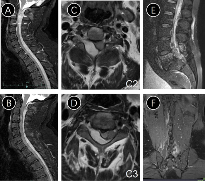
Figure 1: Preoperative magnetic resonance images. (A,B) T2-weighted sagittal magnetic resonance images of the spine showing an extensive epidural abscess in the cervical and thoracic region. (C,D) T2-weighted axial images at the C2 and C3 levels demonstrate an epidural abscess within the spinal canal compressing the spinal cord. (E,F) Contrast-enhanced lumbar spine MRI demonstrated multiple enhancing lesions involving the vertebral bodies and within the spinal canal, along with a subcutaneous abscess. Coronal T2-weighted images revealed a psoas abscess. Please click here to view a larger version of this figure.
The treatment plan centered on emergent decompressive laminectomy to alleviate cord compression, with intraoperative findings confirming purulent material consistent with SEA. Rationale for surgical urgency included progressive flaccid paralysis (indicating impending irreversible cord injury) and sepsis with hemodynamic instability. Continuous epidural irrigation via indwelling catheters was instituted postoperatively to reduce abscess recurrence risk by maintaining local antibiotic concentration. Broad-spectrum intravenous antibiotics were initiated immediately post-diagnosis to cover Gram-positive cocci (including MRSA) and Gram-negative bacilli, pending culture and next-generation sequencing (NGS) results; this regimen was selected based on local resistance patterns and penetration into abscess cavities. Adjunctive hyperbaric oxygen therapy was added to mitigate spinal cord edema through enhanced oxygen diffusion.
Access restricted. Please log in or start a trial to view this content.
Protocol
This protocol follows the guidelines of the Human Research Ethics Committee of First Affiliated Hospital, Zhejiang University School of Medicine. Written informed consent was obtained from the patients for participation in the study. The required consumables and equipment are listed in the Table of Materials.
1. Operative procedure
- Preoperative preparation: Comprehensive laboratory and imaging studies excluded absolute surgical contraindications. The patient was planned to undergo general anesthesia for C3 cervical and T9 thoracic laminectomy with decompression and epidural abscess debridement.
- Anesthesia: Administer intravenous-inhalation combined anesthesia following institutionally approved protocols.
- Incision and exposure:
- Following anesthesia induction, a midline skin incision was made. Subcutaneous fascia and paraspinal muscles were dissected using electrocautery, exposing the laminae.
- Inflammatory connective tissue proliferation with excessive turbid fluid was noted in deep cervical/lumbar musculature; fluid samples were collected for culture.
- Laminectomy: Ultrasonic bone scalpel was utilized to perform osseous laminectomy at C3 and T9 levels.
- Ligamentum flavum resection:
- The ligamentum flavum was meticulously dissected to expose the dorsal dural sac.
- Copious white purulent material within the epidural space was identified; specimens were obtained for culture and next-generation sequencing (NGS).
- Decompression: Adequate neural decompression of cervical/lumbar nerve roots and dural sac was achieved, with restoration of dural pulsation.
- Irrigation:
- The surgical field was copiously irrigated with pulsed lavage using normal saline, povidone-iodine solution, and vancomycin-infused saline (1 g/500 mL).
- The irrigation fluid produced intraoperatively was aspirated through the central surgical suction system.
- The system parameters were set at a negative pressure of -0.03 MPa to -0.07 MPa and a minimum air displacement rate of 30 L/min.
- Irrigation catheter placement and postoperative irrigation:
- Three fenestrated 6Fr (2mm diameter) infant urinary catheter were employed for intraoperative and postoperative continuous antibiotic irrigation. These catheters were then inserted into the spinal epidural space via the laminectomy incisions at C3 and T9 levels, respectively8,10 (Figure 2):
- The first catheter was introduced at the C3 laminectomy site and advanced caudally within the spinal canal, reaching the T9 level.
- The second catheter was inserted at the T9 laminectomy site and advanced cephalad for approximately 20 cm.
- The third catheter was also inserted at the T9 site and advanced caudally for approximately 30 cm, reaching the lumbar region.
- Intraoperatively, the three catheters were irrigated sequentially with normal saline and vancomycin solution to flush out purulent material from the spinal canal.
- Postoperatively, a continuous irrigation regimen was established, infusing 500 mL of normal saline mixed with vancomycin solution through the catheters every 24 h. The irrigant was subsequently drained via a separate paraspinal drainage tube.
- Three fenestrated 6Fr (2mm diameter) infant urinary catheter were employed for intraoperative and postoperative continuous antibiotic irrigation. These catheters were then inserted into the spinal epidural space via the laminectomy incisions at C3 and T9 levels, respectively8,10 (Figure 2):
- Drain placement:
- Two 12-French Jackson-Pratt drains were placed at the C3 level and T9 level, each positioned in the deep muscular layer at the laminectomy sites.
- The drains were placed to bulb suction for the removal of postoperative irrigation fluid, hemorrhage, and tissue exudate.
- Wound closure:
- Incisions were irrigated thoroughly, and the muscular layer was sutured using a barbed suture, followed by the approximation of the subcutaneous tissue with a 2-0 absorbable suture. Finally, the epidermal layer was closed with skin staples.
2. Postoperative treatment
- Initial antibiotic therapy: Broad-spectrum intravenous antibiotics (vancomycin 1000 mg q12h + meropenem 2 g q8h) were initiated postoperatively, achieving fever control (mean temperature 37 °C).
- Antibiotic adjustment based on NGS:
- Specimens from the infected area were collected for NGS. Host DNA was depleted with 1U nuclease and 0.5% Tween 20; microbial DNA/RNA was extracted via DNA/RNA extraction kits, with rRNA removed by an rRNA depletion kit and cDNA synthesized using cDNA synthesis reagents.
- Plasma cell-free nucleic acids were purified using a circulating nucleic acid kit.
- Libraries were built with a DNA library preparation kit, quality-checked via a bioanalyzer and qPCR, then sequenced on a sequencer.
- Raw reads were filtered by fastp, mapped to hg38 (HISAT2) to remove host sequences; microbial analysis used Kraken 2, differential species via rank-sum test, ARGs via DeepARG, and clinical models (AUC, 10-fold validation).
- Differential expression was analyzed using DESeq2, with thresholds set at |log2 Fold Change| ≥ 1.0 and p < 0.05. NGS identified Streptococcus intermedius infection, prompting a regimen change to vancomycin (1000 mg q12h) plus levofloxacin (750 mg q24h), with continued vancomycin-saline epidural irrigation (1 g/500 mL q6h).
- Secondary regimen modification: Following derangements in hepatic and renal function markers, antibiotics were switched to ceftriaxone (2 g q24h) and levofloxacin (500 mg q24h).
- Therapeutic response: After 14 days of targeted therapy, the patient remained afebrile and showed significant improvement in inflammatory markers.
- Psoas abscess drainage: Under ultrasound guidance, percutaneous drainage of the right psoas abscess was performed on postoperative day 7, yielding 100 mL of purulent fluid; cultures confirmed Staphylococcus intermedius (S. intermedius).
- Neurological recovery: Post-drainage assessment noted marked reduction in right lower limb pain and improved motor strength (right LE: 0/5 → 3/5).
- Continuous irrigation protocol: Epidural antibiotic irrigation was maintained for 3 weeks, supplemented by weekly wound dressing changes under sterile conditions.
Access restricted. Please log in or start a trial to view this content.
Results
This patient successfully underwent a laminectomy with decompression and epidural abscess debridement, followed by antibiotics and continuous spinal irrigation. At the 2-week postoperative reassessment, cervical-thoracic-lumbar MRI demonstrated significant resolution of the epidural abscesses but still with small residuals (Figure 3). Subsequent removal of irrigation catheters and surgical drains was performed following normalization of inflammatory markers (CRP 3.2 mg/L, WBC 6.8×10⁹
Access restricted. Please log in or start a trial to view this content.
Discussion
SEA is a severe infection with a global mortality rate of 5%-16%, and fewer than 50% of survivors fully recover. Males are affected more frequently than females, with a ratio of 2:1, for reasons that remain unknown11. SEAs manifest as a multisegmental (3-4 segments) condition because bacteria can spread through the epidural space unhindered by anatomical barriers12. This report presents a challenging case of holocord SEA spanning C2-S1, uniquely originating from a psoas mus...
Access restricted. Please log in or start a trial to view this content.
Disclosures
The authors have nothing to disclose.
Acknowledgements
This study is funded by Shaoxing Health Science and Technology Program (No.2022KY106), National Natural Science Foundation of China (No.82402157), Zhejiang Provincial Natural Science Foundation (No. LQ23H060004), and China Postdoctoral Science Foundation General Funding Program (No.2022M722753).
Access restricted. Please log in or start a trial to view this content.
Materials
| Name | Company | Catalog Number | Comments |
|---|---|---|---|
| Absorbable Suture | ETHICON | VCP739D | |
| Absorbable Suture | ETHICON | SXPP1A404 | |
| AGILENT 2100 Bioanalyzer | AGILENT | 2100 | |
| DeepARG | gaarangoa | https://github.com/gaarangoa/deeparg | |
| Fastp | OpenGene | https://github.com/OpenGene/fastp | |
| Illumina NextSeq 550 Sequencing System | Illumina | https://www.illumina.com/systems/sequencing-platforms/nextseq/specifications.html | Sequencer |
| Kraken2 | DerrickWood | https://github.com/DerrickWood/kraken2 | |
| Maxima Reverse Transcriptase | Thermo Fisher | 18080093 | |
| Nextera XT DNA Library Prep Kit | Illumina | https://www.illumina.com/products/by-type/sequencing-kits/library-prep-kits/nextera-xt-dna.html | DNA library preparation kit |
| Nuclease | Thermo Fisher | EN0321 | |
| QIAamp Circulating Nucleic Acid Kit | QIAGEN | 55114 | Circulating nucleic acid Kit |
| QIAamp UCP Pathogen DNA Kit | QIAGEN | 50214 | DNA/RNA extraction kits |
| QIAamp Viral RNA Kit | QIAGEN | 52904 | |
| Ribo-Zero rRNA Removal Kit | Illumina | https://www.illumina.com/products/by-type/molecular-biology-reagents/ribo-zero-plus-rrna-depletion.html | rRNA depletion kit |
| Skin Stapler | Covidien | 54887 | |
| Tween 20 | SIGMA | P9416 |
References
- Pradilla, G., Ardila, G. P., Hsu, W., Rigamonti, D. Epidural abscesses of the CNS. Lancet Neurol. 8 (3), 292-300 (2009).
- Vakili, M., Crum-Cianflone, N. F. Spinal epidural abscess: A series of 101 cases. Am J Med. 130 (12), 1458-1463 (2017).
- Artenstein, A. W., et al. Spinal epidural abscess in adults: A 10-year clinical experience at a tertiary care academic medical center. Open Forum Infect Dis. 3 (4), ofw191(2016).
- Adogwa, O., et al. Spontaneous spinal epidural abscess in patients 50 years of age and older: A 15-year institutional perspective and review of the literature: Clinical article. J Neurosurg Spine. 20 (3), 344-349 (2014).
- Erşahin, Y. Spinal epidural abscess: A meta-analysis of 915 patients. Neurosurg Rev. 24 (2-3), 156(2001).
- Tetsuka, S., Suzuki, T., Ogawa, T., Hashimoto, R., Kato, H. Spinal epidural abscess: A review highlighting early diagnosis and management. JMA J. 3 (1), 29-40 (2020).
- Akalan, N., Ozgen, T. Infection as a cause of spinal cord compression: A review of 36 spinal epidural abscess cases. Acta Neurochir (Wien). 142 (1), 17-23 (2000).
- Abd-El-Barr, M. M., et al. Extensive spinal epidural abscess treated with "apical laminectomies" and irrigation of the epidural space: Report of 2 cases. J Neurosurg Spine. 22 (3), 318-323 (2015).
- Proietti, L., et al. Extensive spinal epidural abscesses resolved with minimally invasive surgery: Two case reports and review of the recent literature. Acta Neurochir Suppl. 125, 345-353 (2019).
- Xu, T., et al. Extensive spinal epidural abscess resulting in complete paraplegia treated by selective laminectomies and irrigation. Orthop Surg. 14 (9), 2380-2385 (2022).
- Aljawadi, A., et al. Management of pyogenic spinal infection, review of literature. J Orthop. 16 (6), 508-512 (2019).
- Karaja, S., et al. Unusual case of Staphylococcus epidermidis-induced spinal epidural abscess in an adolescent: Clinical insights and diagnostic considerations. Radiol Case Rep. 20 (6), 2699-2703 (2025).
- Jafari Roshan-Zamir, F., Ceaser, S., Chin, K., Mas, D. Streptococcus pneumoniae-related infective endocarditis complicated by development of psoas and epidural abscesses. Cureus. 17 (2), e78691(2025).
- Smith, G. A., et al. Holospinal epidural abscess of the spinal axis: Two illustrative cases with review of treatment strategies and surgical techniques. Neurosurg Focus. 37 (2), E11(2014).
- Tai, C. L., et al. Biomechanical comparison of lumbar spine instability between laminectomy and bilateral laminotomy for spinal stenosis syndrome - an experimental study in porcine model. BMC Musculoskelet Disord. 9, 84(2008).
- Liu, D., Lu, W., Huang, W., Zhai, W., Ling, Q. Extensive spinal epidural abscess due to Streptococcus intermedius: A case report treated conservatively and literature review. Front Neurol. 14, 1237007(2023).
- Kurudza, E., Stadler, J. A. 3rd Pediatric holocord epidural abscess treated with apical laminotomies with catheter-directed irrigation and drainage. Cureus. 11 (9), e5733(2019).
- Urrutia, J., Rojas, C. Extensive epidural abscess with surgical treatment and long term follow up. Spine J. 7 (6), 708-711 (2007).
- Xiang, H., et al. Holocord spinal epidural abscess: Case report and literature review. Orthop Traumatol Surg Res. 102 (6), 821-825 (2016).
- Kumar, K., Hunter, G. Spinal epidural abscess. Neurocrit Care. 2 (3), 245-251 (2005).
- Liu, X., Deng, S., Kang, L. Progress in hyperbaric oxygen therapy for neonatal hypoxic-ischemic encephalopathy: Mechanisms and combination therapies. Brain Inj. , 1-9 (2025).
Access restricted. Please log in or start a trial to view this content.