This study presents a protocol for evaluating the effects of curcumin on retinoblastoma cell behavior through modulation of the Wnt/β-catenin pathway and miR-22 expression.
A subscription to JoVE is required to view this content. Sign in or start your free trial.
Research Article
miR-22-Mediated Regulation of Wnt/β-Catenin Signaling by Curcumin in Retinoblastoma
* These authors contributed equally
In This Article
Summary
Abstract
Retinoblastoma (RB) is a common intraocular malignant tumor affecting infants and children, yet its precise etiology and pathogenesis remain incompletely understood. Curcumin, a bioactive polyphenol, inhibits tumor progression via microRNA-mediated modulation of the Wnt/β-catenin signaling cascade. This study aimed to clarify how curcumin mediates its antitumor effects in RB by investigating its regulation of miRNA-22 (miR-22) expression and exploring the underlying molecular mechanisms. Two validated retinoblastoma models (SO-RB50/WERI-Rb-1) were treated with curcumin at varying concentrations. To delineate miR-22's regulation of Wnt/β-catenin signaling, target cells were transduced with either a miR-22 mimic lentivirus or a non-functional control lentivirus. Xenograft tumor models were established in mice using human RB cells to observe the in vivo effects of curcumin on tumor size, miR-22 expression, and Wnt/β-catenin protein levels. Cellular proliferation, invasion, and apoptosis were assessed using the CCK-8, Transwell, and Annexin V-APC-PI dual staining assay, respectively. miR-22 levels were quantified by RT-PCR, and Wnt1 and β-catenin expression profiles were determined by Western blot analysis. Curcumin treatment resulted in decreased proliferation and invasiveness in RB cells, while enhancing apoptosis and elevating miR-22 expression. Inhibition of miR-22 diminished curcumin's effects on the Wnt/β-catenin signaling pathway. In xenograft studies, curcumin significantly reduced tumor size and enhanced miR-22 expression within the tumors, effectively suppressing Wnt/β-catenin signaling. These findings demonstrate that curcumin inhibits RB cell proliferation and invasiveness while promoting apoptosis, primarily mediated through miR-22 upregulation and subsequent inhibition of the Wnt/β-catenin pathway.
Introduction
Retinoblastoma (RB), a pediatric malignancy with high aggressiveness arising in retinal tissues, poses a substantial health threat, especially to children under the age of five. Genetic mutations, particularly those affecting the RB1 gene, are closely associated with the development of this disease1. Considering the significant prevalence of retinoblastoma (RB) among infants and children, coupled with the fact that approximately 9000 new cases are reported annually, predominantly in low-income and developing countries, a comprehensive understanding of the disease mechanisms is imperative for developing effective therapeutic strategies and improving patient outcomes2. Over the years, research on RB has primarily centered on genomic alterations, specifically in miRNA expression3. Certainly, miR-34a and miR-22 have emerged as vital players in the tumorigenesis of RB4,5. Among these miRNAs, miR-22 has demonstrated its involvement in tumorigenesis across different cancers6,7,8, influencing proliferation, invasion, and migration6,9. Moreover, previous research has indicated reduced miR-22 expression in both mouse and human RB cell lines, further highlighting its potential role in this disease10.
Curcumin, a principal bioactive curcuminoid in Curcuma longa rhizomes, has been a fundamental ingredient in traditional medicine and Asian cuisines11. Its rich pharmacological profile, encompassing antitumor, antimicrobial, anti-inflammatory, antioxidant, and neuroprotective effects, has garnered significant attention in biomedical research12,13,14. Numerous studies have explored its antitumor role in RB, highlighting its influence on different signaling pathways, including NF-κB15, JAK/STAT16, and MRP117.Compared with these previously studied pathways, the curcumin-miR-22/Wnt/β-catenin axis may offer a more targeted and specific approach, potentially improving therapeutic efficacy and reducing off-target effects.
Previous studies have suggested that curcumin achieves antitumor effects via modulating the TET1/Wnt/β-catenin signaling pathway axis in hepatocellular carcinoma18 and colonic cancer cells19. However, whether curcumin can suppress tumor progression in RB cells by affecting the Wnt/β-catenin signaling pathway remains unknown. Recent studies have reported decreased miR-22 expression in retinoblastoma cells and oral squamous cell carcinoma linked to alterations in Wnt/β-catenin signaling pathway activity20,21. Considering practical applicability, curcumin exhibits effective antitumor activity within a defined concentration range (10-40 µM in vitro)15,22 and can be administered in vivo with manageable pharmacokinetic properties, facilitating translational research. Therefore, this study aims to establish an RB model to investigate whether curcumin increases miR-22 levels and subsequently decreases Wnt/β-catenin signaling activity, providing a potentially more efficient and clinically relevant therapeutic strategy compared with conventional NF-κB, JAK/STAT, or MRP1 modulation approaches.
Access restricted. Please log in or start a trial to view this content.
Protocol
All procedures involving animals were reviewed and approved by the Animal Ethics and Welfare Committee (AEWC) of Tianjin Eye Hospital (Approval No. NKYY-DWLL-2023-054). The reagents and the equipment used are listed in the Table of Materials.
1. Cell culture
Cells of the human retinoblastoma lines WERI-Rb-1 and SO-RB50 were cultured in high-glucose Dulbecco's modified Eagle medium supplemented with 10% fetal bovine serum and 1% penicillin-streptomycin. Cells were seeded at an appropriate density in culture flasks and maintained at 37 °C in a humidified atmosphere containing %CO2. Medium was replaced every 2-3 days, and cells were passaged at 70%-80% confluence using standard trypsinization.
2. Animal experiments
A total of 48 specific-pathogen-free Balb/c nude mice (4 weeks old, 16-18 g) were used. Animals were housed in sterile, individually ventilated cages under laminar airflow with controlled environmental conditions of 26-28 °C and 40%-60% relative humidity. Feed, water, and bedding were sterilized prior to use. Mice were allowed free access to food and water and were acclimatized for one week before experiments.
3. Animal grouping and treatment
WERI-Rb-1 and SO-RB50 cells stably transfected with either a miR-22 mimic or a negative control sequence were used for tumor inoculation. Cells in logarithmic growth phase were harvested, resuspended in sterile phosphate-buffered saline, and adjusted to a density of 5 × 10⁷ cells/mL. Each mouse was injected subcutaneously with 0.1 mL of the suspension into the right forelimb. Mice were randomly divided into six groups (n = 6 per group): model group, curcumin group, miR-22 mimic group, miR-22 mimic + curcumin group, miR-22 negative control group, and miR-22 negative control + curcumin group. In groups receiving curcumin, mice were administered intraperitoneal injections of 15 mg/kg curcumin once daily beginning immediately after tumor cell inoculation. The remaining groups received equal volumes of DMSO as vehicle control.
4. Tumor weight and volume measurement
The tumor take rate was approximately 80%-90% in xenografted nude mice. Tumor size was measured weekly using a digital caliper, with the longest diameter recorded as length (L) and the shortest diameter recorded as width (W). Tumor volume was calculated using the formula V = (L × W2)/2. Mice were monitored regularly for tumor growth and health status, and humane endpoints were applied if tumors exceeded 1,500 mm3 or if animals showed signs of distress or weight loss. At the end of week 4, mice were euthanized by CO2 inhalation followed by cervical dislocation to ensure death. Tumors were dissected and weighed immediately after removal.
5. microRNA transfection
Synthetic oligonucleotides, including the miR-22 mimic, negative control, anti-miR-22 (20-methoxy-modified), anti-miR negative control, Wnt1-specific siRNAs, and control siRNA, were used for transfection. Cells at 70%-80% confluence were seeded in 6-well plates one day before transfection. Transfection was performed using a lipid-based reagent according to the manufacturer's instructions. For each well, nucleic acids were diluted in serum-free medium and combined with the transfection reagent at the recommended ratio to form complexes, which were incubated for 15-20 min at room temperature before being added dropwise to cells in complete medium. The final volume per well was 2 mL. After 6 h of incubation at 37 °C with 5% CO2, the medium was replaced with fresh complete medium. Cells were maintained for 48 h before subsequent assays. Transfection efficiency was evaluated by quantitative real-time PCR. Three different siRNAs targeting Wnt1 (si-Wnt1-1: 5'-CCAAGAGTCTGCAACTGGTACTCGA-3'; si-Wnt1-2: 5'-CGAGAAACGGCGTTTATCTTCGCTA-3'; si-Wnt1-3: 5'-GCTTCCTCATGAACCTTCACAACAA-3') were used individually or as an equimolar mixture to minimize off-target effects.
6. Cell counting Kit-8 (CCK-8) assay
Cells were seeded into 96-well plates at a density of 5 × 10³ cells per well in 100 µL of complete medium and allowed to adhere overnight. After treatment with the indicated concentrations of curcumin for the specified duration, 10 µL of CCK-8 working solution was added directly to each well. Wells containing medium alone served as blank controls. Plates were incubated at 37 °C with 5% CO2 for 4 h, after which absorbance was measured at 490 nm using a microplate reader. Cell viability was calculated as:
Viability (%) = 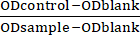 x 100
x 100
Dose-response curves were generated, and the half-maximal inhibitory concentration (IC50) of curcumin was determined accordingly.
7. Transwell assay
For the migration assay, cells were suspended in serum-free medium and seeded into the upper chambers of Transwell inserts (pore size 8 µm) at 3 × 10⁵ cells in 200 µL per insert. The lower chambers were filled with 600 µL of complete medium containing 10% fetal bovine serum as a chemoattractant. Plates were incubated at 37 °C with 5% CO2 for 24 h. After incubation, non-migratory cells on the upper surface of the membrane were gently removed with a sterile cotton swab. Inserts were rinsed twice with phosphate-buffered saline, fixed in 5% glutaraldehyde at 4 °C for 30 min, rinsed again, and stained with 0.1% crystal violet for 30 min at room temperature. Excess dye was removed by washing in PBS. Membranes were allowed to air dry, and migrated cells were visualized under an inverted microscope at 400× magnification. For quantification, three random fields per insert were imaged, and the average number of cells was calculated.
8. Annexin V-APC-PI assay
Cells from each group were harvested, washed twice with pre-cooled phosphate-buffered saline (PBS), and centrifuged at 300 × g for 5 min at 4 °C. The pellet was resuspended in 300 µL of 1× binding buffer at a density of approximately 1 × 10⁶ cells/mL. Annexin V-APC (5 µL) was added, and samples were incubated for 15 min at room temperature in the dark. Immediately before flow cytometry, 5 µL of propidium iodide was added to 200 µL of the cell suspension and incubated for 5 min at room temperature. Samples were analyzed on a flow cytometer using appropriate channels (APC and PI), and data were processed with standard gating strategies to distinguish live, early apoptotic, late apoptotic, and necrotic cells.
9. qRT-PCR Profiling
Total RNA was extracted using a phenol-chloroform method, and concentration and purity were determined by ultraviolet spectrophotometry. For reverse transcription, 1 µg of total RNA was used in a 20 µL reaction with a reverse transcription master mix according to the manufacturer's protocol. Quantitative PCR was performed in 20 µL reactions containing 2 µL of cDNA, 10 µL of SYBR Green master mix, and 0.4 µM of each primer. Reactions were run on a real-time PCR system with the following cycling program: 95 °C for 1 min, followed by 40 cycles of 95 °C for 15 s and 63 °C for 25 s, with fluorescence acquisition at the end of each cycle. A melting curve analysis was performed from 55 °C to 95 °C to verify product specificity. Each sample was analyzed in triplicate, and relative expression was quantified using the 2-ΔΔCt method. U6 served as the reference gene for microRNA quantification, and β-actin was used for mRNA normalization.
The primer sequences were as follows: U6: forward 5'-CTCGCTTCGGCAGCACA-3', reverse 5'-AACGCTTCACGAATTTGCGT-3'; miR-22: forward 5'-GCCTGAAGCTGCCAGTTGA-3', reverse 5'-GTGCAGGGTCGAGGT-3'; Wnt1: forward 5'-CGATGGTGGGGTATTGTGAAC-3', reverse 5'-CCGGATTTTGGCGTATCAGAC-3'; β-actin: forward 5'-AGTGTGACGTGGACATCCGCAAAG-3', reverse 5'-ATCCACATCTGCTGGAAGGTGGAC-3'. Amplification data were processed using the system software. All primers were obtained from a commercial provider.
10. Western blot
Proteins were extracted from cultured cells or tissues using a lysis buffer containing protease inhibitors, and protein concentrations were determined by the bicinchoninic acid method. Equal amounts of protein (40 µg per lane) were separated by 10% SDS-PAGE under constant voltage (80 V for stacking gel and 120 V for resolving gel) and transferred to polyvinylidene difluoride membranes using a wet transfer system at 100 V for 90 min. Membranes were blocked in 5 % skim milk prepared in Tris-buffered saline with 0.1 % Tween-20 (TBST) for 1 h at room temperature. Primary antibodies against Wnt1, β-catenin, and β-actin were diluted 1:1000 in TBST containing 1% skim milk and incubated with the membranes overnight at 4 °C. After washing three times with TBST (10 min each), membranes were incubated with a horseradish peroxidase-conjugated secondary antibody (1:5000 dilution) for 1 h at room temperature. Following another three washes with TBST, protein bands were visualized using an enhanced chemiluminescence detection reagent, and signals were captured with a digital imaging system. Band intensities were quantified using ImageJ software.
11. Luciferase assay
A wild-type reporter plasmid (Wnt1-WT) containing the 3′UTR of Wnt1 with predicted miR-22 binding sites and a mutant reporter (Wnt1-MUT) with disrupted binding sites were constructed. Human retinoblastoma cells were seeded in 24-well plates at a density of 1 × 10⁵ cells per well and co-transfected with either Wnt1-WT or Wnt1-MUT reporter plasmids together with a miR-22 mimic or negative control using a lipid-based transfection reagent. For each well, 500 ng of reporter plasmid and 50 nM of miRNA mimic or control were used. After 48 h of incubation at 37 °C with 5 % CO2, cells were lysed in 100 µL of passive lysis buffer. Firefly and Renilla luciferase activities were measured sequentially using a dual-luciferase reporter assay system according to the manufacturer's protocol. Firefly luciferase activity was normalized to Renilla luciferase activity to control for transfection efficiency. Each condition was tested in triplicate wells and repeated in three independent biological replicates.
12. Statistical analysis
All data are presented as mean ± standard deviation (SD) from at least three independent biological replicates. Statistical analyses were performed using statistical software. Comparisons among multiple groups were conducted by one-way analysis of variance (ANOVA) followed by Tukey's post hoc test. A p-value of <0.05 was considered statistically significant.
Access restricted. Please log in or start a trial to view this content.
Results
Dose-Dependent anti-proliferative and anti-invasive effects of curcumin on human retinoblastoma cells, accompanied by Wnt/β-catenin pathway deactivation
Human RB cells were treated with varying curcumin doses over a 24 h period. The findings revealed a dose-dependent decline in cell viability and invasion, coupled with an increase in apoptosis. Specifically, as the concentration of curcumin increased from 10 to 50 µM, cell proliferation decreased progressively in both SO-Rb50 and WERI-Rb-1 cell lines...
Access restricted. Please log in or start a trial to view this content.
Discussion
Curcumin, a polyphenol derived from Curcuma longa rhizomes, is renowned for its anti-inflammatory, antiangiogenic, anti-proliferative, and antioxidant properties23,24. Additionally, it has antitumor properties, particularly its ability to induce apoptosis in malignant cells25. Although prior research has emphasized the growth-suppressive effects of curcumin on RB cells22,26,
Access restricted. Please log in or start a trial to view this content.
Disclosures
The authors have no conflicts of interest to declare.
Acknowledgements
National Natural Science Foundation of China (82271218); Tianjin Eye Hospital Science Fund (General Project ykyb1908).
Access restricted. Please log in or start a trial to view this content.
Materials
| Name | Company | Catalog Number | Comments |
|---|---|---|---|
| Annexin V-APC/PI dual staining kit | Solarbio | CA1020 | quantitatively assess apoptosis in RB cells via flow cytometry |
| CCK-8 Assay Kit | Beyotime | C0038 | measure the absorbance at 490 nm for evaluating cell proliferative capacity |
| CO2 incubator | Eppendorf | maintain optimal culture conditions (37°C, 5% CO2, and saturated humidity) for RB cell propagation | |
| Curcumin | Sigma | C1386 | Investigating the Anti-Tumor Effects of Curcumin on Retinoblastoma (RB) |
| ECL chemiluminescent substrate | Bioss | C05-07004 | visualize protein bands in Western blot analysis |
| Fetal Bovine Serum (FBS) | VivaCell | C04001-500 C04002-500 | Cell Culture |
| Flow cytometry | Beckman Coulter | Gallios | quantitatively analyze the apoptosis rate of cells |
| ImageJ Software | National Institutes of Health | version 6 | Western blot band analysis,assess protein expression levels |
| Inverted microscope | Olympus | CKX53 | observe and count invasive cells at 400× magnification in the Transwell assay |
| Lipofectamine 3000 | Invitrogen | L3000015 | Cell Transfection |
| Microplate reader | Thermo | K3 | enzyme-linked immunosorbent assay (ELISA) quantification |
| miR-22 mimic Lentivirus | Hanheng Biotechnology | hsa-miR-22 | establish a stably transfected cell line for animal experiments |
| Multiplex real-time PCR system | Bio-Rad | CFX384 | perform quantitative RT-PCR (qRT-PCR) for precise gene expression analysis |
| Penicillin-Streptomycin (PS) | Abbkine | BMC1030 | Added to cell culture media to prevent bacterial contamination |
| Protein Extraction Kit | Solarbio | BC3640-50T | protein isolation from cellular and tissue samples |
| psiCHECK-2 vector | promega | JR20110328 | construct Wnt1-WT (wild-type) and Wnt1-MUT (mutant) reporter plasmids for investigating the interaction between miR-22 and Wnt1 3'UTR |
| PVDF membrane | Merck | ISEQ00010 | protein blotting |
| Rabbit IgG secondary antibody | Cell Signaling Technologies | 14708 | Western blot detection by binding to the primary antibody |
| SPSS statistical software | IBM | SPSS 20.0 | perform statistical analysis on the experimental data |
| SuperScript III First-Strand Synthesis SuperMix Kit | Invitrogen | 11752-050 | reverse transcription of RNA into cDNA for subsequent RT-PCR analysis |
| SYBR Premix Ex Taq II Kit | TaKaRa Bio | RR820A | For RT-PCR to quantitatively detect the expression of Wnt1, β-actin, and other target genes |
| TRIzol reagent | Invitrogen | 12183-555 | RNA extraction from both cellular and tissue samples |
| UV spectrophotometer | Beckman | DU800 | determine the concentration and purity of RNA samples |
| Wnt1 antibody | Abcam | ab15251 | Western blot analysis to detect Wnt1 protein expression |
| β-actin antibody | Abcam | ab8227 | used as an internal reference to normalize protein loading in Western blot analysis |
| β-catenin antibody | Cell Signaling Technologies | 37447 | Western blot analysis to detect β-catenin protein expression |
References
- Dimaras, H., et al. Retinoblastoma. Nat Rev Dis Primers. 1, 15021(2015).
- Pandey, A. N. Retinoblastoma: An overview. Saudi J Ophthalmol. 28 (4), 310-315 (2014).
- Rushlow, D., et al. Detection of mosaic RB1 mutations in families with retinoblastoma. Hum Mutat. 30 (5), 842-851 (2009).
- Yin, W., Gao, F., Zhang, S. MicroRNA-34a inhibits the proliferation and promotes the chemosensitivity of retinoblastoma cells by downregulating Notch1 expression. Mol Med Rep. 22 (2), 1613-1620 (2020).
- Liu, M., Wang, S. M., Jiang, Z. X., Lauren, H., Tao, L. M. Effects of miR-22 on viability, migration, invasion and apoptosis in retinoblastoma Y79 cells by targeting high-mobility group box 1. Int J Ophthalmol. 11 (10), 1600-1607 (2018).
- Guo, S., et al. miR-22 inhibits osteosarcoma cell proliferation and migration by targeting HMGB1 and inhibiting HMGB1-mediated autophagy. Tumour Biol. 35 (7), 7025-7034 (2014).
- Poliseno, L., et al. Identification of the miR-106b~25 microRNA cluster as a proto-oncogenic PTEN-targeting intron that cooperates with its host gene MCM7 in transformation. Sci Signal. 3 (117), ra29(2010).
- Zhang, J., et al. microRNA-22, downregulated in hepatocellular carcinoma and correlated with prognosis, suppresses cell proliferation and tumourigenicity. Br J Cancer. 103 (8), 1215-1220 (2010).
- Chen, H., et al. miR-22 inhibits the proliferation, motility, and invasion of human glioblastoma cells by directly targeting SIRT1. Tumour Biol. 37 (5), 6761-6768 (2016).
- Martin, J., et al. Differentially expressed miRNAs in retinoblastoma. Gene. 512 (2), 294-299 (2013).
- Hewlings, S. J., Kalman, D. S. Curcumin: A review of its effects on human health. Foods. 6 (10), 92(2017).
- He, Y., et al. Curcumin, inflammation, and chronic diseases: how are they linked. Molecules. 20 (5), 9183-9213 (2015).
- Anand, P., Sundaram, C., Jhurani, S., Kunnumakkara, A. B., Aggarwal, B. B. Curcumin and cancer: an "old-age" disease with an "age-old" solution. Cancer Lett. 267 (1), 133-164 (2008).
- Khadrawy, Y. A., Hosny, E. N., Eldein Mohamed, H. S. Assessment of the neuroprotective effect of green synthesized iron oxide nanoparticles capped with curcumin against a rat model of Parkinson's disease. Iran J Basic Med Sci. 27 (1), 81-89 (2024).
- Mu, Y. T., et al. Curcumin suppressed proliferation and migration of human retinoblastoma cells through modulating NF-κB pathway. Int Ophthalmol. 40 (10), 2435-2440 (2020).
- Li, Y., Sun, W., Han, N., Zou, Y., Yin, D. Curcumin inhibits proliferation, migration, invasion and promotes apoptosis of retinoblastoma cell lines through modulation of miR-99a and JAK/STAT pathway. BMC Cancer. 18 (1), 1230(2018).
- Sreenivasan, S., Ravichandran, S., Vetrivel, U., Krishnakumar, S. In vitro and in silico studies on inhibitory effects of curcumin on multi drug resistance associated protein (MRP1) in retinoblastoma cells. Bioinformation. 8 (1), 13-19 (2012).
- Zhu, J., Qu, J., Fan, Y., Zhang, R., Wang, X. Curcumin inhibits invasion and epithelial-mesenchymal transition in hepatocellular carcinoma cells by regulating TET1/Wnt/β-catenin signal axis. Bull Exp Biol Med. 173 (6), 770-774 (2022).
- Lu, Y., Zhang, R., Zhang, X., Zhang, B., Yao, Q. Curcumin may reverse 5-fluorouracil resistance on colonic cancer cells by regulating TET1-NKD-Wnt signal pathway to inhibit the EMT progress. Biomed Pharmacother. 129, 110381(2020).
- Sreenivasan, S., Thirumalai, K., Danda, R., Krishnakumar, S. Effect of curcumin on miRNA expression in human Y79 retinoblastoma cells. Curr Eye Res. 37 (5), 421-428 (2012).
- Yuan, S., et al. miR-22 promotes stem cell traits via activating Wnt/β-catenin signaling in cutaneous squamous cell carcinoma. Oncogene. 40 (39), 5799-5813 (2021).
- Sreenivasan, S., Thirumalai, K., Krishnakumar, S. Expression profile of genes regulated by curcumin in Y79 retinoblastoma cells. Nutr Cancer. 64 (4), 607-616 (2012).
- Park, W., Amin, A. R., Chen, Z. G., Shin, D. M. New perspectives of curcumin in cancer prevention. Cancer Prev Res (Phila). 6 (5), 387-400 (2013).
- Gupta, S. C., Patchva, S., Aggarwal, B. B. Therapeutic roles of curcumin: Lessons learned from clinical trials. AAPS J. 15 (1), 195-218 (2013).
- Reuter, S., Eifes, S., Dicato, M., Aggarwal, B. B., Diederich, M. Modulation of anti-apoptotic and survival pathways by curcumin as a strategy to induce apoptosis in cancer cells. Biochem Pharmacol. 76 (11), 1340-1351 (2008).
- Yu, X., et al. Curcumin exerts antitumor effects in retinoblastoma cells by regulating the JNK and p38 MAPK pathways. Int J Mol Med. 38 (3), 861-868 (2016).
- Sreenivasan, S., Krishnakumar, S. Synergistic effect of curcumin in combination with anticancer agents in human retinoblastoma cancer cell lines. Curr Eye Res. 40 (11), 1153-1165 (2015).
- Jiao, D. M., et al. Exploration of inhibitory mechanisms of curcumin in lung cancer metastasis using a miRNA-transcription factor-target gene network. PLoS One. 12 (2), e0172470(2017).
- Lu, Y., et al. Curcumin increases the sensitivity of Paclitaxel-resistant NSCLC cells to Paclitaxel through microRNA-30c-mediated MTA1 reduction. Tumour Biol. 39 (4), 1010428317698353(2017).
- Suresh, R., Ali, S., Ahmad, A., Philip, P. A., Sarkar, F. H. The role of cancer stem cells in recurrent and drug-resistant lung cancer. Adv Exp Med Biol. 890, 57-74 (2016).
- Yang, Z., et al. Retinoblastoma-binding protein 5 regulates H3K4 methylation modification to inhibit the proliferation of melanoma cells by inactivating the Wnt/β-catenin and epithelial-mesenchymal transition pathways. J Oncol. 2023, 5093941(2023).
- Bai, S., Shao, J., Bi, C., Li, F. β-asarone attenuates the proliferation, migration and enhances apoptosis of retinoblastoma through Wnt/β-catenin signaling pathway. Int Ophthalmol. 43 (5), 1687-1699 (2023).
- Yang, M., Li, Y., Wei, W. MicroRNA-188-5p promotes epithelial-mesenchymal transition by targeting ID4 through Wnt/β-catenin signaling in retinoblastoma. Onco Targets Ther. 12, 10251-10262 (2019).
- Shang, S., Hua, F., Hu, Z. W. The regulation of β-catenin activity and function in cancer: Therapeutic opportunities. Oncotarget. 8 (20), 33972-33989 (2017).
- Lu, Y., Wei, C., Xi, Z. Curcumin suppresses proliferation and invasion in non-small cell lung cancer by modulation of MTA1-mediated Wnt/β-catenin pathway. In Vitro Cell Dev Biol Anim. 50 (9), 840-850 (2014).
- Hao, J., et al. Curcumin suppresses colorectal tumorigenesis via the Wnt/β-catenin signaling pathway by downregulating Axin2. Oncol Lett. 21 (3), 186(2021).
- Shao, J., et al. LincROR mediates the suppressive effects of curcumin on hepatocellular carcinoma through inactivating Wnt/β-catenin signaling. Front Pharmacol. 11, 847(2020).
- Gao, M., Cui, Z., Zhao, D., Zhang, S., Cai, Q. MicroRNA-9 inhibits proliferation and progression in retinoblastoma cells by targeting PTEN. Genes Genomics. 43 (9), 1023-1033 (2021).
- Xu, L., Zhu, S., Tang, A., Liu, W. LncRNA MBLN1-AS1 inhibits the progression of retinoblastoma through targeting miR-338-5p-Wnt/β-catenin signaling pathway. Inflamm Res. 70 (2), 217-227 (2021).
- Fu, C., Wang, S., Jin, L., Zhang, M., Li, M. CircTET1 inhibits retinoblastoma progression via targeting miR-492 and miR-494-3p through Wnt/β-catenin signaling pathway. Curr Eye Res. 46 (7), 978-987 (2021).
- Bi, L. L., Han, F., Zhang, X. M., Li, Y. Y. LncRNA MT1JP acts as a tumor inhibitor via reciprocally regulating Wnt/β-catenin pathway in retinoblastoma. Eur Rev Med Pharmacol Sci. 22 (13), 4204-4214 (2018).
- Lyv, X., et al. Long noncoding RNA ZFPM2-AS1 knockdown restrains the development of retinoblastoma by modulating the MicroRNA-515/HOXA1/Wnt/β-catenin axis. Invest Ophthalmol Vis Sci. 61 (6), 41(2020).
- He, H., Qin, M. Long non-coding RNA LEF1-AS1 is involved in the progression of retinoblastoma through regulating the Wnt/β-catenin pathway. Clin Exp Pharmacol Physiol. 47 (5), 886-891 (2020).
- Yu, F., Pang, G., Zhao, G. RETRACTED: ANRIL acts as onco-lncRNA by regulation of microRNA-24/c-Myc, MEK/ERK and Wnt/β-catenin pathway in retinoblastoma. Int J Biol Macromol. 128, 583-592 (2019).
- Cruickshank, B. M., et al. LncRNA PART1 promotes proliferation and migration, is associated with cancer stem cells, and alters the miRNA landscape in triple-negative breast cancer. Cancers (Basel). 13 (11), 2644(2021).
- Rysz, J., Konecki, T., Franczyk, B., Ławiński, J., Gluba-Brzózka, A. The role of long noncoding RNA (lncRNAs) biomarkers in renal cell carcinoma. Int J Mol Sci. 24 (1), 643(2022).
Access restricted. Please log in or start a trial to view this content.