This protocol sets an objective definition of graft sufficiency based on daily urine output and serum renal function panel values to reproducibly and objectively characterize whether the graft is independently capable of sustaining homeostasis with no reliance on dialysis within the first 7 days post-transplant.
Method Article
A Standardized Porcine Kidney Auto-transplant Protocol for Studying Delayed Graft Function
In This Article
Summary
Abstract
Delayed graft function (DGF) is commonly defined as the need for dialysis within 7 days after kidney transplantation; however, current definitions often rely on subjective clinical judgment and lack standardized criteria describing graft sufficiency accurately. This inconsistency hinders diagnosis and research comparisons. This protocol sets objective thresholds for DGF using quantifiable urine output and serum renal function values to objectively describe graft sufficiency and independent ability to maintain homeostasis. Research in swine models is challenging due to the absence of consensus diagnostic endpoints for DGF and the difficulty of performing dialysis for prolonged durations. To address this, we introduce a clearly defined protocol for defining and inducing DGF in porcine kidney auto-transplantation after 30 min of isolated renal warm ischemia, simulating donor after circulatory death (DCD)-like procurement. We describe non-invasive Foley catheterization, central venous access, and arterial line monitoring methods in female swine, allowing accurate urine output measurement, daily labs, and intraoperative monitoring. These techniques are detailed to ensure transparency and reproducibility, creating a standardized endpoint for DGF studies in swine that recapitulates the clinical phenotype.
Introduction
The U.S. kidney transplant waitlist exceeds 90,000 patients. Due to graft shortage, fewer than 30,000 kidney transplants are performed annually, while a similar cohort of new patients added to the waitlist1. To address this worsening shortage, suboptimal ischemia-injured DCD kidneys are increasingly chosen. In fact, since 2023, 50% of kidneys transplanted in the U.S. came from DCD donors. DCD kidneys are associated with high rates of delayed graft function (DGF). DGF is defined by most as a dialysis requirement within 7 days post kidney transplant. Though DGF is a temporary graft insufficiency, it is associated with increased rates of early and late graft rejection, and a shorter median graft survival of 8-12 years2. Being mostly a subjective clinical definition, DGF is challenging to study in porcine models due to the lack of an objective, standardized definition and the challenges of long-term dialysis in swine. In this work, we developed an objective, standardized protocol for defining graft sufficiency and inducing DGF in porcine kidney auto-transplantation. Due to the xenotransplantation-proven similarities in the function of porcine and human kidneys, this protocol is translatable to human kidney transplant. Porcine kidney auto-transplantation was chosen for this model for the primary reason of isolating the effect of ischemia-reperfusion injury as the variable controlling early graft insufficiency and neutralizing allo-immunity or immunosuppression toxicity as confounders.
Definition of delayed graft function: To overcome the two main challenges of 1) objectively defining graft sufficiency and 2) defining the temporary nature of the phenotype in absence of dialysis support as a bridge to functional graft recovery, we used objective urine output and renal function panel values to define the disease. Standardizing the volume resuscitation, criteria of auto-transplantation, as well as absence of anastomotic compromise (artery, vein, or ureter), rule out a permanent, anatomic cause of graft insufficiency. We defined delayed graft function in swine auto-transplantation as: Early temporary graft insufficiency due to prolonged anuria (<100 mL/day) exceeding 3 days leading to variable levels of BUN, creatinine, and potassium accumulation up to reaching a humane endpoint of K ≥ 8 mEq/L (high risk of cardiac arrythmia and an objective indication for urgent/emergent dialysis) in the absence of anastomotic compromise. Based on this definition, a functional graft produces enough urine output to maintain electrolyte balance and down trending BUN and creatinine. Conversely, an insufficient graft does not produce enough urine to excrete BUN and creatinine and cause life-threatening hyperkalemia necessitating dialysis (or humane euthanasia in its absence), due to reversible parenchymal ischemia-reperfusion injury and not irreversible anastomotic compromise or rejection.
Protocol
This study was approved by the Duke University Institutional Animal Care and Use Committee (IACUC). Female-only (for non-invasive Foley catheter insertion) Yorkshire pigs weighing 40-50 kg (for convenient renal vascular and urethral ostial diameters for cannulation) are used in this model. This protocol could be performed by a team of four personnel: a surgeon, an assistant, an organ perfusionist, and a study manager (for samples, supplies, and data).
1. Postoperative day (POD) -1 (Kidney procurement)
- Anesthesia
- Premedicate the animals with intramuscular (IM) injections of Ketamine (22 mg/kg) and Midazolam (0.5 mg/kg).
NOTE: Starting with a relatively higher dose of ketamine helps safely reduce the intra-op need for isoflurane. - Deliver Isoflurane initially via snout cone at 1-1.5% (max 2.5%) until jaw and upper airway are relaxed to allow intubation. Set the tidal volume to 7-7.5 mL/kg and the ventilation rate at 18-20 breaths per minute to keep physiologic levels of oxygen saturation and PCO2. Gently apply eye ointment and/or tape the eyelids shut for eye protection from scratching or drying.
- Premedicate the animals with intramuscular (IM) injections of Ketamine (22 mg/kg) and Midazolam (0.5 mg/kg).
- Tunneled central line placement
- Place a 9.6 Fr single lumen Hickman tunneled central line exiting the skin from the back of the swine in between the shoulder blades to allow for painless on-demand blood sampling, while allowing them to lie on their sides with minimal distress and with low risk of disruption of the central line catheter (Figure 1).
- After prepping the skin, using an 11-blade scalpel, perform three superficial skin stab incisions through the dermis to the subcutaneous fat layer, between the shoulder blades, at the nape, and at the right anterior aspect of the neck.
- Use the tissue tunneler to create a subcutaneous tunnel that connects the three incisions. While prone, thread the catheter from between the shoulder blades to the right anterior neck.
- To insert the central line catheter in the right external jugular vein (EJ), turn the pig to the supine Trendelenburg position and use an 18 G finder needle to aspirate the EJ using the Flourney and Mani anatomic triangulation technique3. Thread a wire into the lumen using Seldinger's technique4, use the peel-away introducer to dilate the tract, thread the Hickman catheter into the lumen of the EJ, and secure the central line on the back. Following confirmation of blood return, heparin lock the line and repeat after every use5. If accessing the right EJ proves challenging, use the tissue tunneler to extend the subcutaneous tunnel anterior to the trachea and deliver the catheter to the left side of the neck for insertion into the left EJ.
- Place a 9.6 Fr single lumen Hickman tunneled central line exiting the skin from the back of the swine in between the shoulder blades to allow for painless on-demand blood sampling, while allowing them to lie on their sides with minimal distress and with low risk of disruption of the central line catheter (Figure 1).
- Foley placement (three methods of increasing invasiveness in case of failure of insertion)
- Manual approach
- Place the animal supine on the operating table. Prepare the skin around the vaginal vault with betadine and flood the vaginal vault with 10 mL of betadine (to decrease the microbial load and UTI risk).
NOTE: Even when fully inflated, 5 mL balloons tend to slip out. - Insert a 12-14 Fr lubricated Foley catheter stiffened with a wire manually by feeling for the urethral opening 4-5 cm deep into the vaginal opening on the anterior (ventral) vaginal wall.
- Place the animal supine on the operating table. Prepare the skin around the vaginal vault with betadine and flood the vaginal vault with 10 mL of betadine (to decrease the microbial load and UTI risk).
- Speculum approach: If the manual approach fails, use a lubricated speculum to visualize the urethral opening inside the vagina. Use the long DeBakey forceps with sterile gauze to gently dab the Betadine dry and insert the 12-14 Fr, 10 mL balloon Foley catheter with or without a stiffening wire under direct visualization. Inflate the balloon only after verifying urine return.
- Surgical approach: If the speculum approach (step 1.3.2) also fails, insert the Foley catheter surgically over a wire. After laparotomy, insert an 18 G needle into the bladder, insert a 0.035" wire approximately 1 m long into the urethra through the bladder, and thread the Foley catheter over it to the bladder and inflate the balloon. Withdraw the wire through the vagina (to avoid abdominal contamination) and close the small needle hole in the bladder with a single-layer 6-0 Prolene figure-of-8 closure. Suture the body of the Foley and a 1 L urine collection bag to the hide of the swine (Figure 1).
- Manual approach
- Preincision medication
- Administer the first of the daily antiplatelet dose aspirin 81 mg orally inside the cheek pouch, followed by ceftiofur depot IM antibiotic, 1 g cefazolin IV via central line for SSI prevention, IM Buprenorphine (0.005-0.01 mg/kg) for pain control, and start a total of 1 L of balanced electrolyte solution at a constant rate of 250 mL/h infusion.
2. Life-sustaining porcine DCD kidney auto-transplantation
- Kidney procurement
- Prepare and drape the skin of the abdomen. Perform a midline laparotomy from the xyphoid to the pubis.
NOTE: There are 1-2 large veins that cross the midline subcutaneously near the pubis that bleed profusely if cut but are easily controlled with electro-cautery. - Set up a Bookwalter retractor (or two Balfour retractors at 90°) with four body wall blades holding both sides of the abdominal wall upward to prevent small bowel spillage out of the abdomen with subsequent venous and lymphatic compression, and bowel swelling.
- Using two warm saline-moistened surgical towels to retract the bowel to the left side of the abdomen and replace the retractors above them to gently expose the right retroperitoneum containing the right kidney.
NOTE: Some degree of Trendelenburg (head down) position may help with bowel retraction as well. - Open the posterior peritoneum on the lateral side of the kidney (to avoid injuring the right ureter before visualizing it). Dissect the right ureter from the posterior peritoneum and off the right renal artery and divide it between two 2-0 silk ties at the lower pole of the right kidney. Leave a longer suture tail on the remaining ureter for ease of identification during implant (24 h later). Leave the remainder of the ureter covered under its peritoneal lining to reduce scarring and the need for extensive dissection on the second implant operation.
NOTE: Take good care not to injure the two lumbar veins that join the IVC immediately under the take-off of the right renal artery from the aorta (pig-specific anatomy different from human retro-peritoneal anatomy). Tying and dividing those lumbar veins is also a safe option. - Dissect the renal artery away from the renal vein and along its course under the IVC to its aortic origin to obtain maximum available arterial length. Compress the IVC briefly and roll it towards the left retroperitoneum to expose the origin of the right renal artery coursing underneath it after its takeoff from the abdominal aorta.
NOTE: In swine, there are typically 1-2 lymph nodes associated with the renal artery that obscure it; however, the renal artery becomes well-skeletonized after these nodes are dissected and removed. - Dissect the right renal vein away from the right adrenal gland cranially. To obtain a few millimeters of clearance between the right renal vein and the adrenal gland, carefully lift the posterior peritoneal lining off the right renal vein until the larger adrenal draining veins are visualized entering the IVC.
- To simulate the DCD procurement process, administer 500 U/kg of heparin via the central line and allow it to circulate for 5 min, then double tie the renal artery followed by the renal vein at their origin (for maximum length) using 2-0 silk ties or larger (3-0 silk ties are too thin and brittle for this compliant tissue and slip frequently) and start a stopwatch for 30 min of in-situ isolated renal warm ischemic time. During the warm ischemic time, use tenotomy scissors or electrocautery on the cut setting to skeletonize and separate the renal artery and vein from each other for later ease of implant.
NOTE: Skeletonizing the vessels in situ during the warm ischemic time takes advantage of the vascular control in preventing blood loss in case of vascular injury, while allowing the blood-filled vessels to reveal any small vascular leaks that could be repaired through bleeding from the isolated kidney circulation tied off for the simulation of ischemic injury. - At the end of the warm ischemic time, divide the vessels and ureter and cannulate the renal artery with a 3 mm straight cannula and administer 250 mL of ice-cold Belzer University of Wisconsin solution via the renal artery.
NOTE: A 5 mm straight cannula is too large and could lead to intimal damage and flap formation. - Preserve the graft via hypothermic unoxygenated machine perfusion for 24 h. Irrigate the abdomen with 1 L of balanced electrolyte solution and close the fascia with continuous 1-0 Prolene looped sutures. Irrigate the wound and close the skin with staples.
- Extubate the pig and return to their enclosure and recover from anesthesia for 24 h. For pain control, instill Bupivacaine (weight-based dosing) subcutaneously in the wound edges and administer IM extended-release Buprenorphine (weight-based dosing).
NOTE: Acetaminophen could be administered as well without effect on GFR (unlike NSAIDs).
- Prepare and drape the skin of the abdomen. Perform a midline laparotomy from the xyphoid to the pubis.
- Kidney implantation
NOTE: Kidney implantation occurs intraperitoneally following the modified Starzl approach of renal implant directly onto the abdominal aorta and IVC with ureter-ureter anastomosis6. The right kidney is explanted and re-implanted in a lower position, inverted in the right retroperitoneum, following the Simforoosh technique7. The implant surgery time ranges between 3 and 5 h, and the implant sewing time between 20 and 40 min, based on the level of training or expertise and the complexity of the anatomy.- After 24 h of recovery, premedicate, intubate, prepare, and drape the pig's abdomen again in the OR (Figure 2).
- Arterial line placement (three methods in case of failure of insertion)
- Auricular arterial line: To monitor the hemodynamics intraoperatively and guide fluid and pressor resuscitation, especially after reperfusion to support the animal through the reperfusion syndrome and not cause a secondary low-flow acute kidney injury (AKI), place a 22 G angiocath arterial line in the auricular artery through palpation of the pulse of the auricular artery on the posterior side of either ear of the swine.
- Carotid arterial line: If auricular insertion fails, alternatively, create a midline incision in the neck down to the trachea. Dissect the connective tissue adjacent to the trachea and retract it using a Weitlaner or a Gelpi self-retaining retractor. Once visualized, encircle the carotid artery with a 2-0 silk tie. Place a percutaneous 4 Fr Cordis carotid line via Seldinger's technique without the need for arterial dilation. Tie off the distal end of the carotid and leave a 2-0 silk tie to tie off the proximal end at the end of the operation once the arterial line is removed, followed by stapled skin closure.
- Femoral arterial line. Alternatively, a femoral line could be inserted percutaneously or via cut down.
NOTE: A femoral line will be rendered temporarily non-functional during the clamping of the aorta for the arterial anastomosis during implant.
- Arterial line placement (three methods in case of failure of insertion)
- Preincision medication
- Administer the second of the daily antiplatelet dose, aspirin 81 mg orally inside the cheek pouch, followed by 1 g of cefazolin IV via central line for SSI prevention, IM Buprenorphine (0.005-0.01 mg/kg) for pain control, and start a total of 4 L of balanced electrolyte solution at a constant rate of 250 mL/h infusion.
- Start a constant non-titratable dopamine infusion through the central line at 2 µg∙kg-1∙min-1 and norepinephrine infusion titratable to a max of 0.4 µg∙kg-1∙min-1 for a target MAP of 80-100 mmHg.
NOTE: The rate of fluid administration can be temporarily increased to 1 L/h or higher via a pressure bag immediately before reperfusion to provide better hemodynamic support if hypotension during reperfusion syndrome is particularly challenging.
- Re-open the abdomen by removing the skin staples and cutting the fascial sutures, and reintroduce the Bookwalter retractor.
- Dissect the descending colon peritoneal covering off the midline overlying the aorta and IVC (in swine the descending colon is attached via shorter peritoneal covering to the midline overlying the aorta and IVC from the left renal vessels to the inferior mesenteric artery to reveal the aorta and IVC in the retroperitoneum (site of renal graft insertion)8.
NOTE: Take good care to divide only the peritoneal attachment of the colon immediately above the great vessels and not de-vascularize the descending colon by dividing the arched superior and inferior mesenteric arteries coursing closer to the colon wall.
No internal hernia was observed with this maneuver, as evidenced by necropsies, which revealed that this space had already scarred down and closed spontaneously by postoperative day 7. - Dissect the aorta and IVC from their lateral peritoneal attachment to the psoas muscles bilaterally. Afterwards, dissect the aorta and IVC away from each other until the lumbar arteries and veins are visualized. Optional: allow for maximal elevation and lumen exposure of the aorta in the Satinsky clamp during sewing, dissect, tie, and divide the 1-2 vertebral arteries and veins that are usually found in that segment of the great vessels (from left renal vessels to the inferior mesenteric artery) in that segment of the lumbar aorta between 2-0 silk ties. Skeletonize the lumbar section of the aorta from its heavy adventitial covering to prepare it for implantation.
NOTE: No spinal complications were observed with this maneuver. - Administer 100 U/kg of heparin systemically via the central line and allow it to circulate for 5 min. Simultaneously, ask the anesthesia team to increase the rate of balanced electrolyte solution infusion temporarily from 250 mL/h to 1 L/h and start titrating norepinephrine as needed to a maximum of 0.4 µg∙kg-1∙min-1 to achieve a MAP of 80-100 mmHg in preparation for reperfusion syndrome.
- Clamp the IVC with a side-biting Satinsky clamp angled caudally (to not interfere with the sewing surgeon's hands positioned on the left side of the swine) and create a venotomy and suction/flush the inside of the IVC.
- Remove the kidney from ice or from machine perfusion and flush it with 250 mL of ice-cold balanced electrolyte solution and bring it to the operative field, marking the beginning of the second warm ischemic (sewing) time.
- Flip the right kidney autograft upside down (Simforoosh technique) so the ureter is pointed upward, the artery medial, and the vein lateral and placed in the right side of the abdomen facing the infrarenal aorta7,9.
- Venous anastomosis: Use two double-armed 6-0 Prolene sutures to perform a running venous anastomosis. Place a bulldog clamp on the renal vein and remove the Satinsky clamp. Regardless of the timing of the sewing of the vein, keep the Satinsky clamp on the IVC for a standardized 25 min to allow for standardized sewing ischemic injury to the graft.
- Arterial anastomosis: After the 25 min IVC clamp and 5 min hold between vessels, use the Satinsky clamp again to completely clamp the aorta. Make a slit incision and widen it using a 4.0 mm round punch to secure a clean opening. Use a double-armed 6-0 Prolene suture to perform a parachuting or heal-to-toe arterial anastomosis. Similarly, regardless of the speed of the completion of the arterial anastomosis, keep the Satinksy clamp for 25 min on the aorta to standardize a total sewing time of 55 min.
NOTE: The total warm ischemic time (30 min simulating donation and 55 min simulating implant manipulation led to the induction of DGF. - Graft reperfusion: After the total standardized 55 min of sewing warm ischemic time, remove the bulldog clamp on the renal vein first, followed by the Satinksy clamp on the aorta, and check for graft reperfusion and hemostasis. Maintain aggressive fluid and norepinephrine resuscitation to achieve a MAP of 80-100 mmHg for approximately 1 h post reperfusion to support the animal through the reperfusion syndrome.
NOTE: Do not forget to control any 18 G needle biopsy sites-if performed-with pressure and high coagulative surface electrocautery. - Ureter anastomosis: Skeletonize the remaining and the graft ureters by stripping 1 cm of the surrounding fatty tissue. Spatulate 1 cm of the graft ureter and the remnant right ureter. Use two double-armed 6-0 Prolene (PDS when planning longer-term survival to decrease the risk of stone formation) sutures to approximate them. Thread a 7F x 24 cm double-J ureteral stent across the fresh anastomosis and complete the anastomosis with running sutures.
- Contralateral nephrectomy
- To allow for the transplanted kidney to be the life-sustaining kidney, remove the contralateral (Left) kidney.
- Abdominal closure
- After checking for hemostasis and counts, irrigate the abdomen with 1 L of balanced electrolyte solution and close the fascia with 1-0 looped Prolene, followed by skin irrigation and closure with a stapler. Instill 0.25% bupivacaine in the suture line to provide local anesthesia and inject SQ extended-release sufficie for pain control. Extubate the pig and return it to their separate enclosure via glass or plastic barriers to avoid them biting each other's central lines and foley catheters.
- After 24 h of recovery, premedicate, intubate, prepare, and drape the pig's abdomen again in the OR (Figure 2).
- Daily postoperative care
- Check the pigs on rounds QID mainly to ensure the urine collection bag does not get overfilled, causing iatrogenic postrenal obstruction and bladder or ureter ruptures.
- In the morning, administer oral antiplatelet 81 mg of aspirin, draw a blood sample from the central line and hep-lock it, and measure urine output in the urine bag and sample it.
NOTE: No further IV fluids or medications are indicated, as the pigs usually regain their appetite and bowel function by POD1.
- Sacrifice
- At the end of the study on POD7, or earlier for a humane endpoint, place the pig again under general endotracheal anesthesia and examine the graft with ultrasound for anastomotic integrity followed by re-opening the abdomen as described earlier.
- Examine the graft for signs of perfusion and examine the three anastomoses for patency. Remove the graft with the three anastomoses intact and spatulate them along their length to verify their internal integrity and patency.
- Biopsy the parenchyma of the kidney with an 18 G biopsy gun.
Results
Immediate versus delayed graft function. We performed n = 5 porcine 30 min DCD kidney auto-transplants after 24 h of hypothermic unoxygenated machine preservation using (MAP = 40 mmHg) and monitored the phenotype of graft recovery and function in the first week post transplantation, using daily blood samples and total urine output. We observed two posttransplant recovery phenotypes. The first phenotype was of Immediate/early graft recovery/sufficiency. Three out of five pigs implanted with the simulated DCD grafts (60%) had return of urine production (daily urine output > 500 mL/day) by POD 2-3, allowing for continuous electrolyte homeostasis without intervention (dialysis, potassium chelation, or insulin-mediated intracellular shifting) (Figure 3). The second phenotype was delayed graft function (DGF), characterized by sustained anuria past POD 3, associated with the buildup of serum BUN, creatinine, and development of hyponatremia and hyperkalemia, which would be an objective clinical indication for dialysis. Anuria and hyperkalemia progressed to cause EKG changes (peaked T-waves) around K ≥ 8 mEq/L. This represented an urgent indication for dialysis and was thus treated as a humane endpoint. Once this endpoint was reached, euthanasia under general anesthesia was performed to rule out other reasons for graft insufficiency/failure due to anastomotic compromise or other surgical complications. Using this definition of DGF, porcine DCD kidneys with 30 min of in-situ warm ischemia, followed by 24 h of hypothermic unoxygenated machine preservation and 55 min of sewing warm ischemic time developed DGF at a rate of 40% (two out of five pigs). This DGF rate recapitulates the reported rate in human DCD kidney transplantation1.
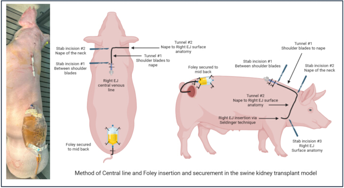
Figure 1: Tunneled central line and Foley catheter placement. Please click here to view a larger version of this figure.
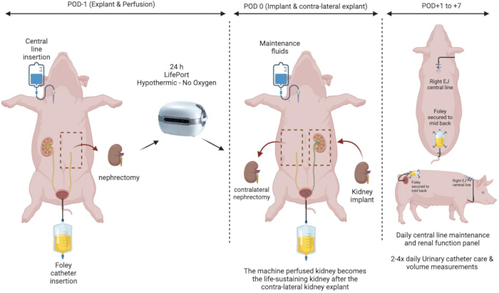
Figure 2: Life-sustaining porcine DCD kidney auto-transplantation. Please click here to view a larger version of this figure.
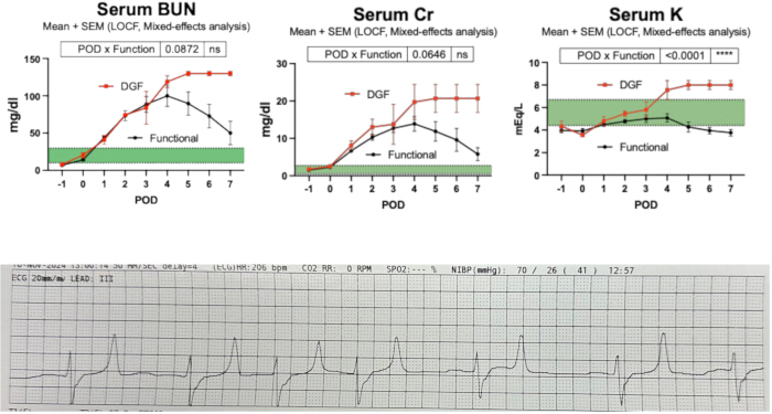
Figure 3: Delayed graft function in the porcine model. The difference in (A) urine output between immediate and delayed function grafts; (B) potassium levels between immediate and delayed function graft; (C) daily serum creatinine levels between immediate and delayed function grafts. All figures are represented as Mean + SEM with values reported as LOCF and analyzed with mixed-model analysis. Abbreviation: LOCF = last observation carried forward. N=5 pigs per arm. Please click here to view a larger version of this figure.
Discussion
Delayed graft function negatively impacts the clinical course following kidney transplantation. In the early posttransplant setting, it is associated with prolonged hospitalization, a higher rate of invasive procedures, and an increased risk of rejection. Longer-term, DGF is also associated with shorter graft survival. DGF is a manifestation of severe ischemia-reperfusion injury (I/RI) as evidenced by its higher prevalence in DCD transplants compared to DBD or living donor kidney transplants. Acute kidney injury (AKI) is secondary to acute tubular necrosis caused by I/RI and is thought to be the most common reason an allograft fails to function immediately. Clinically, DGF is defined by the need for dialysis within the first week of transplant. In large animal models, DGF has been challenging to define and study due to the lack of a standardized definition of graft sufficiency and the inability to perform dialysis to ascertain the temporary nature of the condition with dialysis as a bridge to graft functional recovery.
In this protocol, a porcine model was chosen as it is widely accepted to closely parallel human renal physiology (as proven by recent successful xeno-transplantation)10,11. Auto-transplantation was chosen to eliminate the allo-immune response, rejection, and need for and confounding toxic effects of calcineurin inhibitors. The definition of DGF is based on prolonged anuria (<100 mL of urine per day) past POD3 leading to severe hyperkalemia-associated EKG changes. Foley catheterization enables precise urine collection, which anchors the definition of delayed graft function and sufficiency to the functional outcome of urine quality and quantity. Daily blood collection enables the daily surveillance of traditional renal function panel parameters, providing a comprehensive picture of electrolyte homeostasis.
Establishing a reliable and reproducible animal model of delayed graft function, we believe, would accelerate the study of effective therapies for delayed graft function and result ultimately in improved kidney transplant outcomes. In summary, this protocol establishes a reproducible porcine kidney auto-transplantation model that enables objective characterization of delayed graft function using quantifiable urine output and serum biochemical parameters following controlled warm ischemic injury. By incorporating standardized surgical conditions, non-invasive urine collection, and serial laboratory monitoring, this approach provides a practical framework for studying DGF in the absence of dialysis and facilitates comparison with clinically relevant phenotypes of post-transplant graft dysfunction12,13,14,15.
Disclosures
The authors have no conflicts of interest to declare.
Acknowledgements
We thank the Toronto organ perfusion lab (Top lab) for sharing their expertise during training on porcine kidney transplantation. We also thank Michelle Mendiola Pla for sharing her expertise on non-invasive porcine Foley insertion.
Materials
| Name | Company | Catalog Number | Comments |
|---|---|---|---|
| Supine Foley Insertion | |||
| 12 Fr Foley | Urine collection | ||
| Surgical lube | foley insertion | ||
| 1 L urine bag | urine collection | ||
| 2-0 Nylon suture | foley and central line securement | ||
| Speculum & long debakey & Light | foley insertion | ||
| Urine sampling tubes (50ml conical) | urine sampling | ||
| CG8 + Chem 8 + Piccolo cartridges | daily labs | ||
| Prone-to-supine Central line insertion | |||
| 9.6 Fr Single lumen Hickman catheter tray | daily labs | ||
| 10 blade | stab incisions for CVC insertion | ||
| Central line neddless luer-lock cap | |||
| Hep-saline bag (1L saline + 10 Heparin Vials) | heplock CVC | ||
| Explant | |||
| Laparotomy pack | |||
| Bovie | |||
| Sterile light handles | |||
| Sponge pack | |||
| OR towels - white 17X23 inch (4 per pack) | |||
| 2-0 Silk ties | vascular control | ||
| 1 L Plasmalyte | balanced electrolyte solution | ||
| Sterile general laparotomy instruments tray | |||
| Warm normal saline for wet bowel towels | bowel handling | ||
| needle management box | |||
| Bookwalter retractor | |||
| Backtable | |||
| Sterile organ bags - 2 per kidney | |||
| 1 L crystalloid bag for slushy | |||
| UW solution - 250ml per kidney | |||
| Flush tubing | |||
| 14 gauge angiocath | |||
| Machine drape | |||
| organ weight (Scale + bowl + Sterile organ bag) | |||
| Machine perfusion | |||
| Perfusion machine | Lifeport Kidney transporter | ||
| Disposable organ chamber | Lifeport Kidney transporter | ||
| Perfusion solution | KPS-1 solution | ||
| Implant | |||
| Suction tubing | |||
| Yankauer suction tip | |||
| Suction cannister | |||
| Rubber shods | |||
| 4 mm Aortic punch | arterial anastomosis | ||
| Microsurgery instruments + clamp | |||
| Ureter guide wire | |||
| Ureter stent - 4.8 fr X 12 mm | ureter anastomosis | ||
| Abdominal closure | |||
| 1 looped prolene | |||
| Bupivacaine | |||
| Skin staplers | |||
| Samples | |||
| Sample tray | |||
| 14 G Biopsy gun | |||
| NBF biopsy jars | |||
| Eppendorf tubes | |||
| Green top blood tubes | |||
| 15 mL conicals | |||
| Meds | |||
| Norepinephrine vial | |||
| Dopamine vial | |||
| 1 L plasmalyte | balanced electrolyte solution | ||
| Aspirin 81 mg po qd tablets | |||
| Cefazoline vial | |||
| Exceed | |||
| Heparin 10K units |
References
- Lentine, K. L., et al. OPTN/SRTR 2023 annual data report: kidney. Am J Transplant. 25 (2 Suppl 1), S22-S137 (2025).
- Zwaini, Z., et al. Comparative analysis of risk factors in declined kidneys from donation after brain death and circulatory death. Medicina (Kaunas). 56, (2020).
- Flournoy, W. S., Mani, S. Percutaneous external jugular vein catheterization in piglets using a triangulation technique. Lab Anim. 43, 344-349 (2009).
- Seldinger, S. I. Catheter replacement of the needle in percutaneous arteriography: a new technique. Acta Radiol. 39, 368-376 (1953).
- Abraham, N., et al. Subnormothermic oxygenated machine perfusion (24 h) in DCD kidney transplantation. Transplant Direct. 10 (6), e1633(2024).
- Starzl, T. E., Marchioro, T. L., Morgan, W. W., Waddell, W. R. A technique for use of adult renal homografts in children. Surg Gynecol Obstet. 119, 106-108 (1964).
- Simforoosh, N., et al. Right laparoscopic donor nephrectomy and the use of inverted kidney transplantation: an alternative technique. BJU Int. 100, 1347-1350 (2007).
- Abraham, N., et al. Orthotopic transplantation of the full-length porcine intestine after normothermic machine perfusion. Transplant Direct. 8, e1390(2022).
- Kawamura, M., et al. Normothermic ex vivo kidney perfusion preserves mitochondrial and graft function after warm ischemia and is further enhanced by AP39. Nat Commun. 15, 8086(2024).
- Kawai, T., et al. Xenotransplantation of a porcine kidney for end-stage kidney disease. N Engl J Med. 392, 1933-1940 (2025).
- Montgomery, R. A., et al. Results of two cases of pig-to-human kidney xenotransplantation. N Engl J Med. 386, 1889-1898 (2022).
- Siedlecki, A., Irish, W., Brennan, D. C. Delayed graft function in the kidney transplant. Am J Transplant. 11, 2279-2296 (2011).
- Mannon, R. B. Delayed graft function: the AKI of kidney transplantation. Nephron. 140, 94-98 (2018).
- Kim, D. W., et al. Financial impact of delayed graft function in kidney transplantation. Clin Transplant. 34, e14022(2020).
- Schröppel, B., et al. Peritransplant eculizumab does not prevent delayed graft function in deceased donor kidney transplant recipients: results of two randomized controlled pilot trials. Am J Transplant. 20, 564-572 (2020).
Reprints and Permissions
This article has been published
Video Coming Soon