The study investigated serum LDH levels, which were significantly elevated in patients with large cell neuroendocrine lung cancer (LCNEC) and correlated with tumor size, stage, differentiation, and lymph node metastasis. LDH emerged as an independent prognostic factor, demonstrating high predictive accuracy, indicating its clinical utility in LCNEC prognosis.
Method Article
Clinical Significance of Serum LDH Concentration in the Prognostic Assessment of Large Cell Neuroendocrine Lung Cancer
1Department of Thoracic Surgery, Beijing Chest Hospital, Capital Medical University, Beijing Tuberculosis and Thoracic Tumor Research Institute, 2Department of Metabolism and Endocrinology, Zhangjiajie People's Hospital, 3Department of Internal Medicine, Zhangjiajie Public Hospital, 4Department of Metabolism and Endocrinology, The Third Hospital of Changsha (Changsha Hospital Affiliated to Hunan University)
Corresponding Authors: Ting Zhang <Zhangting0225@139.com>
Opens in a new tab
In This Article
Summary
Loading...
$$\rightleftharpoonup{xx}$$
$$\longleftharp{xx}$$,
$$\longrightharp{xx}$$,
Abstract
Loading...
$$\rightleftharpoonup{xx}$$
$$\longleftharp{xx}$$,
$$\longrightharp{xx}$$,
This study investigates the expression features of serum lactate dehydrogenase (LDH) in patients with large cell neuroendocrine carcinoma of the lung (LCNEC) and evaluates its clinical prognostic significance. A prospective cohort study was conducted, enrolling 80 LCNEC patients admitted between January 2022 and January 2024 (case group) and 80 healthy individuals undergoing medical check-ups during the same period (control group). Serum LDH levels were quantitatively measured by enzyme-linked immunosorbent assay (ELISA). Independent risk factors for poor prognosis were analyzed using a Multifactorial Logistic Regression (MLR) model. The prognostic predictive capability of LDH was assessed with the receiver operating characteristic (ROC) curve. Serum LDH levels in the case group were notably higher than those in the control group (p < 0.05). Subgroup analysis revealed that LDH levels of patients with tumor maximum diameter ≥5 cm, low variation, stage III-IV, and lymph node metastasis were significantly higher than those of the equivalent reference groups (all p < 0.05). The poor prognosis group had higher LDH expression levels and lymph node metastasis rates compared to the good prognosis group (both p < 0.05). MLR revealed that LDH (OR = 2.130, 95% CI: 1.312-3.465) and lymph node metastasis were autonomous predictors of poor prognosis (both p < 0.05). ROC analysis indicated that LDH predicted prognosis with an AUC of 0.825 (95% CI: 0.752-0.898), an optimal critical value of 280.5 U/L, a sensitivity of 75.0%, and a specificity of 78.0%. Serum LDH levels were abnormally elevated in LCNEC patients and significantly correlated with tumor malignancy features, which can act as a valid biological marker for assessing disease prognosis.
Introduction
Loading...
$$\rightleftharpoonup{xx}$$
$$\longleftharp{xx}$$,
$$\longrightharp{xx}$$,
As a highly malignant subtype of special lung cancer (LC), large cell neuroendocrine carcinoma (LCNEC) has exposed a clear trend of frequency development in clinical diagnosis and treatment in recent years and has attracted a great deal of attention in clinical and scientific research fields1. According to statistics, LCNEC accounts for 3% to 5% of all lung cancers. Compared with other LC types, LCNEC has unique biological behaviors and clinical features, such as poorly distinguished tumor cells and rapid development, which frequently lead to poor prognosis and a 5-year survival rate of less than 30%2. However, there are still many challenges in the diagnosis and treatment of LCNEC, particularly the lack of effective predictive indicators, which significantly impacts the advancement of clinical treatment plans and patient prognosis.
Under normal physiological conditions, serum levels of Lactate Dehydrogenase (LDH) are relatively stable and maintained within a narrow range3,4. However, when pathological changes occur5, especially in malignant tumors, the abnormal proliferation of tumor cells is frequently accompanied by a significant increase in metabolic activity, and this pathological change leads to a significant amount of intracellular LDH release, which in turn causes an abnormally high serum LDH level6,7. Recent clinical studies have confirmed that serum LDH levels are closely associated with the biological behaviors of several malignant tumors. Their fluctuations not only indicate tumor invasiveness but also serve as crucial molecular markers for measuring disease progression and predicting patient outcomes8,9,10.
In lung cancer research, the clinical significance of LDH has become increasingly recognized. Data from multiple clinical studies indicate that serum LDH levels in non-small cell lung cancer (NSCLC) and small cell lung cancer (SCLC) patients are significantly higher than those in healthy people. It is important to note that the level of this increase is significantly related to the tumor's clinicopathological features, as patients with higher tumor loads, later stages, and more general metastases tend to display higher serum LDH levels. More importantly, the abnormal changes of this biochemical index have been proven to be closely related to disease prognosis in patients11,12,13. In SCLC, the LDH level is also included in the prognostic assessment model, making it a critical measure for defining the prognosis of patients. These results have laid a compacted basis for using LDH in LC prognosis assessment.
However, relatively few studies have been conducted on LDH in LCNEC patients, and its clinical significance in the prognostic value of LCNEC is still unclear. As a subtype of LC with features of neuroendocrine differentiation, the cell biological behavior and metabolic features of LCNEC may differ from those of other types of lung cancer, and the expression and function of LDH in it may also be unique14. Therefore, the methodical study of serum LDH expression and its clinical value in LCNEC patients has multiple significances, which will not only enrich the important theoretical understanding of LCNEC but also have an essential practical value for improving the clinical management of patients.
Lactate dehydrogenase (LDH) is a systemic biomarker that is economical in terms of cost when compared to other biomarkers, as it reflects tumor burden and glycolytic activity; however, it is not precise because it also increases with liver disease or hemolysis6,12. Other lineage-oriented markers include neuron-specific enolase (NSE), pro-gastrin-releasing peptide (ProGRP). NSE is less consistent and more sensitive and specific to high-grade neuroendocrine tumors. Spatially resolved imaging biomarkers, like measurements of immune dyes (FDG), PET/CT (SUVmax, MTV, TLG), propose prognostic data and are costly and interoperable across platforms. More effective prognostic stratification of LCNEC is achieved by combining LDH with neuroendocrine markers and imaging11. Under clinical practice, LDH measurement is affected by the test conditions, such as the sensitivity of substances used and correction trials. Threshold values indicate inter-laboratory variability, and they need standardized reference ranges. Confounders that are assured to be interpreted include hemolysis, liver dysfunction, or systemic inflammation that can raise LDH per se, and thus affect specificity in oncological prognostication.
Protocol
Loading...
$$\rightleftharpoonup{xx}$$
$$\longleftharp{xx}$$,
$$\longrightharp{xx}$$,
This study strictly adhered to the principles of medical ethics and was approved by the hospital ethics committee of the Department of Thoracic Surgery, Beijing Chest Hospital, Capital Medical University, Beijing Tuberculosis and Thoracic Tumor Research Institute, Beijing, China. Written informed consent was obtained from enrolled participants.
1. Study subjects
- Use a prospective cohort design and select 80 LCNEC-diagnosed patients admitted between February 2022 and January 2024, including 55 male and 25 female LCNEC patients aged 45-75 years (mean 62.5 ± 10.3 years) in the study cohort. Concurrently, recruit 80 healthy individuals, matched for age and gender, as the control group, comprising 48 males (60%) and 32 females (40%), aged 40 to 70 years, with an average age of 60.8 ± 9.5 years.
- Include individuals aged ≥18 years old, diagnosis of LCNEC confirmed by pathohistological test15, not receiving any antitumor therapy (including radiotherapy, chemotherapy, immunotherapy, and targeted therapy) at the initial diagnosis; and having complete clinical data and conditions for follow-up.
- Exclude individuals who have a combination of other primary malignant tumors, severe organic diseases, active infectious diseases, mental disorders or cognitive dysfunction, and pregnant or lactating women.
- For prognostic categorization, sort patients into two groups based on follow-up outcomes: those without recurrence or metastasis, or with stable disease post-treatment, presentation no disease spread and living until the end of the follow-up; and those with poor results, characterized by tumor relapse, distant metastasis, or any-cause mortality (including death due to tumor progression or other causes during follow-up).
2. Research methodology
- Clinical data collection
- Collect data related to clinical cases in a standardized method through a specially designed case report form (CRF), and perform data entry and verification independently by two physicians with the title of attending physician or above. General data include demographic features: age, gender, height, weight (BMI); lifestyle: history of smoking (defined as more than 6 consecutive months with an average of 10 or more cigarettes per day), alcohol drinker (defined as at least 3 drinks in a week for at least one year); and comorbidities: previous hypertension, diabetes mellitus.
- Collect clinicopathological data of LCNEC patients, including tumor features, tumor diameter. Measure the postoperative pathological measurements as the basis; the site of tumor rate; the degree of pathological difference: highly differentiated, discreetly separated, and poorly differentiated; TNM staging: the evaluation of tumor staging strictly follows the 8th edition of the International Association for the Study of Lung Cancer (IASLC) TNM staging criteria16, and adopt a multidisciplinary comprehensive test model; and lymph node metastasis.
- Treatment plan development
- According to the principles of individualization and comprehensiveness, test individualized and comprehensive treatment under the guidance of the multidisciplinary cancer diagnosis and treatment team as described below.
- Early-stage patients (Stage I-II): Prefer radical surgical resection with the surgical modality including lobectomy + systematic lymph node dissection. Make the decision of whether adjuvant chemotherapy (platinum-containing double-drug regimen, such as cisplatin + pemetrexed) or radiotherapy is to be done based on the pathological results after the operation. Perform pemetrexed or radiotherapy for patients with positive margins or lymph node metastases.
- Locally advanced patients (stage III): For these patients, carry out multidisciplinary, complete treatment. For patients with a good prognosis, treat with neoadjuvant chemotherapy (2-4 cycles), followed by evaluation of surgical feasibility and postoperative adjuvant radiotherapy. Treat unresectable patients with simultaneous radiotherapy.
- Advanced patients (Stage IV): Use systemic behavior, including chemotherapy, targeted therapy, immunotherapy, and local radiotherapy if required, to alleviate symptoms. Record the treatment method of all patients in detail in the electronic medical record system, including the treatment cycle, drug dosage, adverse reactions, and dosage changes, to ensure the traceability of the treatment program.
3. Serum LDH detection
- Collect blood specimens from all study subjects and process them according to a standardized procedure. For patients in the LCNEC group, collect 5 mL of venous blood on an empty stomach on the morning of the 2nd day of admission (07:00-08:00). In the control group, perform blood collection on the day of physical investigation.
- Collect blood using vacuum blood collection tubes (K2-EDTA tubes) and gently invert (8x- 10x) to avoid clotting. Immediately centrifuge the specimens at 1,000 x g for 15 min at 4 °C. Then, remove the serum and divide it into two 2 mL cryopreservation tubes (1 mL for each tube), storing them in an ultra-low-temperature refrigerator at -80 °C to avoid repeated freezing and thawing during the entire process.
- At the same time, establish a comprehensive sample data management system to record the location and preservation time of the samples.
- Determine the serum LDH level using the double-antibody sandwich ELISA. Thaw the serum sample on ice and centrifuge at 10,000 x g at 4 °C.
- In each well, add 20 µL of plasma with 200 µL of LDH reagent buffer (pH 7.4) and incubate at 37 °C, 5 min. Spectrophotometrically, monitor NADH oxidation every 2 min at 320 nm with kinetic reads for 25.5 min. Use the NADH molar extinction coefficient (6.22 /mM/cm) to determine the activity of LDH (U/L) by dividing 6.22 mM by the pathlength, with 0.3 cm as the pathlength.
- For samples that were above the linear range, dilute and re-assay to check that they are linear (r 2 0.99). Perform comparative NSE and ProGRP biomarker tests in duplicate with 1:10 diluted plasma by using commercial ELISA kits. Perform a 3x washing procedure with PBST and incubate the reaction for 1 h at room temperature with HRP-conjugated antibody.
NOTE: In this study, serum LDH was quantified by using enzyme-linked immunosorbent assay (ELISA) because it has a greater analytical sensitivity and specificity and is applied more in research studies. ELISA can accurately quantitatively measure even at lower concentrations as compared to routine automated clinical chemistry analyzers, which quantify the overall activity of LDH and not the minute differences amongst different batches. Also, ELISA supports the identification of specific isoforms, should they be necessary, and is also flexible in the processing of samples, hence is adaptable to carefully controlled prospective studies. Although automated analyzers are fast and convenient in clinical practice, ELISA provides the ability to control the reproducibility and consistency of the result when measuring samples in a research-based study, which should ensure that the measurement is not confounded by hemolysis, lipemia, or other serum components, which is essential when comparing LDH levels and clinicopathological features and prognostic information in LCNEC patients. - Develop for 15 min with TMB substrate, followed by stopping the reaction with 2 M H2SO4 before measuring the absorbance at 450 nm (reference 620/650 nm). Use four-parameter logistic regression to create standard curves and keep the intra- and inter-assay CVs to less than 10% and 15% percent, respectively.
- Conduct Kaplan-Meier survival analysis and Cox proportional hazards regression as supplementary analyses to evaluate the prognostic value of LDH. Define continuous variables (LDH, NSE, ProGRP, tumor size, age, BMI) and categorical variables (sex, smoking, alcohol, stage, differentiation, lymph-node status) in SPSS v26.0 by clicking File > Open > Data and in R v4.3.1 (read_csv function).
- Normalize the values of all biomarkers to SI units, LDH to U/L (1 µkat/L = 60 U/L), NSE to ng/mL, and ProGRP to pg/mL. Log transform skewed distributed biomarkers (LN in SPSS, log in R), and then normalize to z scores to permit multivariate analysis, but retaining the raw scores to permit clinical interpretation. Use the SPS and R to conduct the Kaplan-Meier survival analysis, stratified by LDH levels (280.5 U / L), and use the log-rank test to compare the groups.
- Use LDH, lymph-node status, stage, differentiation, tumor size, age, sex, smoking, and alcohol as covariates in Cox proportional hazards regression. Set these in SPSS by clicking Analyze > Survival > Cox Regression, and in R use the coxph function. Use the Cox proportional hazards test (cox.zph in R) to test model assumptions.
- Present all statistical findings in the form of hazard ratios with a 95% confidence interval and treat the case-wise deletion of missing data by deletion, to preserve the level of analytical rigor and reproducibility. Elevated LDH levels were significantly associated with reduced overall survival (log-rank p < 0.01), and Cox regression confirmed LDH as an independent predictor of poor prognosis after adjustment for clinical covariates.
4. Follow-up protocol and prognostic grouping
- Follow-up process
- The start time of follow-up (commenced on February 1, 2022) is the date of diagnosis or admission of patients, and the end time is January 31, 2024, with a total follow-up period of 2 years. Use a multimodal follow-up program to ensure the completeness of the data.
- For postoperative patients, use standardized follow-up: arrange outpatient follow-up every 3 months for the first 2 years, including chest CT, abdominal ultrasound, tumor markers (CEA, NSE), and blood, liver, and kidney function tests; for patients with advanced stages, shorten the follow-up interval to every 2 months.
- For patients with limited mobility, use telephone follow-up every 1-2 months to record the changes in clinical warning signs, treatment response, and survival status. Focus follow-up visits on time to tumor recurrence, distribution of metastases, adjustment of treatment regimen, and cause of death (with distinct distinction for tumor-related deaths).
- Record all the follow-up data in a spreadsheet using the two-person entry method, and establish a regular verification mechanism to ensure the accuracy of the data.
- Prognostic grouping
- According to the results of follow-up, divide patients into 2 groups: no recurrence or metastasis during the follow-up, or recurrence but stable after treatment, with no sign of disease progression, and still alive at the end of the follow-up; poor prognosis group: tumor recurrence, distant metastasis, or all-cause death during the follow-up period (including death from tumor progression and death from other causes).
- Determine the cause of death of the deceased patients by combining clinical data, autopsy report (if any), and confirming it with two physicians with the title of deputy chief physician or above.
5. Statistical analysis
- Use software for statistical analysis. Present measurement data as mean ± standard deviation
 . For group comparisons, apply the independent samples t-test for two-group comparisons, and the analysis of variance (ANOVA) for multiple-group comparisons, with post-hoc multiple comparisons performed by the SNK-q test. Express categorical variables as percentages, and for group comparisons, use the chi-square test; when necessary, employ Fisher's exact probability method.
 - To explore factors influencing the prognosis of LCNEC, build a multivariable logistic regression model. To assess the prognostic predictive ability of serum LDH, use the receiver operating characteristic (ROC) curve, with the area under the curve (AUC) and its 95% confidence interval considered. Determine the optimal cut-off value, along with sensitivity and specificity. All statistical tests were two-sided, and a P-value < 0.05 was used to define statistically significant differences.
- Handle missing data (<5%) using case-wise deletion and exclude subjects with incomplete values for relevant variables from specific analyses. Given the low proportion of missingness, this method minimized bias and preserved statistical validity without significantly affecting sample size, analytical power, or the robustness of study results.
Results
Loading...
$$\rightleftharpoonup{xx}$$
$$\longleftharp{xx}$$,
$$\longrightharp{xx}$$,
Comparison of general information
The results presented that the percentage of smokers in the LCNEC group was markedly higher than that in the control group, and the discrepancy was clinically meaningful (p < 0.05; Table 1). There was no clinically significant difference between the two groups in terms of age, gender, and proportion of alcohol consumption (p > 0.05).
| Characteristic | LCNEC Group (n=80) | Control Group (n=80) | χ²/t | P |
| Age (years) | 62.5 ± 10.3 | 60.8 ± 9.5 | 1.023 | 0.308 |
| Gender (n, %) | 1.235 | 0.266 | ||
| - Male | 55 (68.75%) | 48 (60.00%) | ||
| - Female | 25 (31.25%) | 32 (40.00%) | ||
| BMI (kg/m²) | 24.5 ± 3.2 | 24.2 ± 2.8 | 0.546 | 0.586 |
| Smoking (n, %) | 50 (62.50%) | 30 (37.50%) | 10.25 | 0.001 |
| Alcohol use (n, %) | 35 (43.75%) | 30 (37.50%) | 1.025 | 0.311 |
Table 1: Comparison of general features between the two groups.
Comparison of LDH expression levels in serum
The serum LDH expression level in the LCNEC group reached 285.6 ± 56.3 U/L, while that in the control group stood at 198.5 ± 32.4 U/L (Table 2). The level was notably higher in the LCNEC group compared to the control group, and this difference was statistically significant (t = 10.245, p < 0.05).
Relationship between serum LDH expression levels and clinicopathological features in LCNEC patients
Statistical analysis revealed a significant correlation between serum LDH levels and certain clinicopathological features in LCNEC patients (p < 0.05). Specifically, serum LDH concentrations were remarkably increased in the tumor diameter ≥5 cm group, the poorly differentiated group, the stage III-IV group, and the group with lymph node metastasis than in the corresponding control groups (the tumor diameter <5 cm group, the moderately and highly differentiated group, the stages I-II group, and the group without metastatic liver nodes). Notably, the differences in serum LDH levels among LCNEC patients with different histologic subtypes were not statistically significant (p > 0.05; Table 2).
| Pathological Feature | No. of Cases (n=80) | LDH (U/L) | t | P |
| Tumor Size | 5.632 | <0.001 | ||
| <5 cm | 45 | 256.3 ± 48.5 | ||
| ≥5 cm | 35 | 321.5 ± 62.3 | ||
| Tumor Differentiation | 12.32 | <0.001 | ||
| Well-differentiated | 20 | 225.6 ± 35.7 | ||
| Moderately-differentiated | 30 | 278.9 ± 45.6 | ||
| Poorly-differentiated | 30 | 345.2 ± 58.9 | ||
| TNM Stage | 8.735 | <0.001 | ||
| Stage I-II | 40 | 262.5 ± 42.3 | ||
| Stage III-IV | 40 | 308.7 ± 55.6 | ||
| Lymph Node Metastasis | 7.89 | <0.001 | ||
| No | 45 | 248.6 ± 40.5 | ||
| Yes | 35 | 325.7 ± 60.3 |
Table 2: Relationship between serum LDH expression levels and clinicopathological features.
Comparison of LDH expression levels in the serum of LCNEC patients with different prognoses
When compared with the favorable-prognosis group, serum LDH expression levels in LCNEC patients with low incomes in the unfavorable-prognosis group were high, and the disparity was statistically significant (p < 0.05; Table 3).
| Group | No. of Cases (n=80) | LDH (U/L) |
| Poor Prognosis Group | 30 | 315.6±65.3 |
| Good Prognosis Group | 50 | 265.3±50.4 |
| t | - | 3.846 |
| P | - | 0.027 |
Table 3: Comparison of serum LDH levels between LCNEC patients with different prognoses.
Comparison of clinicopathologic features of LCNEC patients with different prognoses
In comparison to the favorable-prognosis group, the proportion of lymph node metastasis was higher in individuals with low incomes in the unfavorable-prognosis group, and the difference reached statistical significance (p < 0.05; Table 4).
| Pathological Feature | Poor Prognosis Group (n=30) | Good Prognosis Group (n=50) | χ² | P |
| Tumor Size (n, %) | 2.247 | 0.13 | ||
| <5 cm | 15 (50.00%) | 30 (60.00%) | ||
| ≥5 cm | 15 (50.00%) | 20 (40.00%) | ||
| Tumor Differentiation (n, %) | 4.597 | 0.1 | ||
| Well-differentiated | 5 (16.67%) | 15 (30.00%) | ||
| Moderately-differentiated | 10 (33.33%) | 20 (40.00%) | ||
| Poorly-differentiated | 15 (50.00%) | 15 (30.00%) | ||
| TNM Stage (n, %) | 3.276 | 0.07 | ||
| Stage I-II | 12 (40.00%) | 28 (56.00%) | ||
| Stage III-IV | 18 (60.00%) | 22 (44.00%) | ||
| Lymph Node Metastasis (n, %) | 10.21 | 0 | ||
| No | 10 (33.33%) | 35 (70.00%) | ||
| Yes | 20 (66.67%) | 15 (30.00%) |
Table 4: Comparison of clinicopathologic features of LCNEC patients with different prognoses
Logistic regression analysis of elements impacting unfavorable prognosis in LCNEC patients
The prognosis of LCNEC patients served as the dependent variable (poor prognosis = 1, good prognosis = 0). The indices with p < 0.05 (LDH, lymph node metastasis) from Table 2 and Table 4 were selected as autonomous variables. MLR analysis was conducted. The results indicated that LDH and lymph node metastasis were factors influencing the poor prognosis of LCNEC patients (p < 0.05), as presented in Table 5.
| Variable | β | SE | Wald χ² | OR | 95% CI | P |
| LDH | 0.76 | 0.25 | 8.806 | 2.13 | 1.312-3.465 | 0 |
| Lymph Node Metastasis | 0.69 | 0.29 | 5.763 | 1.99 | 1.123-3.546 | 0.02 |
Table 5: Logistic regression analysis of risk factors for poor prognosis in LCNEC patients.
ROC analysis of serum LDH levels for prognostic assessment of LCNEC patients
ROC curve analysis results revealed that the AUC of serum LDH levels for evaluating the prognosis of LCNEC patients was 0.825, with a 95% CI of 0.752-0.898. The cut-off value was 280.5 U/L, featuring a sensitivity of 75.0% and a specificity of 78.0%, as displayed in Table 6.
| Indicator | AUC | 95% CI | Cut-off Value | Sensitivity (%) | Specificity (%) |
| LDH | 0.83 | 0.752~0.898 | 280.5U/L | 75 | 78 |
Table 6: ROC analysis of serum LDH levels for prognostic evaluation in LCNEC patients.
Figure 1 illustrates the Receiver Operating Characteristic (ROC) curve for the predictive model under investigation. The ROC curve proves the trade-off between sensitivity and specificity across different classification thresholds, providing a quantitative assessment of model discriminative ability. The Area Under the Curve (AUC) is indicated, reflecting the overall performance; an AUC closer to 1 indicates excellent predictive accuracy, whereas values closer to 0.5 propose random classification.

Figure 1: ROC curve. The diagnostic efficiency of the proposed classification framework is demonstrated through the Receiver Operating Characteristic (ROC) analysis, which illustrates the trade-off between the true positive rate (sensitivity) and the false positive rate (1-specificity). The curve effectively highlights the discriminative capability of the model, with the area under the curve (AUC) signifying superior performance compared with baseline classifiers. Please click here to view a larger version of this figure.
Figure 2 presents a comparative analysis of tumor characteristics, including diameter, stage, differentiation, and lymph node metastasis, across patient subgroups. Visualizations such as boxplots and subgroup comparisons highlight variations in tumor burden and progression indicators, helping the identification of patterns associated with clinical outcomes. The figure aids in understanding how tumor heterogeneity correlates with prognosis and treatment response. Bonferroni correction was applied to adjust for multiple subgroup analyses. With four comparisons (tumor size, stage, differentiation, lymph-node status), the significance threshold was set at α_adj = 0.0125. LDH differences across all subgroups remained significant after correction, confirming robustness of associations while minimizing type I error risk.
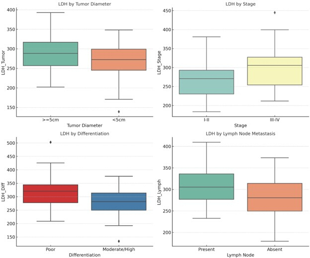
Figure 2: Tumor Analysis. The tumor segmentation results obtained using the proposed hybrid deep learning model are presented, where the segmented tumor regions are precisely delineated against ground-truth annotations. This visualization emphasizes the robustness and accuracy of the segmentation framework in identifying and localizing malignant regions. Please click here to view a larger version of this figure.
Figure 3 displays the mean serum lactate dehydrogenase (LDH) levels stratified by prognosis groups (poor versus favorable). The bar graph includes error bars representing standard deviation, and the results show that patients with a poor prognosis have significantly higher LDH levels compared to those with favorable results. This highlights LDH as a potential prognostic biomarker, reflecting tumor metabolic activity and systemic disease burden.
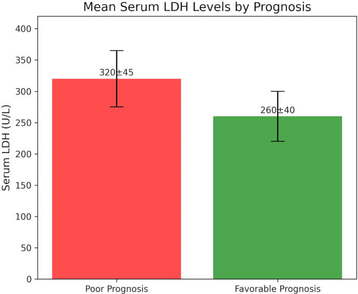
Figure 3: LDH mean levels. The levels present a comparative assessment of the mean Lactate Dehydrogenase (LDH) levels across benign, malignant, and control patient cohorts, revealing significantly elevated LDH levels in malignant samples, thereby indicating metabolic dysregulation associated with tumor progression and aggressiveness. Please click here to view a larger version of this figure.
Discussion
Loading...
$$\rightleftharpoonup{xx}$$
$$\longleftharp{xx}$$,
$$\longrightharp{xx}$$,
Biological mechanism of LDH in the development of LCNEC
Lactate dehydrogenase (LDH), a pivotal enzyme in the glycolysis pathway, makes LCNEC cancer cells prone to the Warburg effect. This entails their superior support on glycolysis for energy, a process that triggers substantial LDH release into the circulation17. In this study, serum LDH levels in LCNEC patients were significantly higher than those in healthy controls (285.6 ± 56.3 U/L versus 198.5 ± 32.4 U/L, p < 0.05), consistent with results from prior research on non-small cell lung cancer (NSCLC) and small cell lung cancer (SCLC)12.
The practical integration of LDH into diagnostic and prognostic workflows for LCNEC lies in its role as a rapid, cost-effective, and commonly accessible serum biomarker. LDH measurement can be incorporated alongside imaging and histopathology at baseline evaluation to provide complementary prognostic data. Elevated LDH may identify high-risk patients who warrant intensified surveillance, earlier initiation of systemic therapy, or enrollment in clinical trials exploring glycolysis-targeted approaches. Furthermore, LDH can be monitored longitudinally to track treatment response or detect relapse and present a dynamic adjunct to conventional follow-up strategies. Its ease of measurement in routine laboratories ensures feasibility, while incorporation into risk models with nodal status or molecular profiling may enhance personalized prognostication in LCNEC.
In terms of molecular mechanisms, the elevation of LDH may involve the following pathways: mutation of the oncogene TP53 and amplification of the oncogene MYC, which are commonly found in LCNEC, can upregulate the expression of the LDH-A subunit through activation of the HIF-1α pathway, promoting lactic acid production and LDH release18. Meanwhile, a hypoxic microenvironment often exists in LCNEC tumor tissues, and hypoxia-inducible factor (HIF-1α) induces the development of LDH-A expression, which promotes the translation of pyruvic acid into lactic acid and maintains the energy supply of tumor cells18.
At the same time, the accumulation of lactic acid decreases the pH value of the tumor microenvironment, promotes the activation of matrix metalloproteinases (MMPs), and enhances the invasion and metastasis of tumor cells19. LCNEC exhibits characteristics of neuroendocrine differentiation, and the abnormal function of intracellular mitochondria in LCNEC may lead to a decrease in aerobic oxidation efficiency, thereby relying on glycolysis for energy. This metabolic characteristic may render LDH a unique metabolic marker for this subtype. Elevated LDH reflects enhanced glycolytic activity (Warburg effect) in LCNEC, indicating metabolic reprogramming that fuels tumor progression. This biochemical signature may guide consideration of glycolysis-targeted therapies, such as LDH inhibitors or metabolic modulators, and present a rationale for personalized treatment methods in patients with persistently high LDH levels20.
Association between LDH and clinicopathological features of LCNEC
Data from this research indicated that serum LDH levels were notably correlated with the tumor burden and malignancy degree of LCNEC. This result closely coincided with that of 20, who determined the optimal preoperative critical value of LDH as 195.5 U/L using ROC curves. It verified that high preoperative LDH levels and the postoperative LDH elevation trend were independent prognostic factors impacting DFS (p < 0.001). Remarkably, the LDH level (345.2 ± 58.9 U/L) in this study was significantly higher in patients with poorly differentiated tumors, consistent with the research21 on prognostic factors of pHGNEC. That study emphasized the correlation between tumor differentiation degree and prognosis and developed a prognostic nomogram incorporating LDH and other indicators through multifactorial analysis.
In terms of tumor metabolic features, the present study found that LDH levels were significantly elevated in patients with tumor diameters ≥5 cm, which is similar to the findings of22 on neuroendocrine carcinomas, which pointed out that the tumor load was positively correlated with the level of LDH, and that the survival of patients with IPNECs, including LCNECs, correlated markedly with LDH and other indices (p < 0.05). Of particular note, in the present study, LDH levels were elevated by 17.6% in phase III-IV versus phase I-II patients, a degree of difference that is superior to that reported in NSCLC (HR 1.24, p = 0.02), possibly reflecting the more aggressive biology of LCNEC23.
Regarding the prognostic value of LDH, the result of the present study contrasts interestingly with the immunotherapy study24, which did not find a significant connection between LDH and PFS (p = 0.83) but recommends the need to incorporate multifactorial analyses when assessing the therapeutic response of LCNEC. In contrast, the misdiagnosed case reported25, side-steps the heterogeneity of LCNEC, emphasizing that neuroendocrine tumor possibilities should be considered in the presence of abnormally elevated LDH.
Clinical significance of LDH as a prognostic indicator in LCNEC
In this study, using MLR analysis, LDH (OR = 2.130, 95% CI = 1.312-3.465, p = 0.003) and lymph node metastasis were identified as independent risk factors for poor prognosis in LCNEC patients. This finding aligns with a study in NSCLC, which demonstrated that elevated baseline LDH was associated with a poor prognosis, regardless of the treatment received (HR = 1.49, p = 0.0001)12.
Notably, the prognostic predictive performance of LDH in this study (AUC = 0.825) was significantly superior to that of the traditional marker NSE. This contrasts with the research26on neuroendocrine-transformed tumors, which reported that NSE had a sensitivity of only 75% in diagnosing neuroendocrine transformation, suggesting that LDH may be more suitable for the overall management of LCNEC.
Regarding the prognostic assessment system, the LDH cutoff value (280.5 U/L) set in this study has complementary value with the LIPI scoring system27. They found that inflammatory markers (LIPI and PIV) were notably associated with the prognosis of LCNEC (p = 0.002 and p = 0.001). In combination with the results of this study, it is recommended that, in the future, it may be valuable to develop a comprehensive scoring method that integrates metabolic markers (LDH) and immunoinflammatory indicators.
Notably, NLR was significantly correlated with the tumor immune microenvironment of LCNEC, as proved by CD8+ TILs (r = -0.648, p = 0.005)19,28. Meanwhile, this study revealed that the LDH level was correlated with tumor burden. These two indicators may individually reflect the biological features of LCNEC from the dimensions of immune status and metabolic activity.
Differences and similarities with similar studies and probable clinical applications
In comparison with studies of LDH in other LC subtypes, the clinical significance of LDH in LCNEC is unique: high LDH in NSCLC is mainly correlated with tumor stage, and it is worth noting that serum LDH levels in patients with LCNEC show a stronger correlation with the degree of tumor variation and the metastatic status of lymph nodes29,30. This phenomenon may stem from the metabolic heterogeneity caused by the unique neuroendocrine differentiation features of LCNEC31.
Although SCLC and LCNEC are both highly invasive neuroendocrine tumors, and both serum LDH levels effectively reflect tumor load, comparative analyses have shown that the degree of LDH elevation is more pronounced in patients with LCNEC, which may be related to their higher rate of glycolysis32.
Potential clinical applications include: (1) auxiliary diagnosis: although LDH lacks tumor specificity, combined with clinical symptoms and imaging features, elevated LDH may recommend the possibility of LCNEC, especially when puncture specimens are limited, and serum LDH can be used as a supplementary indicator33. (2) Therapeutic decision: For patients with significantly elevated LDH, it may indicate active tumor metabolism, and a combination of drugs targeting glycolysis (e.g., 2-deoxy-D-glucose) based on chemotherapy may be considered to improve the therapeutic sensitivity34. (3) Follow-up monitoring: Regular detection of LDH level can detect tumor recurrence or progression at an early stage, and its sensitivity is better than that of traditional imaging (imaging often lags behind metabolic changes by 1-3 months)35.
Study limitations and future directions
This study has the following limitations: (1) Small sample size: only 80 LCNEC patients were included, which can cause bias in the study results and insufficient statistical efficacy, especially in the analysis of rare metastatic sites. (2) Single-center study: The study data came from a single center, and the geographical and population features may affect the generalizability of the results. (3) Lack of dynamic monitoring: only baseline levels of LDH were detected, and the prognostic relationship between dynamic changes in LDH during treatment was not analyzed. (4) Insufficient research on mechanisms: The specific pathways of LDH in LCNEC, such as the co-expression of LDH-A subunit and neuroendocrine markers, have not been explored in depth.
TP53 and RB1 mutation status were not assessed in this study; however, they are acknowledged as relevant complementary biomarkers in LCNEC. Integrating molecular profiling with serum LDH evaluation could enhance prognostic precision and provide a more comprehensive model for risk stratification and therapeutic decision-making in future research.
Future research can be expanded in the following directions: expanding the sample size and establishing a prognostic model of LDH in LCNEC; screening for markers synergistic with LDH (e.g., HIF-1α, MMP-9)35 and constructing a multi-indicator prognostic model to improve the accuracy of prediction; designing clinical studies based on the aberrant expression of LDH to evaluate the therapeutic effects of glycolysis inhibitors (e.g., FX-11) in LCNEC36.
Conclusion
This study revealed that serum LDH concentration is notably elevated in LCNEC patients and closely correlates with tumor problems, differentiation degree, TNM stage, and lymph node metastasis. LDH can serve as an independent risk factor for prognostic evaluation of LCNEC, with its diagnostic efficacy surpassing that of some traditional markers. In the future, it is advisable to conduct a multicenter sample study to validate these results further, explore the molecular mechanism of LDH in LCNEC development, and thus help its translational application in individualized diagnosis and treatment.
Disclosures
Loading...
$$\rightleftharpoonup{xx}$$
$$\longleftharp{xx}$$,
$$\longrightharp{xx}$$,
The authors have no competing interests to disclose.
Materials
| Name | Company | Catalog Number | Comments |
|---|---|---|---|
| Hardware | |||
| 2 TB HDD | Segante | 6G SATA 7.2k 3.5"(LFF) | Storage |
| 8 GB RAM | Kingston Technology | 4Rx4 DDR4 LRDIMM 2666 MT/s. | Storage |
| Intel Core i5 | Intel | Intel Core i7 | Processor |
| NVIDIA GeForce RTX 4060 Ti | Nvidia Corporation | NA | Graphics Card |
| Software | |||
| R | (version 4.1.2) | Programming Language | |
| SPSS | IBM | 30 | Data Visualization Tool |
References
Loading...
$$\rightleftharpoonup{xx}$$
$$\longleftharp{xx}$$,
$$\longrightharp{xx}$$,
- Management of large cell neuroendocrine carcinoma. Frontiers in Oncology. 11, 653162(2021).">Corbett, V., Arnold, S., Anthony, L., et al. Management of large cell neuroendocrine carcinoma. Frontiers in Oncology. 11, 653162(2021).
- Large cell neuroendocrine carcinoma of the lung: current understanding and challenges. Journal of Clinical Medicine. 11 (5), 1461(2022).">Andrini, E., Marchese, P. V., De Biase, D., et al. Large cell neuroendocrine carcinoma of the lung: current understanding and challenges. Journal of Clinical Medicine. 11 (5), 1461(2022).
- Pulmonary large cell neuroendocrine carcinoma. Pathology & Oncology Research. 28, 1610730(2022).">Yang, L., Fan, Y., Lu, H. Pulmonary large cell neuroendocrine carcinoma. Pathology & Oncology Research. 28, 1610730(2022).
- Urinary large cell neuroendocrine carcinoma: a clinicopathologic analysis of 22 cases. American Journal of Surgical Pathology. 45 (10), 1399-1408 (2021).">Wang, G., Yuan, R., Zhou, C., et al. Urinary large cell neuroendocrine carcinoma: a clinicopathologic analysis of 22 cases. American Journal of Surgical Pathology. 45 (10), 1399-1408 (2021).
- Incidence, survival comparison, and novel prognostic evaluation approaches for stage III-IV pulmonary large cell neuroendocrine carcinoma and small cell lung cancer. BMC Cancer. 23 (1), 312(2023).">Huang, L., Feng, Y., Xie, T., et al. Incidence, survival comparison, and novel prognostic evaluation approaches for stage III-IV pulmonary large cell neuroendocrine carcinoma and small cell lung cancer. BMC Cancer. 23 (1), 312(2023).
- Serum lactate dehydrogenase activities as systems biomarkers for 48 types of human diseases. Scientific Reports. 11 (1), 12997(2021).">Wu, Y., Lu, C., Pan, N., et al. Serum lactate dehydrogenase activities as systems biomarkers for 48 types of human diseases. Scientific Reports. 11 (1), 12997(2021).
- Ubiquinol-mediated suppression of mitochondria-associated ferroptosis is a targetable function of lactate dehydrogenase B in cancer. Nature Communications. 16 (1), 2597(2025).">Deng, H., Zhao, L., Ge, H., et al. Ubiquinol-mediated suppression of mitochondria-associated ferroptosis is a targetable function of lactate dehydrogenase B in cancer. Nature Communications. 16 (1), 2597(2025).
- The multiple roles of LDH in cancer. Nature Reviews. Clinical Oncology. 19 (12), 749-762 (2022).">Claps, G., Faouzi, S., Quidville, V., et al. The multiple roles of LDH in cancer. Nature Reviews. Clinical Oncology. 19 (12), 749-762 (2022).
- Rationale for LDH-targeted cancer immunotherapy. European Journal of Cancer. 181, 166-178 (2023).">Miholjcic, T. B. S., Halse, H., Bonvalet, M., et al. Rationale for LDH-targeted cancer immunotherapy. European Journal of Cancer. 181, 166-178 (2023).
- Role of LDH in tumor glycolysis: regulation of LDHA by small molecules for cancer therapeutics. In: Seminars in Cancer Biology. 87, Academic Press. 184-195 (2022).">Sharma, D., Singh, M., Rani, R. Role of LDH in tumor glycolysis: regulation of LDHA by small molecules for cancer therapeutics. In: Seminars in Cancer Biology. 87, Academic Press. 184-195 (2022).
- Analysis of systemic inflammatory biomarkers in neuroendocrine carcinomas of the lung: prognostic and predictive significance of NLR, LDH, ALI, and LIPI score. Therapeutic Advances in Medical Oncology. 12, 1758835920942378(2020).">Galvano, A., Peri, M., Guarini, A. A., et al. Analysis of systemic inflammatory biomarkers in neuroendocrine carcinomas of the lung: prognostic and predictive significance of NLR, LDH, ALI, and LIPI score. Therapeutic Advances in Medical Oncology. 12, 1758835920942378(2020).
- Pre- and on-treatment lactate dehydrogenase as a prognostic and predictive biomarker in advanced non-small cell lung cancer. Cancer. 128 (8), 1574-1583 (2022).">Tjokrowidjaja, A., Lord, S. J., John, T., et al. Pre- and on-treatment lactate dehydrogenase as a prognostic and predictive biomarker in advanced non-small cell lung cancer. Cancer. 128 (8), 1574-1583 (2022).
- Targeting lactate dehydrogenase a improves radiotherapy efficacy in non-small cell lung cancer: from bedside to bench. Journal of Translational Medicine. 19 (1), 170(2021).">Yang, Y., Chong, Y., Chen, M., et al. Targeting lactate dehydrogenase a improves radiotherapy efficacy in non-small cell lung cancer: from bedside to bench. Journal of Translational Medicine. 19 (1), 170(2021).
- Prognostic value of lactate dehydrogenase in non-small cell lung cancer patients with brain metastases: a retrospective cohort study. Journal of Thoracic Disease. 14 (11), 4468(2022).">Wang, S., Lv, J., Lv, J., et al. Prognostic value of lactate dehydrogenase in non-small cell lung cancer patients with brain metastases: a retrospective cohort study. Journal of Thoracic Disease. 14 (11), 4468(2022).
- Recent Advances and Controversies in Minute Pulmonary Meningothelial-like Nodules. Zhongguo Fei Ai Za Zhi. 26 (8), 621-629 (2023).">Li, H., et al. Recent Advances and Controversies in Minute Pulmonary Meningothelial-like Nodules. Zhongguo Fei Ai Za Zhi. 26 (8), 621-629 (2023).
- The IASLC lung cancer staging project: external validation of the revision of the TNM stage groupings in the eighth edition of the TNM classification of lung cancer. Journal of Thoracic Oncology. 12 (7), 1109-1121 (2017).">Chansky, K., Detterbeck, F. C., Nicholson, A. G., et al. The IASLC lung cancer staging project: external validation of the revision of the TNM stage groupings in the eighth edition of the TNM classification of lung cancer. Journal of Thoracic Oncology. 12 (7), 1109-1121 (2017).
- Large cell neuro-endocrine carcinoma of the lung: current treatment options and potential future opportunities. Frontiers in Oncology. 11, 650293(2021).">Ferrara, M. G., Stefani, A., Simbolo, M., et al. Large cell neuro-endocrine carcinoma of the lung: current treatment options and potential future opportunities. Frontiers in Oncology. 11, 650293(2021).
- Molecular subtypes and prognostic factors of lung large cell neuroendocrine carcinoma. Translational Lung Cancer Research. 13 (9), 2222(2024).">Liu, T., Chen, X., Mo, S., et al. Molecular subtypes and prognostic factors of lung large cell neuroendocrine carcinoma. Translational Lung Cancer Research. 13 (9), 2222(2024).
- The prognostic significance of lung immune prognostic index (LIPI) and pan-immune-inflammation value (PIV) in patients with large cell neuroendocrine carcinoma. , (2022).">Menekse, S. The prognostic significance of lung immune prognostic index (LIPI) and pan-immune-inflammation value (PIV) in patients with large cell neuroendocrine carcinoma. , (2022).
- Correlation analysis of serum LDH concentration before and after operation and prognosis of large cell neuroendocrine lung cancer patients. Zhongguo Fei Ai Za Zhi. 24 (5), (2021).">Haocheng, W., Dongfeng, S., Ya, D., et al. Correlation analysis of serum LDH concentration before and after operation and prognosis of large cell neuroendocrine lung cancer patients. Zhongguo Fei Ai Za Zhi. 24 (5), (2021).
- Prognostic factors and nomogram for pulmonary resected high-grade neuroendocrine carcinomas: a 20-year single institutional real-world experience. Orphanet Journal of Rare Diseases. 19 (1), 232(2024).">Liu, L., Zhang, J., Zhao, K., et al. Prognostic factors and nomogram for pulmonary resected high-grade neuroendocrine carcinomas: a 20-year single institutional real-world experience. Orphanet Journal of Rare Diseases. 19 (1), 232(2024).
- Clinical features and prognosis of advanced intra- and extra-pulmonary neuroendocrine carcinomas. Journal of Cancer Research and Therapeutics. 19 (4), 951-956 (2023).">Chen, X. Y., Guo, N. J., Guo, P. L., et al. Clinical features and prognosis of advanced intra- and extra-pulmonary neuroendocrine carcinomas. Journal of Cancer Research and Therapeutics. 19 (4), 951-956 (2023).
- Interrelations between patients' clinicopathological characteristics and their association with response to immunotherapy in a real-world cohort of NSCLC patients. Cancers. 13 (13), 3249(2021).">Callejo, A., Frigola, J., Iranzo, P., et al. Interrelations between patients' clinicopathological characteristics and their association with response to immunotherapy in a real-world cohort of NSCLC patients. Cancers. 13 (13), 3249(2021).
- Preliminary analysis of progression-free survival results for atezolizumab plus platinum etoposide as first-line treatment in metastatic lung large-cell neuroendocrine carcinoma patients. , (2025).">Evangelou, G., Vamvakaris, I., Nikolaidou, V., et al. Preliminary analysis of progression-free survival results for atezolizumab plus platinum etoposide as first-line treatment in metastatic lung large-cell neuroendocrine carcinoma patients. , (2025).
- Pulmonary large-cell neuroendocrine carcinoma, a multifaceted disease-case report and literature review. Diagnostics. 15 (9), 1056(2025).">Constantin, A. A., Cotea, A. A., Mihălțan, F. D. Pulmonary large-cell neuroendocrine carcinoma, a multifaceted disease-case report and literature review. Diagnostics. 15 (9), 1056(2025).
- Serum NSE is early marker of transformed neuroendocrine tumor after EGFR-TKI treatment of lung adenocarcinoma. Cancer Management and Research. 2023, 1293-1302 (2023).">Mao, X., Liu, J., Hu, F., et al. Serum NSE is early marker of transformed neuroendocrine tumor after EGFR-TKI treatment of lung adenocarcinoma. Cancer Management and Research. 2023, 1293-1302 (2023).
- Prognostic impact of peripheral blood neutrophil to lymphocyte ratio in advanced-stage pulmonary large cell neuroendocrine carcinoma and its association with the immune-related tumour microenvironment. British Journal of Cancer. 124 (5), 925-932 (2021).">Shirasawa, M., Yoshida, T., Horinouchi, H., et al. Prognostic impact of peripheral blood neutrophil to lymphocyte ratio in advanced-stage pulmonary large cell neuroendocrine carcinoma and its association with the immune-related tumour microenvironment. British Journal of Cancer. 124 (5), 925-932 (2021).
- Combining lactate dehydrogenase and fibrinogen: potential factors to predict therapeutic efficacy and prognosis of patients with small-cell lung cancer. Cancer Management and Research. 2021, 4299-4307 (2021).">Huang, W., Liu, P., Zong, M., et al. Combining lactate dehydrogenase and fibrinogen: potential factors to predict therapeutic efficacy and prognosis of patients with small-cell lung cancer. Cancer Management and Research. 2021, 4299-4307 (2021).
- Afatinib reverses EMT via inhibiting CD44-Stat3 axis to promote radiosensitivity in nasopharyngeal carcinoma. Pharmaceuticals. 16 (1), 37(2023).">Huang, H., Huang, F. Afatinib reverses EMT via inhibiting CD44-Stat3 axis to promote radiosensitivity in nasopharyngeal carcinoma. Pharmaceuticals. 16 (1), 37(2023).
- M2 macrophage exosome-derived lncRNA AK083884 protects mice from CVB3-induced viral myocarditis through regulating PKM2/HIF-1α axis mediated metabolic reprogramming of macrophages. Redox Biology. 69, 103016(2024).">Zhang, Y., Zhu,, et al. M2 macrophage exosome-derived lncRNA AK083884 protects mice from CVB3-induced viral myocarditis through regulating PKM2/HIF-1α axis mediated metabolic reprogramming of macrophages. Redox Biology. 69, 103016(2024).
- Multitargeted immunomodulatory therapy for viral myocarditis by engineered extracellular vesicles. ACS Nano. 18 (4), 2782-2799 (2024).">Pei, W., Zhang, Y., et al. Multitargeted immunomodulatory therapy for viral myocarditis by engineered extracellular vesicles. ACS Nano. 18 (4), 2782-2799 (2024).
- Regulation of oncoprotein 18/Stathmin signaling by ERK concerns the resistance to Taxol in nonsmall cell lung cancer cells. Cancer Biotherapy and Radiopharmaceuticals. 31 (2), 37-43 (2016).">Lin, X., Liao, Y., et al. Regulation of oncoprotein 18/Stathmin signaling by ERK concerns the resistance to Taxol in nonsmall cell lung cancer cells. Cancer Biotherapy and Radiopharmaceuticals. 31 (2), 37-43 (2016).
- Identification of cuproptosis-related lncRNAs with the significance in prognosis and immunotherapy of oral squamous cell carcinoma. Computers in Biology and Medicine. 171, 108198(2024).">Gong, H., Liu, Z., et al. Identification of cuproptosis-related lncRNAs with the significance in prognosis and immunotherapy of oral squamous cell carcinoma. Computers in Biology and Medicine. 171, 108198(2024).
- Activatable fluorescent probes for imaging and diagnosis of hepatocellular carcinoma. Journal of Innovative Optical Health Sciences. 18 (3), 2530004(2024).">Men, X., Liu, F., et al. Activatable fluorescent probes for imaging and diagnosis of hepatocellular carcinoma. Journal of Innovative Optical Health Sciences. 18 (3), 2530004(2024).
- Deubiquitinase PSMD7 facilitates pancreatic cancer progression through activating Notch1 pathway via modifying SOX2 degradation. Cell & Bioscience. 14 (1), 35(2024).">Luo, C., Yu, Y., et al. Deubiquitinase PSMD7 facilitates pancreatic cancer progression through activating Notch1 pathway via modifying SOX2 degradation. Cell & Bioscience. 14 (1), 35(2024).
- Lnc-TMEM132D-AS1 as a potential therapeutic target for acquired resistance to osimertinib in non-small-cell lung cancer. Molecular Omics. 19 (3), 238-251 (2023).">Wang, N., Zhao, Q., et al. Lnc-TMEM132D-AS1 as a potential therapeutic target for acquired resistance to osimertinib in non-small-cell lung cancer. Molecular Omics. 19 (3), 238-251 (2023).
Reprints and Permissions
Request permission to reuse the text or figures of this JoVE article
Request Permission