Dyslipidemia drives atherosclerosis: it requires mechanistic research, emerging therapies, precision strategies, combined with guidelines and technologies to optimize prevention and treatment.
A subscription to JoVE is required to view this content. Sign in or start your free trial.
Review Article
Precision Management of Dyslipidemia in Atherosclerosis: Mechanisms, Therapeutic Strategies, and Future Directions
In This Article
Summary
Abstract
Dyslipidemia is a central driver in the initiation and progression of atherosclerosis (AS). The chronic inflammation and endothelial injury triggered by dyslipidemia are key pathological events in AS development. Elucidating the molecular network underlying dyslipidemia and developing precise interventions are critical for achieving precision prevention and treatment of AS. Recent studies have demonstrated that sterol regulatory element-binding protein 1 (SREBP1) and lipoprotein(a) [Lp(a)] play pivotal roles in the regulation of lipid synthesis and transport. Additionally, gut microbiota-derived metabolites, such as trimethylamine N-oxide (TMAO) and short-chain fatty acids (SCFAs), can activate inflammatory pathways and promote lipid deposition via inter-organ signaling axes, thereby accelerating the progression of AS.
However, clinical studies have revealed that even when low-density lipoprotein cholesterol (LDL-C) levels are within the recommended range, a significant number of patients continue to experience cardiovascular events. This indicates the widespread presence of "residual risk". Such residual risk is primarily driven by elevated non-high-density lipoprotein cholesterol (non-HDL-C), abnormal levels of Lp(a), and imbalances in the triglyceride to HDL-C (TG/HDL-C) ratio, highlighting the limitations of traditional therapies in comprehensive lipid profile management.
Emerging targeted therapies, including proprotein convertase subtilisin/kexin type 9 (PCSK9) inhibitors, small interfering RNA (siRNA)-based treatments, and Lp(a)-lowering agents like pelacarsen, represent promising strategies for more precise lipid modulation.
With the continuous advancement of related research, the precise management of AS will increasingly rely on deeper mechanistic insights and individualized therapeutic strategies. Current strategies for AS prevention and treatment focus on understanding key pathways, including lipid metabolism, inflammation, and vascular dysfunction, to develop targeted therapies. The integration of the 2023 Chinese Guidelines for Lipid Management, imaging, and AI-assisted decision-making will promote data-driven, precision medicine. Personalized drug selection, efficacy monitoring, and long-term follow-up will optimize clinical outcomes and enhance prevention strategies for high-risk patients.
Introduction
Atherosclerosis (AS) is the primary pathological basis of cardiovascular diseases and is fundamentally recognized as a chronic inflammatory condition1. It leads to arterial narrowing or even occlusion, ultimately resulting in atherosclerotic cardiovascular diseases (ASCVD) such as coronary artery disease (CAD) and stroke2,3. According to global estimates, approximately 17.6 million deaths annually are attributed to ASCVD4. In China alone, the total number of ASCVD patients has reached 330 million, including 11.39 million individuals diagnosed with CAD, with the prevalence increasing at an average annual rate of 20%5. AS-related diseases not only pose a serious threat to public health but also impose a substantial medical and socioeconomic burden. Therefore, establishing a more effective system for the prevention and management of AS has become a critical challenge in the field of global public health6.
Among the many risk factors for AS, dyslipidemia is considered the most critical and modifiable determinant. Although conventional lipid-lowering therapies-such as statins-have shown efficacy in reducing lipid levels, considerable interindividual variability in treatment response and inconsistent clinical outcomes remain prevalent in real-world practice. These limitations hinder the comprehensive inhibition of AS progression. As a result, the concept of precision management, which is grounded in mechanistic understanding and tailored to individual differences, has emerged and is increasingly viewed as a future direction in the prevention and treatment of chronic cardiovascular diseases7,8. Precision management emphasizes the identification of key disease-driving factors at the mechanistic level and the implementation of targeted, personalized interventions to enhance both therapeutic efficacy and safety9.
At the pathological level, dysregulated lipid metabolism forms the foundation for the initiation and progression of AS. Lipid accumulation mediated by low-density lipoprotein cholesterol (LDL-C), along with endothelial injury and chronic inflammatory responses induced by metabolites such as trimethylamine N-oxide (TMAO), constitute key mechanisms driving AS formation, further development, and plaque instability10. These pathological processes are closely associated with various clinical risk phenotypes, including residual cholesterol risk, inflammatory risk, and metabolic risk. Identifying the correspondence between underlying mechanisms and risk profiles facilitates disease-oriented precision therapies, thereby overcoming the limitations of traditional treatment paradigms that focus on single biomarkers. This approach enables the development of more targeted and sustained intervention strategies tailored to individual patients11.
Overall, precision management transforms the traditional prevention and treatment paradigm of AS by deeply integrating molecular mechanisms with the identification of residual risks. This approach not only enhances therapeutic efficacy and safety but also drives the shift from a "one-size-fits-all" strategy toward individualized care, providing both a theoretical foundation and practical guidance for the scientific management of AS12,13.
Access restricted. Please log in or start a trial to view this content.
Review and Perspective
Core Pathological Mechanisms of As:
Key transcription factors in lipid metabolism
A hallmark of AS is abnormal lipid accumulation in the arterial wall, closely linked to dysregulated transcriptional control of lipid metabolism, including cholesterol and fatty acid metabolism. Key regulators like liver X receptors (LXRs), peroxisome proliferator-activated receptors (PPARs), and SREBPs control lipid homeostasis. Dysfunction of these transcription factors can directly contribute to lipid metabolic disorders, promoting AS progression.
Liver X receptors (LXRs)
LXRs, members of the nuclear receptor family, reside in the nucleus and act as ligand-activated transcription factors. Their endogenous ligands, such as 22-hydroxycholesterol, enable LXRs to sense intracellular cholesterol levels. Upon activation, LXRs form heterodimers with retinoid X receptors (RXRs)14, binding to LXR response elements (LXREs) in target gene promoters, exerting dual regulatory effects15.
LXRs activate ATP-binding cassette transporters A1 (ABCA1) and G1 (ABCG1), promoting cholesterol efflux from macrophages to HDL, reducing vascular lipid accumulation, and inhibiting plaque formation16. LXRs also upregulate SREBP-1c and fatty acid synthase (FASN), enhancing hepatic fatty acid synthesis17.
Excessive LXR activation can worsen hepatic lipid accumulation, but its role in cholesterol clearance in the vascular wall remains a promising therapeutic target for AS prevention and treatment16(Figure 1).
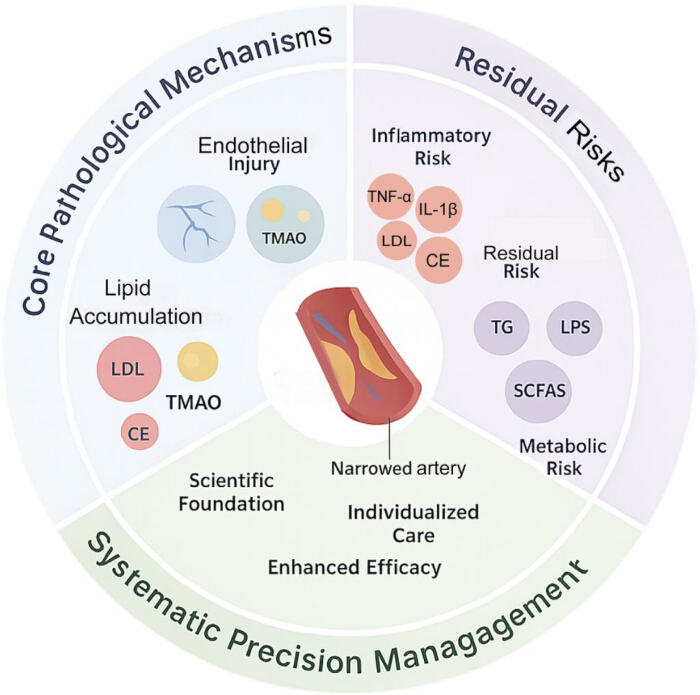
Figure 1: Schematic diagram of core pathological mechanisms and precision management of arterial stenosis. This diagram outlines the central pathological mechanisms underlying atherosclerosis, emphasizing lipid accumulation, endothelial injury, and chronic inflammation as key drivers of disease initiation and progression. It also links these mechanistic insights to clinical risk phenotypes-such as residual cholesterol, inflammatory, and metabolic risks-thereby illustrating how precision management strategies can align targeted interventions with individual patient profiles. Please click here to view a larger version of this figure.
Peroxisome proliferator-activated receptors (PPARs)
PPARs are ligand-activated nuclear transcription factors of the steroid hormone receptor superfamily, primarily expressed in the liver18. The PPAR family includes three isoforms-PPARα, PPARβ/δ, and PPARγ-which regulate fatty acid oxidation, glucose utilization, and lipoprotein metabolism to maintain energy homeostasis.
PPARα, mainly expressed in the liver and skeletal muscle, promotes fatty acid β-oxidation, reduces triglyceride (TG) levels, and improves lipid distribution. PPARβ/δ regulates fatty acid synthesis, with its aberrant activation linked to lipid accumulation in the arterial wall19. PPARγ, enriched in adipocytes, enhances insulin sensitivity and glucose uptake, indirectly inhibiting abnormal lipid accumulation20. Clinically, thiazolidinediones (e.g., rosiglitazone) modulate glucose and lipid levels through PPARγ activation21,22.
PPAR dysfunction is closely linked to obesity, insulin resistance, and AS pathogenesis. Thus, targeting PPARα and PPARγ for drug development offers a promising approach for precision lipid management.
Sterol regulatory element-binding proteins (SREBPs)
The SREBP family comprises key transcription factors involved in cholesterol and LDL receptor (LDL-R) synthesis23. It includes SREBP1 (isoforms 1a and 1c) and SREBP2, which regulate lipid biosynthesis and export. SREBP-1a and SREBP-1c primarily control fatty acid metabolism24, and their overactivation can lead to hepatic lipid accumulation and elevated circulating triglycerides.
SREBP2 regulates cholesterol biosynthesis by activating enzymes like HMG-CoA reductase, enhancing LDL-R expression, and promoting LDL uptake into cells. This elevates intracellular TC in hepatocytes while lowering plasma LDL, maintaining cellular cholesterol homeostasis25. Dysregulation of this pathway increases LDL-C and accelerates AS ( Figure 2).
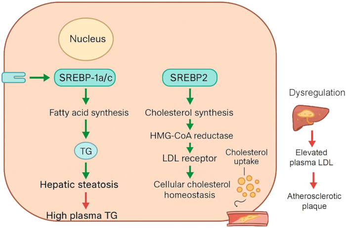
Figure 2: Molecular mechanisms of lipid metabolism regulation by the SREBP family. The schematic depicts how SREBP isoforms regulate lipid metabolism. SREBP-1a and SREBP-1c control fatty acid synthesis, whereas SREBP2 promotes cholesterol biosynthesis through HMG-CoA reductase activation and upregulation of LDL receptor expression. These coordinated actions maintain intracellular cholesterol balance but, when dysregulated, contribute to hyperlipidemia and atherosclerosis. Please click here to view a larger version of this figure.
Studies have shown that SNPs in SREBP1 are linked to coronary AS risk. Case-control studies indicate that individuals with specific SREBP1 variants have a reduced risk of left coronary AS26. These findings suggest that SREBP1 polymorphisms may influence coronary AS pathogenesis through cholesterol synthesis or lipid transport pathways. Carriers of the protective allele have a 30% to 35% lower risk of lipid deposition and plaque formation, highlighting a potential target for genetically-based cardiovascular risk stratification and personalized interventions.
Synergy and dysregulation in transcription factor networks
Multiple studies have shown that transcription factors involved in lipid metabolism do not act independently but coordinate through complex interaction networks. LXR-α, a key regulator of cholesterol homeostasis and fatty acid synthesis, upregulates SREBP-1c expression, promoting fatty acid synthesis27. SREBP-1c activation may also be mediated by enhanced PPARγ activity28. This regulatory effect results from their complex interactions, not simple additive changes in individual transcription factors29.
Mechanisms by which gut microbiota dysbiosis contributes to AS
The gut microbiota, a symbiotic microbial community, is essential for maintaining host homeostasis. Its abundance and composition are dynamically regulated to adapt to both internal and external changes, sustaining physiological balance or triggering pathological disturbances30. The gut microbiota directly influences AS pathology and indirectly promotes disease progression by modulating atherosclerotic risk factors31. Key microbial metabolites, including TMAO, Short-chain fatty acids(SCFAs), secondary BAs, and LPS, are closely linked to AS development and progression32,33,34,35.
TMAO pathway in AS
TMAO, a diet-induced metabolite produced by the gut microbiota, is linked to increased AS risk36. It upregulates scavenger receptor expression on macrophages, enhancing Ox-LDL uptake and foam cell formation37. TMAO also affects hepatic bile acid secretion and reverse cholesterol transport. In endothelial cells, it promotes inflammation, apoptosis, increased permeability, and oxidative stress, contributing to endothelial dysfunction38. Additionally, TMAO enhances platelet reactivity, upregulates tissue factor expression, and promotes vascular cell adhesion, facilitating thrombosis39. These effects on macrophages, liver, endothelial cells, and platelets collectively accelerate AS progression. TMAO also increases plaque instability and promotes rupture, leading to myocardial infarction, stroke, and other cardiovascular events, highlighting its multifaceted role in AS pathogenesis40. The main pathophysiological mechanisms involved in atherosclerotic plaque formation are illustrated in Figure 3.
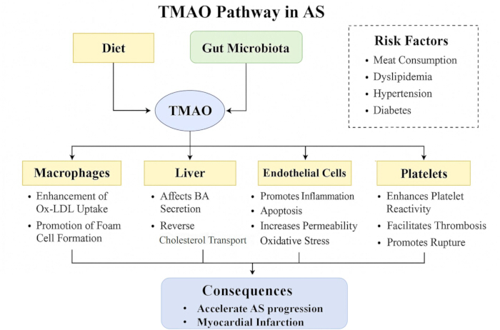
Figure 3: Mechanistic illustration of how TMAO enhances the development of AS and thrombosis. The schematic summarizes the multifaceted actions of TMAO in atherosclerosis. TMAO promotes macrophage scavenger receptor expression and foam cell formation, alters hepatic bile acid metabolism and reverse cholesterol transport, and induces endothelial inflammation, apoptosis, and oxidative stress, leading to vascular dysfunction. It also enhances platelet reactivity, tissue factor expression, and vascular adhesion, collectively accelerating plaque progression, instability, and thrombosis. Please click here to view a larger version of this figure.
Dysregulation of SCFAs and AS
SCFAs, including acetate, propionate, and butyrate, are carboxylic acids produced by gut bacteria through dietary fiber fermentation in the cecum and colon41. As signaling molecules, SCFAs influence metabolic, immune, and inflammatory processes. Their main receptors, GPR43 and Olfr, activate phospholipase C (PLC)-dependent mechanisms, stimulating insulin secretion and improving glucose and lipid metabolism42,43,44. Reduced SCFAs production can lead to dyslipidemia, as SCFAs inhibit hepatic lipogenesis45. SCFAs also upregulate genes involved in cholesterol metabolism, such as SREBP-2, LDL-R, and CYP7A1, enhancing cholesterol uptake and bile acid excretion, thereby lowering serum cholesterol46.
Additionally, SCFAs reduce hepatic lipid-metabolizing enzyme activity and regulate cholesterol distribution between the liver and bloodstream, lowering serum triglyceride and cholesterol levels. They also enhance intestinal barrier function, modulate epithelial permeability, increase satiety, and influence the gut microbial microenvironment47. Gut microbiota dysbiosis reduces fecal SCFAs levels (e.g., butyrate, acetate, propionate), impairing lipid metabolism regulation.
Bile acid (BA) metabolism
BAs are amphipathic steroid molecules that form mixed micelles with lipids, aiding in the solubilization and absorption of dietary lipids. Clinical studies have shown a significant correlation between serum bile acid levels and AS. In coronary artery disease patients, fasting serum total bile acid levels are elevated and positively correlated with disease severity, also independently predicting disease onset risk48.
BAs, in addition to aiding lipid digestion, serve as important signaling molecules. They bind to receptors such as FXR, TGR5, and S1PR249, participating in intracellular signal transduction. BA synthesis is the primary catabolic pathway for cholesterol, with two major biosynthetic pathways: the classical (neutral) pathway and the alternative (acidic) pathway. The classical pathway, catalyzed by CYP7A1, is rate-limiting and responsible for over 75% of total bile acid synthesis under physiological conditions50.
In the intestine, BAs aid in the solubilization and absorption of dietary fats and fat-soluble vitamins51. Studies suggest that the gut microbiota influences lipid metabolism in blood and tissues by modulating secondary bile acid production52. Thus, BAs are potential modulators of AS53(Figure 4).
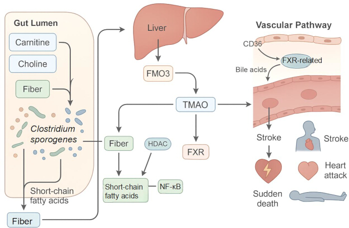
Figure 4: Schematic illustration of the gut microbiota functioning as an endocrine organ via the production of bioactive metabolites that contribute to AS. The diagram highlights the role of bile acids (BAs) as both digestive agents and signaling molecules. BAs, synthesized through classical and alternative pathways, facilitate lipid absorption and act on receptors such as FXR, TGR5, and S1PR2 to regulate cholesterol metabolism and vascular function. Gut microbiota further modulate BA composition by generating secondary bile acids, linking BA metabolism to atherosclerosis progression. Please click here to view a larger version of this figure.
The role of FXR in AS
FXR, a member of the nuclear receptor superfamily, is primarily expressed in the liver and small intestine, where it regulates bile acid, lipid, and glucose metabolism54. FXR is key to maintaining bile acid, cholesterol, and lipid homeostasis, with its dysfunction linked to diseases like hepatic steatosis55, cholesterol gallstones56, and AS57.
Recent studies show that FXR is activated by bile acids. Upon activation, FXR reduces lipid peroxidation by upregulating genes involved in ferroptosis58. Wu et al.59 found that intestinal FXR expression correlates with AS progression. FXR inhibition may reduce atherosclerosis by modulating SMPD360. Additionally, FXR promotes phospholipid transfer between lipoproteins and regulates HDL metabolism by controlling PLTP gene transcription61.
Mechanisms of TGR5 in AS
TGR5, a key mediator of bile acid signaling, is expressed in various tissues and plays a crucial role in metabolic homeostasis, making it a potential target for AS and inflammation prevention62. Its protective effects are mainly mediated through the regulation of immune cells, metabolic organs, and vascular wall cells63.
Macrophage inflammatory activity is categorized into two polarization phenotypes: M1, which is proinflammatory, and M2, which is anti-inflammatory64. TGR5 suppresses inflammation by promoting M2 polarization65. It also reduces chemokine expression in macrophages, inhibiting their migration and infiltration66.
TGR5 reduces macrophage proinflammatory activity in AS via the cAMP-NF-κB signaling pathway67,68. The activation of TGR5 lowers CD36 and SR-A expression in macrophages, suggesting that it may inhibit lipid uptake.
Impairment of TGR5 signaling-due to altered bile acid composition, reduced receptor expression, or desensitization-weakens its anti-inflammatory effects, cholesterol efflux, and metabolic regulation. These deficits, combined with hypercholesterolemia and chronic inflammation, accelerate atherosclerotic plaque formation and destabilization69. Thus, targeted TGR5 activation is a promising therapeutic strategy, offering multi-target intervention for atherosclerotic cardiovascular disease70. Beyond bile acid-related pathways, other metabolic sensors also contribute to vascular protection. In particular, activation of the AMPK/Sirt1/HIF-1α signaling axis has been shown to alleviate mitochondrial dysfunction under hypoxic conditions, thereby offering additional mechanistic insights into the maintenance of vascular homeostasis71. Table 1 lists the key molecular mechanisms and targets in lipid metabolism and atherosclerosis.
| Pathway/Target | Role in Lipid Metabolism / Atherosclerosis | Representative Evidence |
| SREBP1/SREBP2 | Regulate fatty acid and cholesterol synthesis; SREBP2 enhances LDL-R expression, affecting cholesterol uptake | SNPs in SREBP1 linked to coronary AS risk; SREBP2 dysregulation accelerates LDL-C elevation |
| LXRs | Activate ABCA1/G1 to promote cholesterol efflux; excessive activation may worsen hepatic lipid accumulation | Reduce vascular lipid deposition and plaque formation |
| PPARα/γ | PPARα promotes fatty acid oxidation; PPARγ improves insulin sensitivity; dysfunction leads to obesity and AS | Thiazolidinediones act via PPARγ to modulate glucose and lipid metabolism |
| Gut Microbiota (TMAO, SCFAs, BAs) | TMAO promotes foam cell formation and endothelial dysfunction; SCFAs enhance cholesterol uptake/excretion; BAs regulate lipid absorption and vascular signaling | Elevated TMAO linked to plaque instability; SCFAs improve cholesterol metabolism |
| Epigenetic/Inflammatory Pathways | ABCA1-seRNA, mTORC1 regulate lipid efflux; NF-κB activation accelerates inflammatory progression | Genetic polymorphisms and inflammation modulate AS susceptibility |
Table 1: Key molecular mechanisms and targets in lipid metabolism and atherosclerosis. This table summarizes the key molecular mechanisms and targets involved in lipid metabolism dysregulation and atherosclerosis progression. It highlights important pathways such as SREBPs, LXRs, PPARs, and gut microbiota metabolites, which play crucial roles in lipid accumulation, cholesterol homeostasis, and inflammatory responses. The table also includes representative evidence from recent studies linking these molecular mechanisms to the development of atherosclerosis and potential therapeutic targets for precision management.
Clinical Challenges and Combination Drug Therapy:
Monotherapy strategies
Lipid-lowering agents
The pathological basis of AS is the abnormal lipid deposition in the vascular wall, making lipid-lowering therapy essential72. Statins, the first-line treatment, inhibit HMG-CoA reductase, reducing LDL-C by up to 60% and modestly increasing HDL-C. Statins also stabilize plaques and reduce vascular inflammation73. Common statins include atorvastatin, rosuvastatin, and simvastatin, with high-intensity therapy further reducing plaque volume74. While intensified statin therapy offers additional benefits, high-dose statins may cause adverse effects, including hepatotoxicity and myopathy75.
For statin-intolerant patients or those failing to reach target LDL-C levels, non-statin agents are key alternatives76. PCSK9 inhibitors reduce LDL-C by over 50% by preventing LDL-R degradation, lowering cardiovascular risk. Ezetimibe reduces LDL-C by 18% as monotherapy, with enhanced effects when combined with statins. Fibrates, like fenofibrate, activate PPARα, lowering triglycerides by 30-50% and mildly increasing HDL-C, suitable for mixed hyperlipidemia77. Niacin can regulate lipid levels, but is limited by side effects like flushing and increased blood glucose. Long-term niacin use requires monitoring liver enzymes and creatine kinase to detect statin-related adverse effects78.
Antiplatelet agents
In AS progression, plaque rupture triggers platelet activation and thrombosis, increasing the risk of acute cardiovascular events79. Antiplatelet drugs reduce platelet activity and prevent thrombosis by inhibiting activation80. These agents are central to both primary and secondary prevention of atherosclerotic cardiovascular disease, improving prognosis. They inhibit platelet activation, adhesion, and aggregation, but a balance must be struck between preventing thrombosis and managing bleeding risks. Caution is needed in elderly patients or those with peptic ulcers or coagulopathy81.
Limitations of traditional monotherapy
Although statins reduce major adverse cardiovascular events (MACE)82, monotherapy has limitations. By inhibiting HMG-CoA reductase, they enhance LDL-R activity and lower LDL-C83,84, but efficacy is capped at ~60-70% reduction85. Higher doses raise risks of muscle disorders and diabetes86. About 10-15% of patients show statin intolerance87, and long-term use is associated with liver dysfunction, rhabdomyolysis, cognitive impairment, and increased diabetes risk (RR 1.25), possibly via reduced insulin sensitivity or β-cell dysfunction88.
Clinical studies show that the Chinese population has significantly lower tolerance to high-dose statins compared to Western populations89. Each doubling of statin dose reduces LDL-C by only about 6%77. These findings highlight the importance of considering both long-term metabolic risks and population-specific drug tolerance when developing individualized lipid-lowering treatment strategies90.
Antiplatelet agents, like aspirin monotherapy, have clear limitations in managing AS91. While reducing thrombotic risk, they fail to intervene in plaque progression, inhibit plaque inflammation, improve endothelial function, or enhance plaque stability, limiting their clinical benefits92.To address this, combining statins with other medications has been shown to significantly reduce the risk of major adverse cardiovascular events (MACE)93.
Lipid-lowering efficacy of combination therapy
Lipid-lowering efficacy of fenofibrate combined with statins
Simvastatin, an HMG-CoA reductase inhibitor, is commonly used to lower cholesterol by inhibiting its synthesis94. However, its efficacy is limited when used alone95. Fenofibrate, a fibrate-class drug, is often used for lipid-lowering in ASCVD patients96. Combining fenofibrate with simvastatin improves lipid parameters and endothelial function, offering better outcomes than simvastatin monotherapy.
Lipid-lowering efficacy of ezetimibe combined with statins
Ezetimibe inhibits cholesterol absorption by blocking the interaction between the sterol transporter NPC1L1 on enterocytes and the cholesterol-activated adaptor protein complex AP-297. This selectively reduces intestinal cholesterol uptake without affecting fat-soluble nutrients. The decreased cholesterol transfer to the liver triggers increased LDL-R expression, enhancing LDL clearance from the bloodstream98 (Figure 5).

Figure 5: Mechanism diagram of lipid-lowering effects of combination therapies. The schematic compares two combination strategies for lipid lowering. Fenofibrate combined with simvastatin enhances lipid control and endothelial function beyond statin monotherapy. Ezetimibe combined with statins reduces intestinal cholesterol absorption via NPC1L1 inhibition, leading to upregulation of hepatic LDL receptors and improved LDL clearance. These mechanisms highlight complementary approaches to optimize lipid management in ASCVD patients. Please click here to view a larger version of this figure.
A study of 4,252 patients showed that switching from statin monotherapy to combination therapy with ezetimibe resulted in a 31.0% to 41.0% further reduction in LDL-C levels99. A 6-year cohort study found that, among patients with acute coronary syndrome and multiple comorbidities, combination therapy reduced the risk of rehospitalization and revascularization compared to statin monotherapy100.
Lipid-lowering efficacy of PCSK9 inhibitors combined with statins
PCSK9, mainly synthesized in the liver, is also produced by the kidneys, intestine, VSMCs, ECs, and macrophages101. It induces LOX-1 in VSMCs, forming a "PCSK9-LOX-1" loop that drives macrophage activation, foam cell formation, and cholesterol deposition102. By binding LDL-R on hepatocytes, PCSK9 promotes receptor degradation and elevates plasma LDL-C103,104. Inhibiting this interaction preserves LDL-R, lowers LDL-C, and prevents ASCVD105.
PCSK9 inhibitors, as novel lipid-lowering agents, specifically block the binding of PCSK9 to LDL-R, inhibiting the degradation of LDL-R, which results in upregulation of LDL-R expression on hepatocyte membranes and enhances its functional activity98. This mechanism significantly improves the liver's ability to clear plasma LDL-C, thereby reducing the risk of cardiovascular events106,107,108,109. A 6-month clinical study demonstrated that PCSK9 inhibitors significantly reduced total cholesterol, non-HDL-C, LDL-C, triglycerides, apoB, and Lp(a), with all changes reaching statistical significance (p < 0.001). Notably, LDL-C levels were reduced by 69.7% (59.1-78.3%)110. When combined with statin therapy, PCSK9 inhibitors provide further lipid-lowering efficacy beyond statins alone, highlighting their value as an intensive strategy for cardiovascular risk reduction.
Innovative Therapeutic Strategies and Precision Management:
Novel targeted drugs
Inhibition of PCSK9 through RNA silencing
Inclisiran is a novel small-interfering RNA (siRNA) that targets PCSK9103, inhibiting its synthesis from mRNA to protein in hepatocytes111,112. PCSK9 is a key regulatory protein that mediates the degradation of LDL receptors on the hepatocyte surface. Inhibition of this process results in LDL receptors remaining on the cell surface, significantly enhancing LDL-C uptake and thereby reducing serum LDL-C levels113,114. The efficacy of Inclisiran has been validated in multiple clinical trials, particularly in the ORION study series. In the ORION-1 Phase II trial, patients receiving two 300mg doses (on Day 1 and Day 90) showed an average LDL-C reduction of 52.6% (confidence interval: 48.1-57.1%) at Day 180115.
Thus, Inclisiran, a siRNA therapy that inhibits PCSK9 production, is suitable for patients with ASCVD or familial hypercholesterolemia (HeFH) requiring further LDL reduction. It can be used alongside statins at the maximum tolerated dose or in combination with ezetimibe. For patients intolerant to statins, Inclisiran offers an effective alternative, leveraging its unique mechanism of targeted hepatocyte uptake116.
Lp(a) inhibitors
Lp(a) is a key residual risk factor for cardiovascular disease. Mendelian randomization and genetic studies confirm its strong association with ASCVD and calcific aortic valve stenosis117,118. Lp(a) promotes atherothrombosis through its LDL-like particle, oxidized phospholipids (OxPLs), and antifibrinolytic effects105. Elevated levels, like LDL-C, increase ASCVD risk119. International guidelines now recognize high Lp(a) as a major risk factor and recommend its measurement for better risk stratification120. Statins lower LDL-C but have little effect on Lp(a)121, prompting growing interest in therapies specifically targeting Lp(a).
Nicholls et al.122 reported Phase I results of Murapavaparin, the first oral Lp(a) inhibitor. It showed dose-dependent pharmacokinetics with plasma peaks at 2-5 h, half-life 12-67 h, and no accumulation after repeated dosing. A 100 mg or higher dose for 14 days reduced Lp(a) by 63-65%, with effects lasting nearly 2 months, without significant changes in total cholesterol, LDL-C, or apoB. Lp(a) and apoB changes moderately correlated with LDL-C, supporting the safety and efficacy of Lp(a) reduction123. As the first oral agent targeting hepatic Lp(a) biosynthesis, it showed good tolerability with no major adverse effects.
Fibrates for lowering TG
Epidemiological, genetic, and clinical studies provide strong evidence linking elevated plasma triglyceride (TG) levels with an increased risk of ASCVD105. Elevated TG levels are a key biological marker for this risk, particularly as they relate to atherogenic residual particles.
Fibrates are effective in lowering TG levels and modestly increasing HDL-C levels. They activate PPARα and lipoprotein lipase (LPL), both of which contribute to lowering serum TG and raising HDL-C levels. Commonly used fibrates include fenofibrate, bezafibrate, and gemfibrozil. However, gemfibrozil should be avoided in combination with statins due to potential side effects124.
Clinical trials on fibrates have shown variable outcomes, but most suggest a reduction in cardiovascular (CV) events. A meta-analysis of 18 trials with 45,058 participants found that fibrates reduced the relative risk (RR) of coronary events by 13% (p<0.0001)125. Table 2 shows the clinical therapies for dyslipidemia in atherosclerosis.
| Therapy | Mechanism of Action | Clinical Benefits | Limitations |
| Statins | Inhibit HMG-CoA reductase → ↓ cholesterol synthesis, ↑ LDL-R | ↓ LDL-C up to 60%, plaque stabilization | Statin intolerance, residual risk, diabetes risk |
| Ezetimibe | Blocks NPC1L1, ↓ intestinal cholesterol absorption | Further ↓ LDL-C by 18% (monotherapy) or ~31% when combined with statins | Limited as monotherapy |
| PCSK9 inhibitors (Evolocumab, Alirocumab) | Prevent LDL-R degradation, ↑ LDL clearance | ↓ LDL-C 50–70%, reduce CV events | High cost, injection route |
| Inclisiran (siRNA) | Inhibits PCSK9 synthesis in hepatocytes | Long-acting, good compliance | Long-term safety still under study |
| Fibrates | Activate PPARα, ↓ TG, ↑ HDL-C | Reduce CV risk in hypertriglyceridemia | Variable outcomes; gemfibrozil + statins unsafe |
| Lp(a) inhibitors (Pelacarsen, Muvalaplin) | Target Lp(a) biosynthesis | ↓ Lp(a) by >60%, reduce residual risk | Still in clinical trials |
Table 2: Clinical therapies for dyslipidemia in atherosclerosis. This table compares various lipid-lowering therapies used in the treatment of dyslipidemia associated with atherosclerosis. It includes conventional therapies such as statins and ezetimibe, as well as emerging treatments like PCSK9 inhibitors and Inclisiran (siRNA). The table provides a summary of their mechanisms of action, clinical benefits, and limitations, offering a comprehensive overview of current and potential future treatment options for patients with ASCVD.
Clinical Management Strategies for Blood Lipids:
China's blood lipid management strategy
The Chinese Guidelines for Prevention and Treatment of Dyslipidemia in Adults (2016 Revised Edition)126previously defined the following as target values for blood lipid levels: TC < 6.2 mmol/L, TG < 2.3 mmol/L, HDL-C > 1.03 mmol/L, and LDL-C < 2.6 mmol/L. However, with the advancement of large-scale clinical research and the accumulation of high-quality clinical evidence in recent years, the "Chinese Guidelines for Blood Lipid Management (2023)" has evolved. The new guidelines have redefined target LDL-C values for individuals at different risk levels, aiming to effectively reduce the risk of ASCVD, thus updating and iterating the blood lipid management standards127(Table 3 and Figure 6).
| Risk Category | LDL-C Target (mmol/L) | Special Considerations |
| Low Risk | <3.4 | Lifestyle modification emphasized |
| Medium/High Risk | <2.6 | Statins as first-line therapy; consider combination if target unmet |
| Very High Risk | <1.8 and reduction > 50% from baseline | PCSK9 inhibitors or Inclisiran; pharmacokinetics in Chinese patients important |
Table 3: LDL-C target values across risk categories in the Chinese Blood Lipid Management Guidelines (2023). This table presents the LDL-C target values recommended by the 2023 Chinese Guidelines for Lipid Management, stratified by cardiovascular risk categories. It details the specific LDL-C targets for low-risk, medium/high-risk, and very high-risk individuals, emphasizing the importance of individualized treatment approaches. The table also highlights special considerations for the Chinese population, such as pharmacokinetic variations and personalized therapeutic strategies.
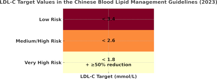
Figure 6: Recommended LDL-C target values across risk categories in the Chinese Blood Lipid Management Guidelines (2023). This heatmap visually illustrates the stepwise reduction in LDL-C targets with increasing cardiovascular risk. Low-risk individuals are advised to maintain LDL-C <3.4 mmol/L, medium/high-risk groups <2.6 mmol/L, and very high-risk patients <1.8 mmol/L together with a ≥50% reduction from baseline. Data from the Chinese Society of Cardiology Guidelines (2023)127. Please click here to view a larger version of this figure.
Epidemiological studies have shown that for every 1 mmol/L reduction in low-density lipoprotein cholesterol (LDL-C) levels, the risk of cardiovascular (CV) events is reduced by approximately 21%128.
International blood lipid management strategies
Numerous international studies show that lowering LDL-C to very low levels markedly reduces ASCVD events without adverse effects, underscoring its dual benefits for efficacy and safety. Post-hoc analyses of the FOURIER and ODYSSEY OUTCOMES trials confirm the safety of PCSK9 inhibition in high-risk patients, achieving LDL-C < 10 mg/dL with continued event reduction. However, not all patients with recurrent cardiovascular events require such ultra-low LDL-C levels129.
The Lipid Association of India (LAI) recommends LDL-C as the primary lipid target, non-HDL-C as a co-primary target, and Apo-B as secondary. Non-HDL-C, measurable in the non-fasting state, should complement LDL-C in management. Treatment goals differ by risk: for low risk, LDL-C < 100 mg/dL, non-HDL-C < 130 mg/dL, Apo-B < 90 mg/dL; for moderate risk, LDL-C < 100 mg/dL (optional < 70 mg/dL), non-HDL-C < 130 mg/dL (optional < 100 mg/dL), and Apo-B < 90 mg/dL, with progressively lower thresholds in higher-risk groups130.
Future Challenges and Directions:
Residual cardiovascular (CV) risk
Given the strong association between low-density lipoprotein cholesterol (LDL-C) and ASCVD incidence and mortality, current guidelines emphasize LDL-C reduction as a central strategy for risk management131,132. Although LDL-C has long been the primary focus, many studies show that even within target ranges, patients may still experience atherosclerosis progression and cardiovascular events. Thus, LDL-C measurement alone is insufficient for assessing residual risk, prompting interest in alternative lipid markers133. Evidence further indicates that even at recommended targets (<1.4 mmol/L), substantial residual vascular risk persists134. Beyond LDL-C, non-high-density lipoprotein cholesterol (non-HDL-C) and lipoprotein(a) [Lp(a)] are strongly linked to residual risk135,136. Non-HDL-C reflects all atherogenic lipoproteins except HDL-C, including very-low-density lipoprotein (VLDL) and intermediate-density lipoprotein (IDL)137,138, which play key roles in atherosclerosis and coronary artery disease, especially when LDL-C is well controlled. Elevated Lp(a) is another inadequately addressed factor, contributing to disease progression, plaque instability, and thrombogenesis through its LDL-like particle, oxidized phospholipids, and antifibrinolytic activity139.
Accordingly, non-HDL-C and Lp(a) are considered two critical residual risk factors beyond LDL-C. A growing body of research supports incorporating these markers into clinical risk assessment to establish more precise treatment goals alongside conventional LDL-C management.
Significance of the TG/HDL-C ratio
Recent studies indicate that the triglyceride-to-HDL cholesterol (TG/HDL-C) ratio is strongly associated with AS and predicts its progression better than LDL-C. Among lipid parameters, it shows the strongest correlation with CAD severity140. Even with strict LDL-C control, residual risk persists, often linked to elevated TG/HDL-C141,142. Multiple studies demonstrate its superiority over other lipid markers in predicting ASCVD risk143. As the ratio rises, LDL particles become smaller and denser, accelerating AS progression144. Weinstein et al.140 reported that higher TG/HDL-C is associated with diverse cardiovascular conditions and adverse outcomes, supporting its role as a biomarker. A prospective cohort of 9,368 participants followed for 20 years further confirmed this positive association with ASCVD risk. A key limitation, however, is the absence of a universally accepted cutoff145. While some studies propose >2, others recommend >2.5 or >3, restricting its clinical applicability as a standardized biomarker141,146.
Beyond epidemiology, an elevated TG/HDL-C ratio indicates an atherogenic phenotype with triglyceride-rich remnants, impaired HDL efflux, and smaller, denser LDL particles prone to arterial penetration and oxidation147. Clinically, it identifies residual risk even when LDL-C and non-HDL-C are controlled, being associated with events, plaque progression, and adverse outcomes, thus improving secondary prevention risk stratification148. Practically, it can be measured in a non-fasting state and easily calculated from routine lipid panels, supporting its use as a complementary marker with LDL-C and Lp(a) in daily practice.
Access restricted. Please log in or start a trial to view this content.
Conclusions
Dysregulation of lipid metabolism is closely associated with the initiation and progression of atherosclerosis (AS), and dyslipidemia remains one of the most modifiable risk factors for this condition. This review has outlined the molecular mechanisms underlying AS, key targets involved in lipid abnormalities, and diverse clinical intervention strategies. Current research highlights the pivotal roles of gene regulation (e.g., SREBP1, LPA) and epigenetic pathways (e.g., ABCA1-seRNA, mTORC1...
Access restricted. Please log in or start a trial to view this content.
Disclosures
The authors declare that they have no conflicts of interest. The authors disclose using an artificial intelligence language model (ChatGPT) only for language polishing in preparing this review manuscript. All outputs were thoroughly reviewed and edited by the authors. ChatGPT was strictly confined to improving linguistic clarity and expression, without involvement in idea generation, data analysis, interpretation, or conclusion drafting. The authors conducted all literature citations, evidence synthesis, and conclusions independently based on the referenced studies, ensuring both accuracy and compliance with academic standards. Thus, the use of ChatGPT was limited solely to language optimization, while the scientific content and intellectual contribution remain entirely the responsibility of the authors.
Acknowledgements
The authors declare they have no funding or financial support for this work.
Access restricted. Please log in or start a trial to view this content.
References
- Wei, Y., et al. GSDME-mediated pyroptosis promotes the progression and associated inflammation of atherosclerosis. Nat Commun. 14 (1), 929(2023).
- Xiao, S. J., Zhao, M. Arteriosclerosis and immunity. Chin J Arterioscler. 30 (4), 277-286 (2022).
- Carpenter, H. J., et al. Optical coherence tomography based biomechanical fluid-structure interaction analysis of coronary atherosclerosis progression. J Vis Exp. 179, e62933(2022).
- Nayor, M., Brown, K. J., Vasan, R. S. The molecular basis of predicting atherosclerotic cardiovascular disease risk. Circ Res. 128 (2), 287-303 (2021).
- Summary of China Cardiovascular Health and Disease Report 2022. Chin J Intervent Cardiol. 31 (7), 485-508 (2023).
- Báez-Díaz, C., Crisóstomo, V. Large animal models of cardiovascular disease: From training to translation. J Vis Exp. (193), e64983(2023).
- Li, J. L., et al. Drug literacy status and influencing factors of elderly cardiovascular disease patients in rural Hunan. J Hunan Norm Univ Med Sci. 20 (1), 119-124 (2023).
- Huang, Y., Zhao, X. Analysis of health management effect in AS patients. Chin Community Phys. 40 (29), 135-137 (2024).
- Ding, E. C., Lu, D. Y., Wei, L. J., Shen, J. Research progress on noninvasive molecular imaging of vulnerable plaques in atherosclerosis. Chin J CT MRI. 20 (12), 184-185 (2022).
- Wang, J., et al. Microbiome-metabolome analysis insight into the effects of the extract of Phyllanthus emblica L. on high-fat diet-induced hyperlipidemia. Metabolites. 14, 257(2024).
- Ren, C. X., et al. Identification and mechanism elucidation of medicative diet for food therapy XQCSY in NAFLD prevention: an integrative in silico study. Food Med Homol. 1, 9420015(2024).
- Ramos, L., et al. Hydra, a computer-based platform for aiding clinicians in cardiovascular analysis and diagnosis. J Vis Exp. (139), e58132(2018).
- Xu, D. L., et al. Application of computer-aided drug design in the research of natural anti-tumor drugs. Journal of Ningxia University (Natural Science Edition). 44, 44-52 (2023).
- Wang, L. Q., Li, C. P. Research progress of liver X receptor in nonalcoholic fatty liver disease. Mod Clin Med. 50 (6), 466-470 (2024).
- Li, J., et al. A nanodrug system overexpressed circRNA_0001805 alleviates nonalcoholic fatty liver disease via miR-106a-5p/miR-320a and ABCA1/CPT1a axis. J Nanobiotechnol. 19 (1), 363(2021).
- Butler, L. M., et al. Lipids and cancer: Emerging roles in pathogenesis, diagnosis and therapeutic intervention. Adv Drug Deliv Rev. 159, 245-293 (2020).
- Bovenga, F., Sabbà, C., Moschetta, A. Uncoupling nuclear receptor LXR and cholesterol metabolism in cancer. Cell Metab. 21 (3), 517-526 (2015).
- Hao, X. T., et al. The food and medicinal homological resources benefiting patients with hyperlipidemia: categories, functional components, and mechanisms. Food Med Homol. 1, 9420003(2024).
- Zhao, X. L., Gao, X. Y., Zhang, C. Y., Li, M. Q., Ma, Y. H. Effect of PPARγ on liver fibrosis progression and its ligand drug development. Chin J Pharmacovigil. 21 (6), 716-720 (2024).
- Qi, L., et al. Structural characterization of red yeast rice-derived polysaccharide and its promotion of lipid metabolism and gut function in high-fat diet-induced mice. Int J Biol Macromol. 282 (Pt 1), 136744(2024).
- Mirza, A. Z., Althagafi, I. I., Shamshad, H. Role of PPAR receptor in different diseases and their ligands: Physiological importance and clinical implications. Eur J Med Chem. 166, 502-513 (2019).
- Wagner, K. D., et al. Vascular PPAR β/δ promotes tumor angiogenesis and progression. Cells. 8 (12), 1623(2019).
- Li, L., Li, X., Fang, Y. F., Zhou, Y. K., Ma, S. J. Research progress on lipid metabolism regulation via SREBPs, PPARs and PCSK9 pathways. Inf Tradit Chin Med. 39 (9), 85-89 (2022).
- Wang, K. X., Zhang, S. Y., Ji, H. M., Hu, C. C. Research progress of lipid metabolism in glioma. J Clin Neurosurg. 19 (6), 715-720 (2022).
- Romani, P., et al. Extracellular matrix mechanical cues regulate lipid metabolism through Lipin-1 and SREBP. Nat Cell Biol. 21 (3), 338-347 (2019).
- Shilenok, V., Kobzeva, K., Bushueva, O. SERBP1 (Hero45) is a novel link with ischemic heart disease risk: Associations with coronary arteries occlusion, blood coagulation and lipid profile. Cell Biochem Biophys. 83, 3557-3569 (2025).
- Danielewski, M., et al. The impact of anthocyanins and iridoids on transcription factors crucial for lipid and cholesterol homeostasis. Int J Mol Sci. 22 (11), 6074(2021).
- Zheng, S., et al. Yin-xing-tong-mai decoction attenuates atherosclerosis via activating PPARγ-LXRα-ABCA1/ABCG1 pathway. Pharmacol Res. 169, 105639(2021).
- Danielewski, M., et al. Cornelian cherry fruit extract lowers SREBP-1c and C/EBPα in liver and alters PPAR/ LXR target genes in cholesterol-rich diet rabbit model. Int J Mol Sci. 25 (2), 1199(2024).
- Qi, L., et al. Salvia miltiorrhiza bunge extract improves the Th17/Treg imbalance and modulates gut microbiota of hypertensive rats induced by high-salt diet. J Funct Foods. 117, 106211(2024).
- Cao, L., et al. Cornus officinalis vinegar alters the gut microbiota, regulating lipid droplet changes in nonalcoholic fatty liver disease model mice. Food Med Homol. 1, 9420002(2024).
- Qi, L., et al. Microbiome-metabolome analysis insight into the effects of high-salt diet on hemorheological functions in SD rats. Front Nutr. 11, 1408778(2024).
- Wang, J., et al. Effects of three Huanglian-derived polysaccharides on the gut microbiome and fecal metabolome of high-fat diet/streptozocin-induced type 2 diabetes mice. Int J Biol Macromol. 273, 133060(2024).
- Gao, F., et al. Gut microbiota and atherosclerosis: Current status of traditional Chinese medicine intervention. China J Tradit Chin Med Pharm. 40 (2), 775-779 (2025).
- Hou, Y., et al. Identification and validation of core microbes for isoflavone conversion and flavor formation in fermented sojae semen praeparatum. LWT. 229, 118231(2025).
- Papandreou, C., Moré, M., Bellamine, A. Trimethylamine N-oxide in relation to cardiometabolic health-Cause or effect. Nutrients. 12 (5), 1330(2020).
- Luo, Z., et al. Trimethylamine N-oxide promotes oxidative stress and lipid accumulation in macrophage foam cells via the Nrf2/ABCA1 pathway. J Physiol Biochem. 80 (1), 67-79 (2024).
- Brunt, V. E., et al. Trimethylamine-N-oxide promotes age-related vascular oxidative stress and endothelial dysfunction in mice and humans. Hypertension. 76 (1), 101-112 (2020).
- Zhu, W., et al. Gut microbial metabolite TMAO enhances platelet hyperreactivity and thrombosis risk. Cell. 165 (1), 111-124 (2016).
- Canyelles, M., et al. Gut microbiota-derived TMAO: A causal factor promoting atherosclerotic cardiovascular disease. Int J Mol Sci. 24 (3), 1940(2023).
- Ratajczak, W., et al. Immunomodulatory potential of gut microbiome-derived short-chain fatty acids. Acta Biochim Pol. 66 (1), 1-12 (2019).
- Kimura, I., et al. Free fatty acid receptors in health and disease. Physiol Rev. 100 (1), 171-210 (2020).
- Zhang, Y., Wang, D. J. Research progress on relationship between gut microbiota, short-chain fatty acids and coronary heart disease. Mod Pract Med. 37 (3), 320-323 (2025).
- Zheng, H., et al. Association of dietary patterns with cognitive function in older people: Results from the Chinese longitudinal healthy longevity survey. Med Adv. 1, 246-259 (2023).
- Gerdes, V., et al. How strong is the evidence that gut microbiota composition can be influenced by lifestyle interventions in a cardio-protective way. Atherosclerosis. 311, 124-142 (2020).
- Vourakis, M., Mayer, G., Rousseau, G. The role of gut microbiota on cholesterol metabolism in atherosclerosis. Int J Mol Sci. 22 (15), 8074(2021).
- Pathak, P., et al. Small molecule inhibition of gut microbial choline trimethylamine lyase activity alters host cholesterol and bile acid metabolism. Am J Physiol Heart Circ Physiol. 318 (6), H1474-H1486 (2020).
- Li, W., et al. Fasting serum total bile acid level is associated with coronary artery disease, myocardial infarction and severity of coronary lesions. Atherosclerosis. 292, 193-200 (2020).
- Yang, W. Y., Zhang, X. Y., Yu, Y. Role of bile acid metabolism in metabolic cardiovascular diseases. Chin J Pathophysiol. 41 (3), 569-576 (2025).
- Rafieian-Kopaei, M., et al. Atherosclerosis: Process, indicators, risk factors and new hopes. Int J Prev Med. 5 (8), 927-946 (2014).
- Mantovani, A., et al. Plasma bile acid profile in patients with and without type 2 diabetes. Metabolites. 11 (7), 453(2021).
- Schoeler, M., Caesar, R. Dietary lipids, gut microbiota and lipid metabolism. Rev Endocr Metab Disord. 20 (4), 461-472 (2019).
- Cao, H., Zhu, Y., Hu, G., Zheng, L. Gut microbiome and metabolites, the future direction of diagnosis and treatment of atherosclerosis. Pharmacol Res. 187, 106586(2023).
- Henry, Z., Meadows, V., Guo, G. L. FXR and NASH: An avenue for tissue-specific regulation. Hepatol Commun. 7 (5), e0127(2023).
- Xu, W., et al. Hepatocellular cystathionine γ lyase/hydrogen sulfide attenuates nonalcoholic fatty liver disease by activating farnesoid X receptor. Hepatology. 76 (6), 1794-1810 (2022).
- Zhang, Z., et al. Glucagon-like peptide 1 analogue prevents cholesterol gallstone formation by modulating intestinal farnesoid X receptor activity. Metabolism. 118, 154728(2021).
- Byun, S., et al. Phosphorylation of hepatic farnesoid X receptor by FGF19 signaling-activated Src maintains cholesterol levels and protects from atherosclerosis. J Biol Chem. 294 (22), 8732-8744 (2019).
- Tschuck, J., et al. Farnesoid X receptor activation by bile acids suppresses lipid peroxidation and ferroptosis. Nat Commun. 14 (1), 6908(2023).
- Wu, Q., et al. Suppressing the intestinal farnesoid X receptor/sphingomyelin phosphodiesterase 3 axis decreases atherosclerosis. J Clin Invest. 131 (8), e142865(2021).
- Gu, Q., Liu, J., Shen, L. L. FXR activation reduces macrophage foam cell formation and atherosclerotic plaque, possibly by downregulating hepatic lipase in macrophages. FEBS Open Bio. 15 (2), 311-323 (2025).
- Jiang, X. C., Jin, W. J., Hussain, M. M. The impact of phospholipid transfer protein on lipoprotein metabolism. Nutr Metab. 9 (1), 75(2012).
- Gou, X., et al. Research progress of takeda G protein-coupled receptor 5 in metabolic syndrome. Molecules. 28 (15), 5870(2023).
- Yang, X., et al. Research progress on role of TGR5 in cardiovascular diseases. Med J Chin PLA. 49 (6), 711-717 (2024).
- Lu, C. H., et al. Involvement of M1 macrophage polarization in endosomal Toll-like receptors activated psoriatic inflammation. Mediators Inflamm. 2018, 3523642(2018).
- Högenauer, K., et al. G-protein-coupled bile acid receptor 1 agonists reduce proinflammatory cytokine production and stabilize alternative macrophage phenotype. J Med Chem. 57 (24), 10343-10354 (2014).
- Shi, Y., et al. TGR5 regulates macrophage inflammation in nonalcoholic steatohepatitis by modulating NLRP3 inflammasome activation. Front Immunol. 11, 609060(2021).
- Zhu, H., et al. Hyodeoxycholic acid inhibits LPS-induced microglia inflammatory responses through regulating TGR5/AKT/NF-κB pathway. Psychopharmacology. 236 (7), 849-859 (2022).
- Chen, J., et al. Cyclin-dependent kinase 1 (CDK1) in cancers: from upstream regulation to downstream substrates and therapeutic inhibitors. Eur J Med Chem. 299, 118090(2025).
- Miyazaki-Anzai, S., et al. Simultaneous inhibition of FXR and TGR5 exacerbates atherosclerotic formation. J Lipid Res. 59 (9), 1709-1713 (2018).
- Zhong, Y. L., Li, G. Y., Huang, W. Research progress of bile acid reflux-induced intestinal metaplasia and gastric cancer. Chin J Gastroenterol Hepatol. 30 (10), 1086-1090 (2021).
- Wang, X., et al. Mechanistic insights into salidroside's mitochondrial protection via AMPK/Sirt1/HIF-1α pathway in hypoxic HT22 cells. J Vis Exp. (218), e66923(2025).
- Yang, T., et al. Correlation between triglyceride-to-HDL cholesterol ratio and other unconventional lipid parameters with prediabetes and type 2 diabetes in coronary heart disease. Cardiovasc Diabetol. 21 (1), 93(2022).
- Ji, H. Y., et al. Statins increase low molecular weight apolipoprotein(a) phenotype and lipoprotein(a) levels in Uygur CHD patients. Chin J Cardiovasc Res. 21 (7), 607-612 (2023).
- Madasamy, S., et al. Differential effects of lipid-lowering drugs in modulating morphology of cholesterol particles. J Vis Exp. (129), e56596(2017).
- Cai, R. Y. Can Chinese ACS patients tolerate high-intensity statin therapy based on clinical cases. Strait Pharm J. 30 (9), 240-243 (2018).
- Shi, H. Y. How to use statins for hyperlipidemia patients. Sci Life. (5), 132-133 (2025).
- National Committee of Cardiovascular Metabolic Medicine. Chinese expert recommendations on primary-level lipid management technology and quality control. Chin Circ J. 40 (3), 213-218 (2025).
- Chen, X. Y., Chen, J. L., Shen, C. B. Research progress in diagnosis and treatment of dyslipidemia. J Front Med. 14 (31), 35-37 (2024).
- Chen, Y., Ye, M. Risk factors and their correlation with severity of cerebral microbleed in acute large artery atherosclerotic cerebral infarction patients. Clin Neurol Neurosurg. 221, 107380(2022).
- Liu, G. Q., Li, H. H. Long-term management of coronary heart disease: Drug and lifestyle coordination. Health Must-Read. 14, 105(2025).
- Zheng, Z. H., Li, W. Y. Elderly heart health: Scientific use of antiplatelet drugs. Med People. 4, 80-81 (2025).
- Jia, Y., et al. Effect of redundant clinical trials from mainland China evaluating statins in coronary artery disease. BMJ. 372, n48(2021).
- Brown, M. S., Goldstein, J. L. The SREBP pathway: Regulation of cholesterol metabolism by proteolysis of a membrane-bound transcription factor. Cell. 89 (3), 331-340 (1997).
- Goldstein, J. L., Brown, M. S. The LDL receptor. Arterioscler Thromb Vasc Biol. 29 (4), 431-438 (2009).
- Hu, Y. C., Liu, Y. H., Zou, Q. S., Xie, D. Y. Research progress in lipid-lowering therapy for coronary heart disease. J Gannan Med Univ. 45 (1), 37-44 (2025).
- Mansi, I., et al. Association of statin therapy initiation with diabetes progression. JAMA Intern Med. 181 (12), 1562-1574 (2021).
- Banach, M., et al. Statin intolerance - an attempt at a unified definition. Expert Opin Drug Saf. 14 (6), 935-955 (2015).
- Xia, Y., Li, G. Q. Lipid-lowering efficacy of combined lipid-lowering therapy and its effect on glucose metabolism. Adv Clin Med. 13 (2), 1567-1573 (2023).
- Chinese Society of Cardiology, Atherosclerosis and Coronary Heart Disease Working Group, Editorial Board of Chinese Journal of Cardiology. Chinese expert consensus on lipid management in very high-risk atherosclerotic cardiovascular disease patients. Chin J Cardiol. 48 (4), 280-286 (2020).
- Lü, X. X. Cholesterol-lowering drugs may damage liver and kidney. Healthy Life. 1, 31-33 (2025).
- Yang, W. Effect of different doses of atorvastatin on vascular endothelial function in atherosclerosis. Jilin MedJ. 42 (2), 411-413 (2021).
- Chen, J. J., Wu, S. Y. Trends in functional food development from cardiovascular disease prevention perspective. China Food. 4, 87-89 (2025).
- Ajala, O. N., Everett, B. M. Targeting inflammation to reduce residual cardiovascular risk. Curr Atheroscler Rep. 22 (11), 66(2020).
- Li, X. P., Nie, D. P., Li, H. P. Effect of fenofibrate combined with simvastatin on hyperlipidemia. Chin J Conval Med. 35 (8), 21-23 (2023).
- Chou, Y., et al. Emerging insights into the relationship between hyperlipidemia and the risk of diabetic retinopathy. Lipids Health Dis. 19 (1), 241(2020).
- Yu, K. F., Cao, W. X., Xu, B., Shi, W. Z., Zhao, Z. G. Blood-eye barrier and systemic drug delivery for ocular diseases. Clin Med J. 20 (9), 24-27 (2022).
- Urbano, F., et al. Atorvastatin but not pravastatin impairs mitochondrial function in human pancreatic islets. Sci Rep. 7, 11863(2017).
- O'Donoghue, M., et al. Long-term evolocumab in established atherosclerotic cardiovascular disease. Circulation. 146 (15), 1109-1119 (2022).
- Iwen, K., et al. Cold-induced brown adipose tissue activity alters plasma fatty acids and improves glucose metabolism. J Clin Endocrinol Metab. 102 (11), 4226-4234 (2017).
- Balaz, M., et al. Inhibition of mevalonate pathway prevents adipocyte browning. Cell Metab. 29 (4), 901-916 (2019).
- Ding, Z., et al. Cross-talk between PCSK9 and damaged mtDNA in vascular smooth muscle cells. Antioxid Redox Signal. 25 (18), 997-1008 (2016).
- Badimon, L., et al. PCSK9 and LRP5 in macrophage lipid internalization and inflammation. Cardiovasc Res. 117 (9), 2054-2068 (2021).
- Shapiro, M. D., Tavori, H., Fazio, S. PCSK9: From basic science discoveries to clinical trials. Circ Res. 122 (10), 1420-1438 (2018).
- Zhou, Z., et al. Associations between lipids and lipid-lowering drug target genes and osteomyelitis: A Mendelian randomization analysis. Med Adv. 3, 80-87 (2025).
- Kim, K., Ginsberg, H. N., New Choi, S. H. novel lipid-lowering agents for reducing cardiovascular risk. Diabetes Metab J. 46 (4), 517-532 (2022).
- Murphy, S. A., et al. Effect of evolocumab on total cardiovascular events in cardiovascular disease. JAMA Cardiol. 4 (7), 613-619 (2019).
- Sabatine, M. S., et al. Evolocumab and clinical outcomes in cardiovascular disease. N Engl J Med. 376 (18), 1713-1722 (2017).
- Steffens, D., et al. PCSK9 inhibitors and cardiovascular outcomes. Expert Opin Biol Ther. 20 (1), 35-47 (2020).
- Lü, R. X., Gu, W. Safety of PCSK9 inhibitors when reducing LDL-C to very low levels. J Cardiovasc Pulm Dis. 44 (3), 311-315 (2025).
- Levstek, T., et al. Interplay between microRNAs, serum proprotein convertase Subtilisin/Kexin Type 9 (PCSK9), and lipid parameters in patients with very high lipoprotein(a) treated with PCSK9 inhibitors. Genes. 14, 632(2023).
- Soffer, D., Stoekenbroek, R., Plakogiannis, R. Small interfering ribonucleic acid for cholesterol lowering - inclisiran. J Clin Lipidol. 16 (5), 574-582 (2022).
- Fittipaldi, S., Vasuri, F. Research methodologies in vascular disease. J Vis Exp. (186), e64588(2022).
- Katzmann, J. L., et al. Targeting RNA with antisense oligonucleotides in dyslipidemias. J Am Coll Cardiol. 76 (5), 563-579 (2020).
- Di Fusco, S. A., et al. Inclisiran: A new pharmacological approach for hypercholesterolemia. Rev Cardiovasc Med. 23 (11), 375(2022).
- Ray, K. K., et al. et al. Inclisiran in patients at high cardiovascular risk with elevated LDL cholesterol. N Engl J Med. 376 (15), 1430-1440 (2017).
- Albosta, M. S., et al. Inclisiran: A new strategy for LDL-C lowering and ASCVD prevention. Vasc Health Risk Manag. 19, 421-431 (2023).
- Kronenberg, F., et al. Responses to lipoprotein(a) consensus statement. Atherosclerosis. 374, 107-120 (2023).
- Li, J. J., et al. Lipoprotein(a) and cardiovascular disease in Chinese population. JACC Asia. 2 (6), 653-665 (2022).
- Kronenberg, F., et al. Lipoprotein(a) in atherosclerotic cardiovascular disease and aortic stenosis. Eur Heart J. 43 (39), 3925-3946 (2022).
- Reyes-Soffer, G., et al. Lipoprotein(a): A causal risk factor for ASCVD. Arterioscler Thromb Vasc Biol. 42 (1), e48-e60 (2022).
- de Boer, L. M., et al. Statin therapy and lipoprotein(a) levels. Eur J Prev Cardiol. 29 (5), 779-792 (2022).
- Nicholls, S. J., et al. Muvalaplin, an oral inhibitor of lipoprotein(a) formation. JAMA. 330 (11), 1042-1053 (2023).
- Hooper, A. J., Fernando, P. M. S., Burnett, J. R. Potential of muvalaplin as a lipoprotein(a) inhibitor. Expert Opin Investig Drugs. 33 (1), 5-7 (2024).
- Feingold, K. R. Triglyceride Lowering Drugs. , Endotext. South Dartmouth, MA. (2024).
- Geier, R. R., Tannock, L. R. Risk of fasting and non-fasting hypertriglyceridemia in CVD. , Endotext. South Dartmouth, MA. (2022).
- Joint Committee on Revision of Chinese Guidelines for Dyslipidemia. Chinese guidelines for dyslipidemia in adults (2016 revision). Chin J Gen Pract. 16 (1), 15-35 (2017).
- China Cholesterol Education Program. Chinese guidelines on lipid management (2023). Chin J Cardiol. 51 (3), 221-255 (2023).
- Baigent, C., et al. Efficacy and safety of cholesterol-lowering treatment. Lancet. 366 (9493), 1267-1278 (2005).
- Schwartz, G. G., et al. Transiently achieved very low LDL-C levels by statin and alirocumab reduce cardiovascular risk. Eur Heart J. 44 (14), 1408-1417 (2023).
- Puri, R., et al. Lipid Association of India consensus statement IV. J Clin Lipidol. 18 (3), e351-e373 (2024).
- Grundy, S. M., et al. AHA/ACC cholesterol management guideline. J Am Coll Cardiol. 73 (24), e285-e350 (2019).
- Mach, F., et al. ESC/EAS guidelines for dyslipidaemias. Eur Heart J. 41 (1), 111-188 (2020).
- Baba, M., et al. Impact of blood lipids on arterial stiffness. J Cardiovasc Dev Dis. 10 (3), 127(2023).
- Zhang, P., et al. Genetic association of lipids and lipid-lowering targets with vascular calcification. Atherosclerosis. 403, 119136(2025).
- Gomez-Delgado, F., et al. Residual cardiovascular risk: When to treat. Eur J Intern Med. 120, 17-24 (2024).
- Raposeiras-Roubin, S., et al. Triglycerides and residual atherosclerotic risk. J Am Coll Cardiol. 77 (24), 3031-3041 (2021).
- Sniderman, A. D., et al. Discordance among apoB, non-high-density lipoprotein cholesterol, and triglycerides: implications for cardiovascular prevention. Eur Heart J. 45, 2410-2418 (2024).
- Glavinovic, T., et al. Physiological bases for the superiority of apolipoprotein B over low-density lipoprotein cholesterol and non-high-density lipoprotein cholesterol as a marker of cardiovascular risk. J Am Heart Assoc. 11, e025858(2022).
- Boffa, M. B., Koschinsky, M. L. Lipoprotein(a) and cardiovascular disease. Biochem J. 481, 1277-1296 (2024).
- Weinstein, S., et al. Non-interventional weight changes are associated with alterations in lipid profiles and in the triglyceride-to-HDL cholesterol ratio. Nutrients. 16, 486(2024).
- Kosmas, C. E., et al. TG/HDL-C ratio as risk marker for metabolic syndrome and CVD. Diagnostics. 13 (5), 929(2023).
- Dhindsa, D. S., et al. Evolving approach to residual cardiovascular risk management. Front Cardiovasc Med. 7, 88(2020).
- Zhao, Z., et al. Non-traditional lipid parameters predict carotid plaque vulnerability in stroke. Neurol Sci. 44 (2), 835-843 (2023).
- Güzainuer, R., Peng, P. Research progress on TG/HDL-C and cardiovascular diseases. Adv Clin Med. 13 (4), 6115-6120 (2023).
- Zhou, L., et al. Triglyceride to high-density lipoprotein cholesterol ratio and risk of atherosclerotic cardiovascular disease in a Chinese population. Nutr Metab Cardiovasc Dis. 30 (10), 1706-1713 (2020).
- Sultani, R., et al. Elevated TG/HDL ratio predicts long-term mortality in high-risk patients. Heart Lung Circ. 29 (3), 414-421 (2020).
- Goldberg, D., Khatib, S. Atherogenesis, transcytosis, and the transmural cholesterol flux: A critical review. Oxid Med Cell Longev. 2022, 2253478(2022).
- Akin, F., et al. Associations of non-HDL-C and triglyceride/HDL-C ratio with coronary plaque burden and plaque characteristics in young adults. Bosn J Basic Med Sci. 22, 1025-1032 (2022).
- Yellapu, G. D., et al. Development and clinical validation of Swaasa AI platform for screening and prioritization of pulmonary TB. Sci Rep. 13, 4740(2023).
- Fauveau, V., et al. Discovery Viewer (DV): Web-based medical AI model development platform and deployment hub. Bioengineering. 10, 1396(2023).
- Odutola, P. O., et al. Effectiveness of remote glucose monitoring versus conventional care in diabetes management: A systematic review and meta-analysis. Med Adv. 3, 28-36 (2025).
- Fan, S., et al. PRMT5 encourages cell migration and metastasis of tongue squamous cell carcinoma through methylating ΔNp63α. Cell Death Differ. , (2025).
Access restricted. Please log in or start a trial to view this content.