This work aims to describe a simple method of plaque assay to aid in the screening of new bioactive molecules against the tachyzoite stage of the protozoan Toxoplasma gondii.
A subscription to JoVE is required to view this content. Sign in or start your free trial.
Method Article
Antiproliferative Plaque Assay for Screening in vitro Bioactive Molecules against Toxoplasma gondii Tachyzoites
* These authors contributed equally
In This Article
Summary
Abstract
Toxoplasmosis is a parasitic infection commonly related to ocular lesions and neonatal malformations. Since the early 1950s, the first-line treatment of this disease has relied on the combination of sulfadiazine, pyrimethamine, and folinic acid. Over those years, only a few alternative regimens have been introduced. This highlights the need for discovering new bioactive molecules against Toxoplasma gondii. Given that this pathogen is an obligate intracellular parasite, traditional drug screening in the laboratory typically requires expensive materials and equipment, such as assays using fluorescent proteins or β-galactosidase-expressing parasites. Additionally, methods like optical microscopy quantification can be time-consuming. The plaque assay is a method that evaluates the intensity of intracellular pathogen proliferation by measuring the number of regions and area of destruction in a cell monolayer damaged by the parasite's lytic cycle. This work describes an optimized plaque assay protocol designed for screening active molecules against intracellular tachyzoites of T. gondii in vitro. This protocol utilizes inexpensive materials and a straightforward laboratory setup, yielding rapid and reproducible results that facilitate the identification of new active molecules against this parasite by various research groups.
Introduction
Toxoplasmosis is a zoonotic disease with a heterogeneous distribution worldwide, affecting approximately one-third of the global population1,2. The symptoms of this disease are associated with the degree of host immunity and the replication of the parasite3,4. The acute phase of the disease, characterized by the replication of the tachyzoite stage, is generally asymptomatic in immunocompetent individuals5,6. In contrast, immunocompromised individuals present with more frequent symptoms, including nonspecific symptoms such as general malaise, fever, as well as encephalitis and retinochoroiditis6,7,8. Despite its clinical importance, the current treatment available is ineffective against the Toxoplasma gondii bradyzoite stage (in the chronic phase) and does not result in a parasitological cure. Adverse events to current drugs also reduce treatment adherence9,10,11. It is therefore necessary to find new, safer, and more effective treatments for toxoplasmosis.
The development of new drugs involves a long period of time, billions of dollars in investment, and multiple research stages. As the cost of drug development is high, the pharmaceutical industry needs to select the best drug candidate in advance12,13,14,15. For drug screening, several approaches can be employed, including in silico methods, the use of artificial intelligence, and the most common approach, biological methods16,17,18,19. In vitro biological screening involves the use of libraries of molecules (chemolibraries) to make molecules available for the identification of biological activity20,21,22. As these chemolibraries contain a diversity of molecules, it is necessary to optimize resources and time. Therefore, the plaque assay can be a method to help fast-screen new bioactive molecules against various pathogens, such as T. gondii.
The plaque assay is an in vitro method that utilizes the lytic cycle of intracellular pathogens to quantify the concentration of pathogens in a cell monolayer, resulting in the formation of necrotic plaques over time23. The first study using the plaque assay technique dates back to the beginning of the second half of the 20th century, when Renato Dulbecco's team used monolayers of chicken fibroblasts infected with Western Equine Encephalomyelitis virus to demonstrate the proportionality between the number of necrotic plaques formed and the concentration of virus16. The use of this technique in T. gondii has also been well described, as previously elucidated by Ufermann and collaborators23.
One of the first studies to use the plaque assay to observe a response in the inhibition of T. gondii against different concentrations of molecules was carried out by Roberts et al. in 197617. Although the plaque assay is widely used for virus titration16,24, this method has been employed by only a few research groups to evaluate the anti-T. gondii activity of molecules21,22,25,26,27. Seeking an assay that could allow testing compound libraries with high throughput and using inexpensive materials and a simple laboratory setup, the plaque assay was explored as an alternative for routine lab drug screening. That led to the establishment of an optimized protocol that enables the simultaneous evaluation of the activity of different molecules using simple materials and an image acquisition system, yielding fast and reproducible results. Thus, we describe herein the detailed protocol, validated in our laboratory, to aid other groups in screening new bioactive molecules against tachyzoites of T. gondii.
Access restricted. Please log in or start a trial to view this content.
Protocol
The manipulation of parasite and cell cultures, as well as waste disposal and destination, were performed in accordance with Brazilian legislation, as required by the National Agency of Sanitary Vigilance (ANVISA) RDC nº 222/2018. All procedures were supervised by the Internal Biosafety Committee (CIBio) from ICB-UFMG. This study does not involve the use of animals, humans, or genetically modified organisms. Grants from CNPq, CAPES, and FAPEMIG supported this work. ESMD (CNPq 308082/2023-0) and RWAV (CNPq 305574/2021-3) are CNPq productivity fellows. The reagents and the equipment used are listed in the Table of Materials.
1. Preparation of supplemented RPMI-1640 medium
NOTE: RPMI-1640 medium was originally developed for culturing human leukemia cells in both suspension and monolayer. However, subsequent studies have showed its suitability for a variety of mammalian cell cultivations.
- To the bottle containing 250 mL of RPMI-1640, add 2.5 mL of 200 mM L-glutamine (100x).
- Add 2.5 mL of the Antibiotic-Antimycotic (100x) to the medium.
- Next, add 5.0 mL or 25 mL of fetal bovine serum (FBS) to the bottle with RPMI-1640 (2% or 10% FBS).
- Store in a refrigerator (2-8 °C).
2. Preparation of PBS (1x)
NOTE: To prepare 1 L of phosphate-buffered saline (PBS) at pH 7.2 and 25 °C, follow the steps below.
- Measure out the following quantities of salts separately: 1.4 g of Na2HPO4, 0.24 g of KH2PO4, and 8.5 g of NaCl.
- Add the weighted salts separately into a 1 L beaker containing 800 mL of deionized water. Stir the solution until all salts are completely dissolved.
- Measure the pH of the solution using a pH meter. It should be 7.2. If necessary, use 6 M hydrochloric acid or 3 M sodium hydroxide to bring the pH to 7.2.
- Using a glass rod, quantitatively transfer the solution above to a 1 L volumetric flask.
- Add deionized water to the volumetric flask until the total volume reaches 1 L, ensuring that the meniscus is at the correct mark.
- Mix the solution in the volumetric flask, gently inverting it.
- Divide the final PBS solution into smaller glass bottles (100-250 mL).
- Autoclave the bottles at 121 °C, 1.1 kg/cm2 for 15 min.
- Store PBS solution in the refrigerator (2-8 °C).
NOTE 1: Use PBS at 37 °C for all experiments.
3. Preparation of 4% formaldehyde solution
- Mix 35.68 mL of 1x PBS and 4.32 mL of 37% P.A. formaldehyde.
- Store the solution in a plastic flask at room temperature (25 °C).
4. Preparation of the Stock Solution of the Active Molecules
NOTE: Check the solubility of the molecules in DMSO, DMF, ethanol, or water, and select the most suitable solvent. To minimize potential diluent toxicity effects on parasites, the stock solution should be at least 1000 times more concentrated than the highest concentration of the compound to be tested. For example, if the highest concentration to be tested is 1 µM, the stock solution should be at least 1 mM. To prepare a 10 mM stock solution, follow the steps below:
- Weigh the compound to be tested using an analytical balance with a precision of 0.0001 g.
- Determine the volume of the diluent needed to obtain a 10 mM solution using the following formula:
V (mL) = (Y MM) x 100
Where:
Y: mass of compound in milligrams (mg)
MM: molar mass of the compound
V (mL): volume of the solvent - Add the diluent to the recipient containing the weighted compound.
- Homogenize using a vortex mixer.
- Divide the solution in small aliquots and store them in a freezer at -20 °C.
- Before use, allow the aliquot solution to thaw completely and homogenize again using a vortex mixer.
NOTE: To prepare sub-stock solutions with other concentrations, use the following formula
V (µL) = x mM × y µL/10 mM
V (µL) ∶ volume of the stock solution needed to prepare the new solution
x mM: desired concentration of the new solution.
y (µL): desired volume of the new solution.
5. Cell culture
- Use NHDF cells by gently swirling the vial in the 37 °C water bath.
NOTE: NHDF cells can be replaced by Human Foreskin Fibroblasts (HFF) or other cell lines that form a stable monolayer. Plaque assay only works with cell lines that form a stable monolayer, i.e., cells whose growth stops by contact. Cell lines that do not exhibit proliferation inhibition, such as tumoral lines, cannot be used, as the cells compete with the parasites for nutrients in the culture medium and replenish the plaque area formed by the parasites. - Immediately transfer thawed cells to a 15 mL conical tube containing 10 mL supplemented RPMI-1640 medium (seestep 1).
- Centrifuge the cells at 200 x g for 5 min at room temperature. When the spin cycle is complete, a cell pellet should be visible at the bottom of the tube.
- Carefully discard the supernatant to preserve the cell pellet.
- Resuspend the cell pellet with 12 mL supplemented RPMI-1640 medium containing 10% FBS and seed three 25 cm2 culture flasks or one 75 cm2 flask. After 2-3 days, examine the culture flask(s) using an inverted phase contrast microscope to determine the recovery of the cell proliferation (cells should show at least 40%-50% confluence).
- When cells reach a complete monolayer (100% confluence), remove the medium from the flask with a Pasteur pipette.Discard the medium and the pipette.
- Add 1-2mL of PBS 1x to the flask, wait for 1 min, and remove the PBS.
- Add 1 mL of 0.05% trypsin-versene solution to the flask and place it in the incubatorwith 5% CO2, humidity, and a temperature of 37 °C for 2 min.
- After 2 min, observe the cells using the inverted microscope to confirm that the cells have detached.
- Resuspend the detached cells with 24 mL supplemented RPMI-1640 medium with 10% FBS using a 10 mL serological pipette and an automatic pipette dispenser.
NOTE: Each 25 or 75 cm² culture flask with a confluent monolayer is capable of replicating in 5 or 15 other 25 cm² culture bottles, respectively. - After adding the necessary volume of medium (24 mL), homogenize the cell suspension with the serological pipette.
- Transfer 4 mL of the cell suspension in medium to each 25 cm2 flask or 12 mL to 75 cm2 flasks. Remember to leave 4 mL of cell suspension in the initial flask.
- Place the flasks in an incubator with 5% CO2, humidity, and a temperature of 37 °C.
- Wait until a complete monolayer of cells is obtained (100% of confluence).
NOTE: Identify each flask with the date and the passage number.
6. Culture plate preparation
- Inspect the integrity of the cell monolayer (100% confluence) in the culture flask using an inverted microscope.
- Follow the trypsinization procedure as described above (steps 5.7-5.9).
- Using a 5 mL serological pipette, resuspend the detached cells with 4 mL RPMI medium and transfer them to a 15 mL conical tube.
- Centrifuge at 200 x g for 5 min at room temperature for 5 min.
- Carefully discard the supernatant.
- Resuspend the cell pellet with 1 mL of fresh RPMI-1640 medium.
- Homogenize using a vortex mixer.
- Transfer 10 µL to a 1.5 mL tube containing 490 µL of a 4% formaldehyde solution (1:50 dilution) using a micropipette with a sterile tip.
NOTE: Factor of dilution = (Total Volume (µL)) / (Sample volume (µL)) - Set up a Neubauer chamber.
- Homogenize the fixative solution with the parasites (prepared in item 6.8) using a vortex mixer.
- Add 10 µL of the solution with the fixed parasites into the Neubauer chamber.
- Count the four quadrants of the Neubauer chamber, each consisting of 16 squares.
- Determine the number of cells per mL:
Number of cells (per mL) = [(n° cells per square)/4] x Fd x 104
Where:
Fd (Factor of dilution) = 50 - Seed each well of a 12 or 6-well culture plate, with 5 × 10⁴ or 1 x 10⁵ cells, respectively, resuspended in 1 or 2 mL of RPMI-1640 supplemented medium with 10% FBS.
- Place the plates in an incubator with 5% CO2, a humid atmosphere, and a temperature of 37 °C.
- Wait until the cell monolayer reaches 100% confluence.
7. Tachyzoite in vitro cultivation
NOTE: For the plaque assay, we recommend the RH strain of T. gondii.
- Discard the old medium from a 25 cm2 flask containing a complete monolayer (100% confluence) of NHDF cells using a Pasteur pipette. Next, add 4 mL of fresh RPMI-1640 complete medium to the flask.
- Transfer 5 µL of a supernatant containing fresh egressed tachyzoites from a previous flask with a lysed infected culture. The volume of 5 µL allows for a lytic cycle of 6-7 days per 25 cm² flask.
- Place the new infected flask in an incubator with 5% CO2, a humid atmosphere, and a temperature of 37 °C.
8. Tachyzoite quantification
- Remove the medium from the culture flask containing newly egressed parasites using a Pasteur pipette and transfer to a 15 mL conical tube.
- Centrifuge the tube at 760 x g at room temperature for 2 min. When the spin cycle finishes, a visible pellet is seen at the bottom of the tube.
- Carefully discard the supernatant.
- Resuspend the parasite pellet with 1 mL of fresh RPMI-1640 medium with 2% FBS.
- Homogenize the parasite suspension using a vortex mixer.
- Transfer 10 µL of the parasite suspension to a 1.5 mL tube containing 490 µL of 4% formaldehyde solution (1:50 dilution) with a pipette with a sterile tip.
- Follow the steps described in 6.10 to 6.12.
- Determine the number of tachyzoites per 1 mL.
NOTE: Number of tachyzoites (per mL) = [(n° tachyzoites of square)/4] x Fd x 104
Where:
Fd (Factor of dilution) = 50
9. Plaque assay preparation for s creening active molecules from a chemolibrary
NOTE: This assay aims to identify molecules that reduce T. gondii proliferation by more than 50% at a concentration of 1 µM. Before initiating this assay, a cytotoxicity assay (e.g., MTS method or MTT method) must be performed to ensure that the compound concentrations used in the plaque assay are not toxic to the host cells.
- Confirm that cell cultures have reached 100% confluence in the 12 or 6-well plates.
- Discard the old medium and add 1 or 2 mL of fresh supplemented RPMI-1640 medium with 2% FBS to each well in the 12 and 6-well plates, respectively.
- Add the compounds to be tested into each well at the desired concentration. For the initial screening, a concentration of 1 µM is recommended.
NOTE: Use the following formula to calculate the volume to add to each well.
V (µL) = (Final concentration in the well (µM) x Volume inside well (µL))/(Stock solution (µM)) - Add 600 tachyzoites (for the 12-well plate) or 1,000 tachyzoites (for the 6-well plate) of the RH strain to each plate well.
NOTE: Variations in parasite proliferation may occur due to differences in the suppliers of culture medium or FBS, or to the cell lines used. Therefore, it is recommended to conduct a pilot experiment to determine the optimal number of parasites for each lab condition. Adjust the amount of parasite used for this experiment, if necessary. - Place the plates in anincubator at 37 °C with 5% CO2 and a humid atmosphere for 7 days without any disturbance.
CAUTION: The plates should not be moved or touched during the 7-day incubation period. If this occurs, the experiment will be ruined.
10. Plate staining with crystal violet and image acquisition
- After 7 days, remove the medium from each well.
- Wash each well carefully with 1x PBS at room temperature.
- Discard the PBS and carefully add 1 mL of 70% ° alcohol to each well. Leave at room temperature for 15 min, then discard.
- Add the 0.5% crystal violet aqueous solution and let it stand for 30 min at room temperature.
- Remove the crystal violet solution.
- Wash each well carefully once with deionized water.
CAUTION: When removing the medium or PBS during the wash steps, do not touch the bottom of the well with the tip of the pipette, as this can damage the integrity of the monolayer.During the wash steps, do not throw a jet of PBS or alcohol directly onto the cells. Add them thoroughly along the well's border. - Place the plate face down on absorbent paper to drain the excess water and let it dry at room temperature for 24 h.
- Photograph the plates using a high definition flatbed scanner28,29 or an imaging documentation system configured for the Coomassie blue gel option21,22,30.
11. Reversibility assay
NOTE: The primary objective of the reversibility assay with T. gondii tachyzoites is to determine whether the proliferation capacity of treated parasites is completely abolished or reduced after the removal of the compounds from the culture medium31.
- Seed a 6-wellplate with NHDFcells as described in step 6. For each well, add 1 x 10⁵ cells resuspended in 2 mL of supplemented RPMI-1640 with 10% FBS.
NOTE: Once the cell monolayer reaches 100% confluence in each well, the plate is ready for the plaque assay. - Discard the medium and add 1 x 10⁴ tachyzoitesresuspended in 2 mL fresh medium with 2% FBS into each well.
- After allowing the tachyzoites to interact with the NHDF cells for 1 h, add the selected compounds at a final concentration close to the 90% inhibitory concentration (IC90) value. A well containing only the drug diluent is used as the positive control.
- Treat the tachyzoites for 4 days in an incubator at 37 °C with 5% CO2 and a humid atmosphere.
- After the 4-day treatment period, wash the plate three times with 1x PBS at 37 °C to ensure the complete removal of the compounds.
- Add to each well 2 mL of fresh medium without the compounds and maintain the plate in the incubator for an additional 7 days.
- Stain the plate with crystal violet dye and photograph it as described in step 10.
12. Image analysis using ImageJ software
- Open the images using the ImageJ software32.
- Select the area corresponding to one well using the oval selection tool (Figure 1A,B).
NOTE: When making the selection, ignore regions at the well borders that exhibit defects. These are commonly present at the edges of the well. - Copy and create a new image: copy the selected area by pressing Ctrl + C; create a new image file by pressing Ctrl + N; paste the copied image into the new black window using Ctrl + V.
NOTE: When creating the new image, use the default settings: 8-bit, fill with black, 512 x 512 pixels, and one slice (Figure 1C). - Navigate to: Image > Adjust > Threshold, or press Ctrl + Shift + T (Figure 1D).
- In the Threshold window, select the Huang and Red options, then click on Auto (Figure 1E).
- Click on Analyze, then select Analyze Particles (Figure 1F). Keep the default options in the window that appears and click on OK (Figure 1G).
- A new window will open. Copy the value displayed in %Area (highlighted by a red rectangle in Figure 1H) to calculate the percentage of parasite proliferation or inhibition.
- Repeat these steps for each well on the plate.
- To calculate the percentage of proliferation, use the following formula:
% of proliferation = (%Area treated well/%Area control well) × 100 - To calculate the percentage of inhibition, use the following formula:
% of inhibition = [1- (%Area treated well/%Area control well)] × 100
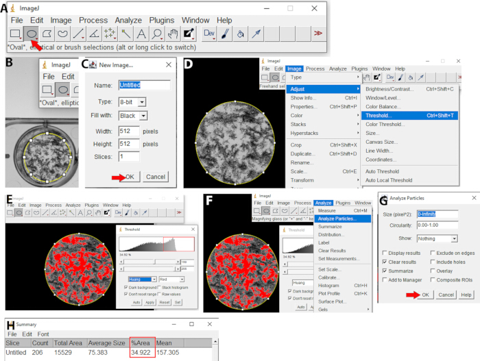
Figure 1: Step-by-step analysis of results in the ImageJ software. (A) "Oval" tool selection. (B) Selection of the area of interest in the image. (C) New image window (Ctrl + N) with recommended settings. (D) Steps for "Threshold analysis after pasting the area of interest in the new image window. (E) Huang and red options selection in the Threshold window, resulting in all white areas being filled in red. (F) Sequence of the steps used for the "Analyze Particles". (G) The setting used foranalyzing theparticles. (H) The result of the percentage of total destroyed area is shown in the summary window. Please click here to view a larger version of this figure.
13. Statistical analy sis
- Organize the data in an Excel spreadsheet within the Office software after calculating the percentage of proliferation inhibition.
- Open GraphPad Prism 5.0 software, and click on XY (Below New Table & Graph).
- In the Y section (below "X:error bar"), enter the number of data repetitions (typically three biological replicates). This number may vary depending on the number of repetitions performed.
- Select Mean only next to the plot and DO NOT select "Enter and plot error values already calculated elsewhere", then click on Create.
- Place the concentration data on the X-axis and the proliferation inhibition percentage data in columns A, A:Y1, A:Y2, A:Y3, A:Y4, A:Y5.
- Click on = Analyze and then click on Transform, and then OK. A new window will open. In this new window, click on Transform X values using X = Log(X), then click on Create a new graph of the results, and then OK.
- Click on = Analyze and then on Normalize. A new window will open. In the section How is 0% defined?, click and set Y = 0.0. In the section How is 100% defined?, click and set Y = 100; select Percentages and click on Create a new graph of the results.
- Click on the symbol: Fit a curve with nonlinear regression located above = Analyse. Click on Dose-response - Inhibition, then on log (inhibitor) vs. normalized response - Variable slope, and then click OK.
NOTE: A window will open displaying the following results: the IC50 (Inhibitory Concentration of 50%), the 95% confidence interval, and the R2 value. These values are crucial and should be included in a results table within the article. - To estimate other inhibitory concentrations, such as IC25, IC90, or IC95, use the link (https://www.graphpad.com/quickcalcs/Ecanything1.cfm), Quick Calcs - GraphPad; EC: effective concentration. Report the IC50 and the HillSlope obtained by the Prism software for each drug tested.
Access restricted. Please log in or start a trial to view this content.
Results
For evaluating the plaque assay after treatment, both the positive (cells infected with tachyzoites and untreated) and negative (uninfected cells with 0.1% DMSO) controls are crucial. At the end of the experiment, the positive control well should exhibit well-defined empty spaces with remnants of adhered cells (Figure 2). In contrast, the negative control well should display an intact monolayer with no empty spaces. The negative control is crucial for confirming the integrity of the monolaye...
Access restricted. Please log in or start a trial to view this content.
Discussion
The standardization of new protocols for antiproliferative tests is a fundamental step for advancing scientific research on the identification of future drugs. The protocol presented here offers an alternative approach to in vitro assessing the activity of bioactive molecules against tachyzoites of T. gondii. Given that T.gondii is an obligate intracellular parasite that locomotes by gliding motility, when the parasites egress from a lysed cell, they will invade the cells located in the vicinit...
Access restricted. Please log in or start a trial to view this content.
Disclosures
The authors declare that they have no conflict of interest.
Acknowledgements
Authors also thank the funding by the Conselho Nacional de Desenvolvimento e Pesquisa (CNPq), Fundação de Amparo à Pesquisa de Minas Gerais (FAPEMIG), and CAPES/PROEX. The authors would like to thank Pró-Reitoria de Pesquisa of the Universidade Federal de Minas Gerais for supporting this research. ESMD (CNPq 308082/2023-0) and RWAV (CNPq 305574/2021-3) are CNPq productivity fellows.
Access restricted. Please log in or start a trial to view this content.
Materials
| Name | Company | Catalog Number | Comments |
|---|---|---|---|
| 0.05 % Trypsin-EDTA | Life Technologies | 25300-062 | |
| 10 mL serological pipette | Olen | K17-110 | |
| 15mL falcon tube centrifuge | Fanem | Model: 206 | |
| 5 mL serological pipette | Olen | K17-115 | |
| 6-well culture plate | Sarstedt | 833920 | |
| Analytical balance | Mettler Toledo | Model: AB204 Serial No: 1116473813 | |
| Antibiotic-Antimycotic (100X) | Life Technologies | 15240-062 | |
| Automatic pipette | Kasvi | K1-AID | |
| Centrifuge | Eppendorf | Model 5415 R Serial No: 0023591 | |
| CO2 Incubator | PHC Corporation | Model No : MCO-230AICUVL-PA Serial No: 210360090 | |
| Culture bottle 25 cm2 | Sarstedt | 833910 | |
| Dimethyl sulfoxide (DMSO) P.A. | Merck | 1,02,95,21,000 | |
| Ethanol P.A | Merck | 1,00,98,31,000 | |
| Fetal Bovine Serum (FBS) | Life Technologies | 12657-029 | |
| Formaldehyde 37-38% | Panreac | 131328 | |
| GraphPad Prism | GraphPad Software | - | Version 8.0.1 |
| ImageJ software | - | - | Version 1.52e |
| Imaging documentation systems | Bio-Rad | Model: ChemiDoc MP Imaging System Serial No: 734BR2249 | |
| Laminar flow hood | Veco do Brasil | Model: VLFS-09 | |
| L-glutamine 200 mM (100X) | Life Technologies | 25030-081 | |
| Micro tube 0.5 mL | Sarstedt | 72,699 | |
| Neonatal Normal Human Dermal Fibroblast (NHDF) | Lonza | CC-3132 | Generously provided by Dr. Sheila Nardelli, from Fiocruz Paraná, Brazil |
| Neubauer chamber 0.100 mm 0.0025 mm2 | New Optics | 7301-1 | |
| pHmeter | Micronal | Model: B374 Serial No: 30/37 | |
| Pipette 20-200 µL | Kasvi basic | 22032893 | |
| Pipette 2-20 µL | Labmate Pro | 656630226 | |
| Pipette 0.2-2 µL | Labmate Pro | 556610114 | |
| Pipette 1000-5.000 µL | Uniscience | YM4A021482 | |
| Pipette 100-1000 µL | Eppendorf Research plus | Q34354C | |
| Potassium Phosphate Monobasic (KH2PO4) P.A | Synth | 01F2002.01.AF | |
| RPMI-1640 medium | Sigma-Aldrich | R0883 | |
| Sodium chloride (NaCl) P.A. | Cromoline Química Fina | - | |
| Sodium phosphate dibasic (Na2HPO4) P.A. | CRQ Produtos Quimicos Eireli | R2715920500 | |
| Tip 1.000 µL | Sarstedt | 7,01,186 | |
| Tip 10 µL | Sarstedt | 7,03,010 | |
| Tip 5.000 µL | Sarstedt | 70,11,83,001 | |
| Tip 200 µL | Sarstedt | 7,03,030 | |
| Transfer pipette (3.5 mL) | Sarstedt | 86,11,71,001 | |
| Tube 15 mL | Sarstedt | 6,25,54,502 | |
| Violet Crystal | Merck | 101408 | |
| Vortex mixer | Phoenix | Model: AP56 Serial No: 6633 | |
| Water Bath | Hemoquimica do Brasil | Model: HM1003 Serial No: 700001556 |
References
- Molan, A., Nosaka, K., Hunter, M., Wang, W. Global status of Toxoplasma gondii infection: systematic review and prevalence snapshots. Trop Biomed. 36 (4), 898-925 (2019).
- Bigna, J. J., et al. Global, regional, and country seroprevalence of Toxoplasma gondii in pregnant women: A systematic review, modelling and meta-analysis. Sci Rep. 10 (1), 12102(2020).
- Dian, S., Ganiem, A. R., Ekawardhani, S. Cerebral toxoplasmosis in HIV-infected patients: A review. Pathog Glob Health. 117 (1), 14-23 (2023).
- Wang, Z. -D., et al. Toxoplasma gondii Infection in Immunocompromised Patients: A Systematic Review and Meta-Analysis. Front Microbiol. 8, 389(2017).
- Layton, J., et al. Clinical spectrum, radiological findings, and outcomes of severe toxoplasmosis in immunocompetent hosts: A systematic review. Pathogens. 12 (4), 543(2023).
- Weiss, L. M., Dubey,, Jitender, P. Toxoplasmosis: A history of clinical observations. Int J Parasitology. 39 (8), 895-901 (2009).
- Fabiani, S., et al. Ocular toxoplasmosis, an overview focusing on clinical aspects. Acta Trop. 225, 106180(2022).
- Butler, N. J., Furtado, J. M., Winthrop, K. L., Smith, J. R. Ocular toxoplasmosis II: Clinical features, pathology and management. Clin Exp Ophthalmol. 41 (1), 95-108 (2013).
- Dunay, I. R., Gajurel, K., Dhakal, R., Liesenfeld, O., Montoya, J. G. Treatment of Toxoplasmosis: Historical perspective, animal models, and current clinical practice. Clin Microbiol Rev. 31 (4), e00057-e00117 (2018).
- Wei, H. -X., Wei, S. -S., Lindsay, D. S., Peng, H. -J. A systematic review and meta-analysis of the efficacy of anti-Toxoplasma gondii medicines in humans. PloS One. 10 (9), e0138204(2015).
- Shammaa, A. M., Powell, T. G., Benmerzouga, I. Adverse outcomes associated with the treatment of Toxoplasma infections. Sci Rep. 11 (1), 1035(2021).
- Hay, M., Thomas, D. W., Craighead, J. L., Economides, C., Rosenthal, J. Clinical development success rates for investigational drugs. Nat Biotechnol. 32 (1), 40-51 (2014).
- Paul, S. M., et al. How to improve R&D productivity: The pharmaceutical industry's grand challenge. Nat Rev Drug Discov. 9 (3), 203-214 (2010).
- Andrade, E. L., et al. Non-clinical studies required for new drug development - Part I: Early in silico and in vitro studies, new target discovery and validation, proof of principles and robustness of animal studies. Braz J Med Biol Res. 49 (11), e5644(2016).
- Brodniewicz, T., Grynkiewicz, G. Preclinical drug development. Acta Poloniae Pharmaceutica. 67 (6), 578-585 (2010).
- Dulbecco, R. Production of plaques in monolayer tissue cultures by single particles of an animal virus. Proc Natl Acad Sci U S A. 38 (8), 747-752 (1952).
- Roberts, C. O., Chaparas, S. D., McLaughlin, D. The use of the plaque assay in chemotherapeutic and dermal hypersensitivity studies on Toxoplasma gondii. Trans Am Microsc Soc. 95 (3), 470(1976).
- Mo, J., et al. Effect of the Pseudomonas metabolites HQNO on the Toxoplasma gondii RH strain in vitro and in vivo. Int J Parasitol Drugs Drug Resist. 21, 74-80 (2023).
- Barbosa, B. F., et al. Enrofloxacin is able to control Toxoplasma gondii infection in both in vitro and in vivo experimental models. Vet Parasitol. 187 (1-2), 44-52 (2012).
- Wall, G., Lopez-Ribot, J. L. Screening repurposing libraries for identification of drugs with novel antifungal activity. Antimicrob Agents Chemother. 64 (9), e00924-e01020 (2020).
- dos Santos, M., et al. Medicines for malaria venture pandemic box in vitro screening identifies compounds highly active against the tachyzoite stage of Toxoplasma gondii. Trop Med Infect Dis. 8 (12), (2023).
- Costa, A. L. O., et al. Antiproliferative and morphological analysis triggered by drugs contained in the medicines for malaria venture COVID-box against Toxoplasma gondii tachyzoites. Microorganisms. 12 (12), 2602(2024).
- Ufermann, C. -M., Müller, F., Frohnecke, N., Laue, M., Seeber, F. Toxoplasma gondii plaque assays revisited: Improvements for ultrastructural and quantitative evaluation of lytic parasite growth. Exp Parasitol. 180, 19-26 (2017).
- Condit, R. C. Principles of virology. Fields Virology. I, 25-57 (2007).
- Montazeri, M., et al. In vitro and in vivo evaluation of kojic acid against Toxoplasma gondii in experimental models of acute toxoplasmosis. Exp Parasitol. 200, 7-12 (2019).
- Ke, O. Y., Krug, E. C., Marr, J. J., Berens, R. L. Inhibition of growth of Toxoplasma gondii by qinghaosu and derivatives. Antimicrob Agents Chemother. 34 (10), 1961-1965 (1990).
- Liu, X., et al. Anti-Toxoplasma gondii effects of lipopeptide derivatives of Lycosin-I. Toxins. 15 (8), 477(2023).
- Emi, A., et al. Development of an automated plaque-counting program for the quantification of the Chikungunya virus. Sci Rep. 15 (1), 12429(2025).
- Sullivan, K., et al. High throughput virus plaque quantitation using a flatbed scanner. J Virol Methods. 179 (1), 81-89 (2012).
- Pereira Filho, A. A., et al. In vitro activity of essential oils from piper species (Piperaceae) against tachyzoites of Toxoplasma gondii. Metabolites. 13 (1), 95(2023).
- Martins-Duarte, E. S., et al. In vitro activity of N-phenyl-1,10-phenanthroline-2-amines against tachyzoites and bradyzoites of Toxoplasma gondii. Bioorg Medl Chem. 50, 116467(2021).
- Schneider, C. A., Rasband, W. S., Eliceiri, K. W. NIH Image to ImageJ: 25 years of image analysis. Nat Methods. 9 (7), 671-675 (2012).
- Subramanian, G., et al. Targeted phenotypic screening in Plasmodium falciparum and Toxoplasma gondii reveals novel modes of action of medicines for malaria venture malaria box molecules. mSphere. 3 (1), e00534(2018).
- Dans, M. G., et al. Screening the medicines for malaria venture pathogen box for invasion and egress inhibitors of the blood stage of Plasmodium falciparum reveals several inhibitory compounds. Int J Parasitol. 50 (3), 235-252 (2020).
Access restricted. Please log in or start a trial to view this content.