This study investigated the effect of electroacupuncture (EA) on high-salt-induced vascular dysfunction. EA reversed vascular damage and gut dysbiosis, possibly by modulating specific gut microbes.
Research Article
Electroacupuncture Orchestrates Intestinal Flora Remodeling: A Step Forward in Alleviating High Salt-Induced Vasculopathy
* These authors contributed equally
Opens in a new tab
In This Article
Summary
Loading...
$$\rightleftharpoonup{xx}$$
$$\longleftharp{xx}$$,
$$\longrightharp{xx}$$,
Abstract
Loading...
$$\rightleftharpoonup{xx}$$
$$\longleftharp{xx}$$,
$$\longrightharp{xx}$$,
This study aims to investigate the therapeutic potential and mechanisms of electroacupuncture (EA) in alleviating vascular dysfunction caused by high-salt diets, with a focus on gut microbiota modulation. We randomly divided 21 rats into Normal Control, High-Salt Model, and EA Intervention groups (n = 7 per group). The "high-salt blood stasis syndrome" model in Wistar rats was induced by intragastric perfusion of 12% NaCl. Treatments were performed between 17:00 and 19:00 Beijing time: rats were restrained with a soft cloth, followed by 20-min EA stimulation at BL23 and KI3. After the experiment, rat blood, blood vessels, and fecal samples were collected. Vascular effects were evaluated via coagulation function test, hemorheological analysis, and arterial histopathological assessment. 16S rDNA sequencing was used to analyze the composition and abundance changes of gut microbiota. The findings revealed that chronic high-salt intake disrupted blood viscosity, damaged vascular endothelial integrity, and caused gut microbial dysbiosis. Remarkably, EA treatment effectively reversed these pathological changes. Correlation analysis further identified specific gut microbiota (e.g., Lachnospiraceae and Ruminococcaceae) that strongly correlated with altered blood viscosity parameters (P < 0.05). Therefore, the present study concludes that high-salt diet-induced vasculopathy can be reversed by EA treatment, which may be attributed to EA's ability to regulate gut microbiota involved in bile acid metabolism and short-chain fatty acid synthesis.
Introduction
Loading...
$$\rightleftharpoonup{xx}$$
$$\longleftharp{xx}$$,
$$\longrightharp{xx}$$,
Dietary salt plays a crucial role in maintaining fluid balance and cellular homeostasis, yet excessive consumption represents a well-established risk factor for cardiovascular and cerebrovascular disease1. The World Health Organization recommends limiting daily sodium intake to less than 5 grams for healthy adults2. Research has demonstrated that a high salt diet (HSD) impairs vascular function and that cardiovascular disease (CVD), the most prevalent disease in the world, is often accompanied by vasculopathy3. These findings underscore that repairing vascular endothelial damage caused by HSD constitutes a critical strategy for preventing CVD progression. "Blood stasis syndrome" (characterized by stagnant blood circulation and internal blood accumulation) aligns with quantifiable coagulation dysfunction in modern medicine. Key validation biomarkers include significantly shortened prothrombin time (PT) and activated partial thromboplastin time (APTT), as well as markedly elevated fibrinogen (FIB) and thrombin time (TT). These indicators reflect a hypercoagulable state, corresponding directly to the pathological features of blood stasis.
Gut microbiota, residing throughout the gastrointestinal tract, serve as key regulators of immunometabolic processes and intestinal homeostasis while influencing diverse physiological and pathological pathways4. Emerging evidence indicates that excessive salt consumption induces gut dysbiosis and nutritional imbalance, triggering pro-inflammatory immune activation that manifests as localized inflammation while promoting microcirculatory damage, vascular remodeling, and systemic endothelial dysfunction1,5. Chronic HSD has been reported to cause a relative increase in the levels of Bacteroides intestinalis, Aspergillus, and Prevotella spp. in mice, and has been associated with contributing to the development of hypertension and vascular inflammation6.
Acupuncture, a cornerstone of Traditional Chinese Medicine (TCM), utilizes precise needle insertion at specific acupoints as an evidence-based complementary and alternative therapy in clinical practice. The National Institutes of Health (NIH) and World Health Organization (WHO) recognize acupuncture as a clinically validated intervention for multiple pathological conditions. Emerging evidence demonstrates that acupuncture has a dual therapeutic mechanism7,8, modulating gut microbiota composition while suppressing vascular inflammatory mediators, thereby collectively mitigating pathological progression in related disorders. Importantly, acupuncture is effective in lowering blood pressure and restoring endothelial function in damaged blood vessels9.
EA was selected over manual acupuncture for its standardized and quantifiable stimulation parameters, which reduce operator-dependent variability and enhance the reproducibility of results. Therefore, we investigated the effect of EA treatment on the gut microbiota in rats with high-salt blood stasis syndrome (HSBS) model and assessed the efficacy of EA on high salt-induced vasculopathy by blood rheology and vascular histopathological changes. To further understand the role of gut microbiota in the improvement of vascular lesions by EA therapy, we performed a correlation analysis between different genera of gut microbiota and whole blood cut rate and blood sedimentation.
We therefore hypothesized that EA alleviates HSD-induced vasculopathy by restoring gut microbiota homeostasis. To test this, we aimed to: (i) quantify the protective effects of EA on vascular function and structure, and (ii) identify the specific gut microbial taxa modulated by EA that correlate with these improvements. Furthermore, Pearson correlation analysis identified significant microbiota-vascular relationships, particularly between differentially abundant genera fluctuations and hemorheological abnormalities. To our knowledge, this is the first study to explore the protective mechanisms of EA in improving high-salt diet-induced vasculopathy from the perspective of gut microbiota. The experimental design of this study is shown in Figure 1.
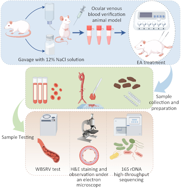
Figure 1: Experimental technical diagram. Please click here to view a larger version of this figure.
Access restricted. Please log in or start a trial to view this content.
Protocol
Loading...
$$\rightleftharpoonup{xx}$$
$$\longleftharp{xx}$$,
$$\longrightharp{xx}$$,
The entire experiment was conducted at the Experimental Center of the Basic Medical School of Chengdu University of Traditional Chinese Medicine. All animal welfare and experimental procedures were approved by the Laboratory Animal Welfare Ethics Committee of Chengdu University of Traditional Chinese Medicine (Ethics No. 2024078). Experiments were performed strictly following the NIH Guide for the Care and Use of Laboratory Animals (1986 revision, Publication 86-23).
Animals and diet
Male and female specific pathogen-free (SPF) Wistar rats, aged 6-8 weeks, weighed 200 ± 20 g. All rats were housed in well-ventilated rooms with a 12-h light-dark cycle and had free access to food and water.
Models and groups
Animal models were induced with reference to previous research experience10. After a 7-day acclimation period, 21 Wistar rats (50% male/female distribution) were randomly assigned to three experimental groups (n = 7/group) using the random number generator in Excel, with the number of male and female rats in each group kept roughly balanced to minimize the impact of gender on the results11. Normal Control (NC): Received daily gastric gavage (1 mL/100 g) of purified water. High-Salt Model (HS) and EA Intervention (EA): Administered 12% NaCl solution (1 mL/100 g) via daily gavage. All interventions were maintained for 30 consecutive days.
EA treatment
Treatment was initiated the day after successful modeling. EA was selected over manual acupuncture for its standardized and quantifiable stimulation parameters, which reduce operator-dependent variability and enhance the reproducibility of results. The locations of BL23 (Shenshu, bladder meridian) and KI3 (Taixi, kidney meridian) were determined based on the rat acupuncture point localization criteria of the Chinese Acupuncture and Moxibustion Society12. BL23 is situated 6 mm lateral to the dorsal midline, inferior to the second lumbar vertebra. KI3 was located at the midpoint depression between the highest point of the medial malleolus and the posterior border of the Achilles tendon. The local hair at the acupoints was removed and sterilized. Disposable sterile acupuncture needles (0.18 mm × 13 mm) were used to vertically puncture the acupoints, with insertion depths of 4-6 mm for BL23 and 3-5 mm for KI3.The positive electrode clip of the electroacupuncture device was connected to the needle handle of the acupuncture needle at BL23 on one side, and the negative electrode clip to that at KI3 on the same side. Parameters of the electroacupuncture device: Waveform: Dense-sparse wave (10/50 Hz); Frequency: 5 s for sparse wave, 10 s for dense wave; Intensity: 1-2 mA, adjusted according to the rats' convulsive and painful responses (screaming, resistance behaviors); Pulse width: ≤ 1 MS. Stimulation protocol: Intervention was performed daily from 17:00 to 19:00 (Beijing time), lasting 20 min each time. Cycle: One treatment cycle consisted of 7 days, with a total of 4 cycles and 1 day of rest between each cycle. The specific process is shown in Figure 2 (Created with BioGDP.com13).
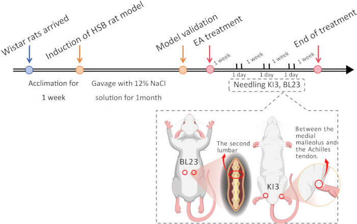
Figure 2: Experimental timeline. Please click here to view a larger version of this figure.
Sample collection and preparation
One month after gavage, orbital venous blood was collected from rats prior to EA intervention. Plasma samples were extracted by centrifugation at 1,000 × g for 15 min at 4 °C. At the end of treatment, fresh feces were collected from each group of rats. Rats were anesthetized with 10% emulsified isoflurane (30 mg/kg) and euthanized. The abdominal cavity was opened, and approximately 5 mL of blood was collected from the abdominal aorta for measurement of whole blood shear rate viscosity (WBSRV) within 4 h. Abdominal aortic tissues were collected for histopathological analysis. All samples were stored at -80 °C for subsequent use.
WBSRV test
After modeling was completed, approximately 4-5 mL of whole blood was collected from the abdominal aorta of rats using a blood collection needle and placed into heparin sodium anticoagulant tubes. The tubes were gently shaken to ensure thorough mixing of the blood samples with the anticoagulant. Within 4 h, the WBSRV and erythrocyte sedimentation rate (ESR) were measured using a fully automated hemorheology analyzer.
Vascular histopathology
Vascular tissues were fixed in 4% paraformaldehyde solution for at least 24 h and sectioned at a thickness of 3 µm for hematoxylin-eosin (HE) staining. Images were captured at 100× and 400× magnifications.
16S rDNA sequencing
Genomic DNA was extracted from fecal samples of each group. The V3-V4 primer sequences (341F: 5'-CCTACGGGNGGCWGCAG-3'; 805R: 5'-GACTACHVGGGTATCTAATCC-3') were used for PCR amplification of total DNA. The PCR amplification program was as follows: pre-denaturation at 98 °C for 30 s, denaturation at 98 °C for 10 s, annealing at 54 °C for 30 s, and extension at 72 °C for 45 s, with a total of 32 cycles, followed by a final extension at 72 °C for 10 min. PCR products were purified and quantified. Paired-end sequencing (2×250 bp) was performed on the NovaSeq 6000 sequencing platform. Paired-end reads were merged and filtered to obtain high-quality data. Using the DADA2 plugin in QIIME 2, length filtering and denoising were performed. This process generated amplicon sequence variants (ASVs) and ASV abundance tables, followed by the removal of singleton ASVs. Taxonomic annotation and abundance profiling were performed based on the ASV sequence file using the SILVA database to investigate species differences and community composition across samples. Species diversity and changes in relative abundance were evaluated via α-diversity and β-diversity analyses, and data visualization was performed on this website (https://www.omicstudio.cn/home).
Statistical analyses
Statistical analyses were performed using SPSS 26.0. A value of P < 0.05 was considered statistically significant. For data with homogeneous variances, one-way ANOVA was used for intergroup comparisons, followed by post-hoc least significant difference (LSD) tests if significant differences were found. For data with heterogeneous variances, the non-parametric Kruskal-Wallis one-way ANOVA by ranks was used. Mann-Whitney U test analysis was applied to assess changes in gut microbiota composition and community structure. Pearson rank correlation analysis was used to evaluate correlations between differentially abundant gut bacteria and other indicators.Pearson correlation analysis was employed because the data conformed to the assumptions of normality and linearity, which were validated by the Shapiro-Wilk test and Durbin-Watson test. Statistical graphs and heatmaps were generated using GraphPad Prism 10.
Access restricted. Please log in or start a trial to view this content.
Results
Loading...
$$\rightleftharpoonup{xx}$$
$$\longleftharp{xx}$$,
$$\longrightharp{xx}$$,
Validation of the animal model
The four coagulation parameters play a pivotal role in vascular lesion research by monitoring coagulation function abnormalities closely linked to vascular pathological changes. Post-modeling evaluation of retro-orbital venous plexus blood demonstrated significant alterations in coagulation parameters (PT, APTT, FIB, TT) between HS/EA groups versus NC controls (Figure 3). Specific test values are provided in Supplementary Table 1
Access restricted. Please log in or start a trial to view this content.
Discussion
Loading...
$$\rightleftharpoonup{xx}$$
$$\longleftharp{xx}$$,
$$\longrightharp{xx}$$,
Vascular lesions are a critical factor in the development and pathological manifestations of various CVD and metabolic disorders. Their detrimental effects include vascular dysfunction, abnormal blood clotting, lipid deposition, and plaque formation, which can lead to elevated blood pressure, thrombosis, and arterial plaque development, ultimately resulting in various CVD14,15,16. Therefore, timely intervention to halt the progr...
Access restricted. Please log in or start a trial to view this content.
Disclosures
Loading...
$$\rightleftharpoonup{xx}$$
$$\longleftharp{xx}$$,
$$\longrightharp{xx}$$,
The authors declare no conflicts of interest.
Acknowledgements
Loading...
$$\rightleftharpoonup{xx}$$
$$\longleftharp{xx}$$,
$$\longrightharp{xx}$$,
This research was funded by the Sichuan Provincial Department of Science and Technology (Grant No. 2024NSFSC0717). Thanks to Figdraw for the assistance provided in figure preparation.
Access restricted. Please log in or start a trial to view this content.
Materials
| Name | Company | Catalog Number | Comments |
|---|---|---|---|
| Acupuncture needles | Suzhou Medical Supplies Factory Co., Ltd. | 242059 | 0.18 × 13 mm |
| Activated Partial Thromboplastin Time (APTT) Assay Kit (Clotting Method) | Beijing Succeeder Technology Inc. | R21D24C4 | |
| AMPure XP beads | Beckman Coulter Genomics | A63881 | |
| Automatic Coagulation Analyzer | Beijing Succeeder Technology Inc. | SF-8050 | |
| Fibrinogen (FIB) Assay Kit (Clotting Method) | Beijing Succeeder Technology Inc. | R31D24D2 | |
| Fully Automatic Blood Rheology Tester | Beijing Succeeder Technology Inc. | SA-5600 | |
| GraphPad Prism 10 | GraphPad Software, Inc. | ||
| H&E staining solution | Wuhan Saiweier Biotechnology Co., Ltd | G1005 | |
| Illumina library quantification kit | Kapa Biosciences | 7960166001 | |
| Isoflurane | RWD Life Science | R510-22-10 | 100 mL |
| Magnetic bead-based stool genomic DNA extraction kit | Biotek | AU46111-96 | |
| Nikon Eclipse Ci-L upright white light microscope | Nikon | ||
| QIIME 2 | https://qiime2.org/ | ||
| SDZ-II Huatuo electronic acupuncture device | Suzhou Medical Supplies Factory Co., Ltd. | SDZ-II | |
| SILVA database | Release 138, https://www.arb-silva.de/ | ||
| Sodium chloride | Chengdu Jinshan Chemical Reagents Co., Ltd. | 20231008 | 500 g |
| SPF Wistar rats | SPF (Beijing) Biotechnology Co., Ltd. | SCXK (Beijing) 2024-0001 | |
| Thrombin Time (TT) Assay Kit (Clotting Method) | Beijing Succeeder Technology Inc. | R4D24E1 |
References
Loading...
$$\rightleftharpoonup{xx}$$
$$\longleftharp{xx}$$,
$$\longrightharp{xx}$$,
- He, F. J., Tan, M., Ma, Y., MacGregor, G. A. Salt reduction to prevent hypertension and cardiovascular disease: JACC state-of-the-art review. J Am Coll Cardiol. 75 (6), 632-647 (2020).
- Tian, J., et al. Protective effect of Paecilomyces cicadae TJJ11213 exopolysaccharide on intestinal mucosa and regulation of gut microbiota in immunosuppressed mice. Food Res Int. 165, 112477(2023).
- Wang, Y., et al. Different changing patterns for stroke subtype mortality attributable to high sodium intake in China during 1990 to 2019. Stroke. 54 (4), 1078-1087 (2023).
- Piewngam, P., De Mets, F., Otto, M. Intestinal microbiota: The hidden gems in the gut. Asian Pac J Allergy Immunol. 38 (4), 215-224 (2020).
- Balan, Y., Packirisamy, R. M., Mohanraj, P. S. High dietary salt intake activates inflammatory cascades via Th17 immune cells: Impact on health and diseases. Arch Med Sci. 18 (2), 459-465 (2022).
- Ferguson, J. F., et al. High dietary salt-induced dendritic cell activation underlies microbial dysbiosis-associated hypertension. JCI Insight. 5 (13), e126241(2019).
- Zhang, Y., et al. Manual acupuncture benignly regulates blood-brain barrier disruption and reduces lipopolysaccharide loading and systemic inflammation, possibly by adjusting the gut microbiota. Front Aging Neurosci. 14, 1018371(2022).
- Zhang, Q., et al. Electroacupuncture and human iPSC-derived small extracellular vesicles regulate the gut microbiota in ischemic stroke via the brain-gut axis. Front Immunol. 14, 1107559(2023).
- Leung, S. B., Zhang, H., Lau, C. W., Lin, Z. X. Attenuation of blood pressure in spontaneously hypertensive rats by acupuncture was associated with reduction oxidative stress and improvement from endothelial dysfunction. Chin Med. 11 (1), 38(2016).
- Qi, L., et al. Microbiome-metabolome analysis insight into the effects of high-salt diet on hemorheological functions in SD rats. Front Nutr. 11, 1408778(2024).
- Gohar, E. Y., et al. Acclimation to a high-salt diet is sex dependent. J Am Heart Assoc. 11 (5), e020450(2022).
- China Association of Acupuncture and Moxibustion. Nomenclature and location of acupuncture points for laboratory animals part 2: Rat. World J Acupunct Moxibustion. 35 (2), 162-163 (2021).
- Jiang, S., et al. Generic diagramming platform (GDP): A comprehensive database of high-quality biomedical graphics. Nucleic Acids Res. 53 (D1), D1670-D1676 (2025).
- Steven, S., et al. Vascular inflammation and oxidative stress: Major triggers for cardiovascular disease. Oxid Med Cell Longev. 2019, 7092151(2019).
- Tiwari, A., Elgrably, B., Saar, G., Vandoorne, K. Multi-scale imaging of vascular pathologies in cardiovascular disease. Front Med (Lausanne). 8, 754369(2021).
- Aguilar, V. M., Paul, A., Lazarko, D., Levitan, I. Paradigms of endothelial stiffening in cardiovascular disease and vascular aging. Front Physiol. 13, 1081119(2022).
- Culshaw, G., et al. The acute pressure natriuresis response is suppressed by selective ETA receptor blockade. Clin Sci (Lond). 136 (1), 15-28 (2021).
- Nishimoto, M., Griffin, K. A., Wynne, B. M., Fujita, T. Salt-sensitive hypertension and the kidney. Hypertension. 81 (6), 1206-1217 (2024).
- Yan, D., et al. Eucommia ulmoides bark extract reduces blood pressure and inflammation by regulating the gut microbiota and enriching the Parabacteroides strain in high-salt diet and N(omega)-nitro-L-arginine methyl ester-induced mice. Front Microbiol. 13, 967649(2022).
- Zhu, H., et al. Biological deciphering of the "kidney governing bones" theory in Traditional Chinese Medicine. Evid Based Complement Alternat Med. 2022, 1685052(2022).
- Mattson, D. L., Dasinger, J. H., Abais-Battad, J. M. Gut-immune-kidney axis: Influence of dietary protein in salt-sensitive hypertension. Hypertension. 79 (11), 2397-2408 (2022).
- Zhang, H. J., Wang, M. M., Cheng, S. Q. Application of experimental animal models in study of treating cancer with acupuncture and moxibustion. Shanghai Univ Tradit Chin Med. 38 (1), 82-88 (2024).
- Patel, S., et al. Age-related changes in hematological and biochemical profiles of Wistar rats. Lab Anim Res. 40 (1), 7(2024).
- Song, Y., Chen, Y., Li, J., Sun, W., Jin, F. Manual acupuncture enhanced therapeutic efficacy in vascular dementia rat model: Systematic review and network meta-analysis. Syst Rev. 14 (1), 80(2025).
- Zhang, Y., et al. Acupuncture, an effective treatment for post-stroke neurologic dysfunction. Brain Res Bull. 215, 111035(2024).
- Zhang, H., et al. Intervention of hypertension by acupuncture-related therapies: A network meta-analysis. Int J Older People Nurs. 19 (3), e12613(2024).
- Li, X., et al. Visual analysis of acupuncture point selection patterns and related mechanisms in acupuncture for hypertension. Technol Health Care. 32 (1), 397-410 (2024).
- Wang, J., Lin, X., Luo, X., Huang, J., Tang, C. Effect of acupuncture at Taixi point on the medulla TNF-α and IL-6 protein expression in SHR. J Jinan Univ (Nat Sci Med Ed). 37 (4), 327-330 (2016).
- Bi, K. Y. To explore the neuroprotective effect and mechanism of acupuncture regulating ACE2-Ang(1-7)-MasR axis on vascular dementia rats [Master's Thesis]. , Anhui University of Chinese Medicine. (2024).
- Li, S. Observations on the efficacy of selective acupuncture in the treatment of essential hypertension (liver-kidney yin deficiency type) [Master's Thesis]. , Liaoning University of Traditional Chinese Medicine. (2020).
- Liao, L., et al. Gut microbiota in Chinese and Japanese patients with cardiovascular diseases: A systematic review and meta-analysis. Ann Saudi Med. 43 (2), 105-114 (2023).
- Lv, Z., et al. Acupuncture ameliorates breast cancer-related fatigue by regulating the gut microbiota-gut-brain axis. Front Endocrinol (Lausanne). 13, 921119(2022).
- Ostrowska, J., Samborowska, E., Jaworski, M., Toczylowska, K., Szostak-Wegierek, D. The potential role of SCFAs in modulating cardiometabolic risk by interacting with adiposity parameters and diet. Nutrients. 16 (2), 266(2024).
- He, M., et al. Gut microbial metabolites SCFAs and chronic kidney disease. J Transl Med. 22 (1), 172(2024).
- Liu, A., et al. Human umbilical cord mesenchymal stem cells ameliorate colon inflammation via modulation of gut microbiota-SCFAs-immune axis. Stem Cell Res Ther. 14 (1), 271(2023).
- Zheng, X., et al. Study on the mechanism of Bifidobacterium animalis subsp. lactis F1-3-2 regulating bile acid metabolism through TMA-TMAO pathway to improve atherosclerosis. Probiotics Antimicrob Proteins. 17 (6), 4851-4866 (2024).
- Xu, H., et al. Gut microbiota-bile acid crosstalk regulates murine lipid metabolism via the intestinal FXR-FGF19 axis in diet-induced humanized dyslipidemia. Microbiome. 11 (1), 262(2023).
- Lin, X., et al. Isolation of potentially novel species expands the genomic and functional diversity of Lachnospiraceae. Imeta. 3 (2), e174(2024).
- Liu, T. H., et al. Hypertension inhibition by Dubosiella newyorkensis via reducing pentosidine synthesis. NPJ Biofilms Microbiomes. 11 (1), 34(2025).
- Qi, S., et al. Effect of acupuncture at Back-Shu points on gut microbiota in insomnia model rats based on metagenomic sequencing technology. Front Microbiol. 16, 1541958(2025).
- Landgraaf, R. G., et al. Acupuncture as multi-targeted therapy for the multifactorial disease obesity: A complex neuro-endocrine-immune interplay. Front Endocrinol (Lausanne). 14, 1236370(2023).
- Jeon, S. H., et al. Study protocol for a prospective, investigator-initiated clinical trial on the vascular effects of acupuncture in the abdomen and lower limbs for patients with diarrhea-predominant irritable bowel syndrome. Int J Colorectal Dis. 40 (1), 103(2025).
- Qi, Q. M., Quan, G. L., Qi, Y. F. Study on the mechanism of TCM treatment of chronic heart failure based on signaling pathway. J Clin Med Res. 5 (4), 637-639 (2025).
- Huang, P. Y., Liu, H. M., Ko, Y. R., Chang, Z. Y., Lee, T. Y. Electroacupuncture relieves portal hypertension by improving vascular angiogenesis and linking gut microbiota in bile duct ligation rats. Front Microbiol. 14, 1207137(2023).
- Liu, X., et al. Electroacupuncture improves lipid metabolism via proteome and gut microbiota profiling in obese rats. Am J Transl Res. 17 (5), 4008-4022 (2025).
- Sun, M. Z., et al. Electroacupuncture at Tianshu (ST25) and Zusanli (ST36) alleviates stress-induced irritable bowel syndrome in mice by modulating gut microbiota and corticotropin-releasing factor. J Tradit Chin Med. 42 (5), 732-740 (2022).
- Kiessling, S., Liu, S., Haller, D., Thaiss, C. A. Origins and functions of microbiome rhythms. Cell Host Microbe. 33 (6), 808-819 (2025).
- Huang, X. M., et al. Effects of chronotherapy of benazepril on the diurnal profile of RAAS and clock genes in the kidney of 5/6 nephrectomy rats. J Huazhong Univ Sci Technolog Med Sci. 33 (3), 368-374 (2013).
- Wan, R., et al. Differential placebo effect of sham acupuncture for chronic pain: A network meta-analysis of randomized controlled trials. BMC Complement Med Ther. 25 (1), 323(2025).
- Liu, J. P., et al. Differential effects of electroacupuncture and manual acupuncture on spontaneously hypertensive rats: Insights from intestinal microbiota and metabolomics. Front Mol Biosci. 12, 1619356(2025).
Access restricted. Please log in or start a trial to view this content.
Reprints and Permissions
Request permission to reuse the text or figures of this JoVE article
Request Permission