We present a standardized procedure for subperiosteal drain insertion and drain anchorage to be used after surgical evacuation of a chronic subdural hematoma via a single burr hole craniostomy.
Method Article
Subperiosteal Drain Insertion and Anchorage after Single Burr Hole Evacuation of Chronic Subdural Hematoma
In This Article
Summary
Abstract
Symptomatic chronic subdural hematoma is treated by surgical evacuation followed by drain insertion for post-operative drainage. There is no international consensus on the location of the drain (subdural or subperiosteal), the type of drainage (passive or active suction), or the duration of drainage (hours or days). However, a growing body of literature highlights the risk of iatrogenic brain injury during subdural drain insertion, causing increased interest in the subperiosteal drainage technique, which has been suggested to be equally effective. There is no consensus on the optimal subperiosteal drain insertion technique, resulting in numerous technical variations in the published literature. Additionally, drain anchoring is crucial to prevent the drain from displacing away from the burr hole. To address both issues, this article presents a standardized method for subperiosteal drain insertion and a novel anchorage technique for drains. All necessary drain entry-, exit-, and anchorage points are clearly defined and marked prior to placement of local anesthesia and skin incision. The stepwise insertion and anchorage of the drain are thoroughly described and illustrated, as well as the removal of the drain after post-operative drainage is complete.
Introduction
Symptomatic chronic subdural hematoma (CSDH) is treated by surgical evacuation followed by drain insertion for post-operative drainage, which reduces recurrence and mortality rates1. Currently, there is no international consensus on the optimal location of drainage (subdural or subperiosteal), the type of drainage (passive or active suction), or the duration of drainage (hours or days)2. In Denmark, all neurosurgical departments employ 24-h passive subdural drainage using a standardized drain insertion technique3,4,5.
However, the subdural drain is placed in close contact with the brain, posing a risk of iatrogenic brain parenchymal injury6, rupture of cortical and bridging veins, and seizure7, and active drain suction is avoided for the same reason. This has increased interest in the subperiosteal drain technique, which carries minimal risk of contact between the drain and brain, as the drain is placed extracranially above the burr hole. Furthermore, active suction subperiosteal drainage has been suggested to be at least equally effective in terms of CSDH recurrence and mortality compared to subdural drainage6,8.
There is, however, no consensus on the optimal subperiosteal drain insertion technique. Previous studies describe varying and, in general terms, that the distal tip of the drain should be inserted in the subperiosteal "space" over the burr hole, and that the proximal end of the drain should be tunneled away from the incision and brought out via a separate skin opening, where it is anchored to the skin by a suture2,7,9,10,11,12,13,14,15,16,17,18,19,20,21,22,23. The varying descriptions allow for many subjective technique variations, which may affect drain positioning. This may also affect the drainage, as the effective suction differs along fenestrated drains, typically highest at the proximal fenestrae and lowest towards the distal tip24. Additionally, with only one anchorage point at the proximal end of the drain, the drain tip can be displaced away from the burr hole due to traction on the drain, movement of the scalp muscles, or during head movement. This may also affect the drainage25.
To ensure reproducible subperiosteal drain insertion and drain anchorage above and straight across the burr hole, a standardized insertion method and a novel drain anchorage technique are warranted and presented in this article. The technique is designed for single burr hole evacuation of CSDH in either local or general anesthesia.
Protocol
The procedure will be performed as part of a multicentre randomized non-inferiority clinical trial comparing active subperiosteal to passive subdural 24-h drainage following evacuation of chronic subdural hematoma via single burr hole craniostomy (the SUPERDURA trial, ClinicalTrials.gov identifier NCT06621407). The SUPERDURA trial has been approved by the National Committees on Health Research Ethics with the number N-202400009, 13 December 2024. The reagents and the equipment used are listed in the Table of Materials.
1. Preparation of materials
- Prepare a CSDH evacuation kit similar to the equipment listed in the Table of Materials.
2. Preparation of the patient for surgery
- Place the patient in a supine position with the head on a vacuum pillow.
- Mark the center of the intended burr hole point on the scalp above the maximum width of the hematoma depicted on the pre-surgery CT-scan.
- Rotate the head of the patient so this mark is at the highest possible point relative to gravity.
NOTE: This head position is kept throughout the procedure to reduce post-operative pneumocephalus26. - Mark the approximately 5 cm long intended scalp incision with the intended burr hole point at its center (Figure 1A).
- Place the drain in a straight line along the incision line with the most proximal fenestrae overlying the marked burr hole center. Follow the drain proximally to the black drain skin-exit marking (Figure 2) and mark this point on the skin representing the proximal exit point of the drain (Figure 1A).
NOTE: The incision line and the proximal drain exit point must be aligned along the same axis. If the drain used has no black drain skin-exit marking, the proximal exit point should be 4.5 cm away from the most proximal fenestrae. - Infiltrate local anesthetic subcutaneously along the planned skin incision line, extending proximally in the same axis to the proximal drain exit point, and distally in the same axis an additional 1 cm beyond the incision line to include the intended distal drain anchorage site.
NOTE: This reduces pain associated with the skin incision and the subsequent creation of a subperiosteal tunnel using a rongeur at each anchorage point.
3. Surgical evacuation of the hematoma
- Perform a straight skin incision guided by the scalp incision marking down to the calvarium using a scalpel.
- Coagulate bleeding from the scalp using bipolar diathermy.
- Expand the opening using a surgical retractor.
- Drill a burr hole below the previously marked intended burr hole center, above the maximum extent of the hematoma.
NOTE: In the current setting, the burr hole is made by a 13 mm perforator. - Open the dura mater and the outer hematoma membrane with a cruciate incision using a scalpel.
- Wash out the subdural collection using a syringe with 37 °C isotonic saline solution.
NOTE: This is done until the outflowing liquid is clear
4. Post-operative drainage
- Insert the proximal end of the drain through the skin incision and puncture the scalp from below using the drain trocar at the drain exit mark (Figure 1B).
- Pull gently on the proximal end of the drain until the most proximal fenestrations align with the border of the burr hole (Figure 1B).
NOTE: The black skin-exit marking on the drain will be near the proximal drain exit point for a 13 mm burr hole. - Cut the distal fenestrated end of the drain to a length where the distal tip is just further than the end of the skin incision ( Figure 1B).
- Insert a 2-0 absorbable suture through the skin just further than the end of the skin incision down to the subperiosteal space, and through the distal hole of the drain and the most distal fenestra (Figure 1C and Figure 3).
NOTE: The entry of the suture needle should be directly above the intended final position of the distal drain tip, on the same axis as the skin incision. - Pass the absorbable suture back from the subperiosteal space to the skin (Figure 1D).
NOTE: The exit of the suture needle should be on the same axis, directly above the distal drain tip, a few millimeters from the suture needle entry point, and on the incision side. - Insert the distal end of the drain subperiosteally across and further than the burr hole, while tightening and tying the absorbable suture (Figure 1E).
- Pull gently on the proximal end of the drain so the drain is tight and located straight across the burr hole.
NOTE: If the distal drain tip is not held in place at the distal drain tip anchorage mark, or the most proximal fenestrae do not align with the border of the burr hole, remove the absorbable suture and return to step 4.4. - Anchor the proximal end of the drain to the scalp at the proximal drain exit point using a simple interrupted 2-0 absorbable suture (Figure 1F).
- Assess the integrity of the drain anchorage by gently pulling the drain away from each anchorage point in both directions above the burr hole.
NOTE: If the drain is not held in place at both anchorage points, remove the suture at the proximal drain exit point and return to step 4.7. - Close the skin incision above the burr hole in 2 layers.
- Connect the drain to the active suction system of choice, and secure the system above the collar bone of the patient3.
NOTE: The patient is allowed to ambulate freely after this step.
5. Removal of the drain after
NOTE: This step was performed after 24 h of drainage.
- Cut and remove the suture at the distal drain tip.
- Cut and remove the suture at the proximal drain exit.
- Pull the drain out through the proximal exit point and cover the site with a sterile dressing.
Results
Following this protocol will result in a drain firmly anchored above and straight across the center of the burr hole, with the most proximal drain fenestrae starting at the edge of the burr hole and covering the entire burr hole. Correct anchorage will prevent the drain from displacing away from the burr hole. This result is confirmed both visually and by manual traction prior to skin incision closure. If the drain is significantly displaced by gently pulling on it away from each anchorage point, or if the drain is not positioned above and straight across the center of the burr hole, the protocol clearly states how to correct this by returning to a prior step.
The position of the drain above and straight across the burr hole can also be evaluated at any time after incision closure by manual palpation, as the drain is easily palpable through the scalp, and the center of the burr hole is approximately underneath the center of the closed skin incision.
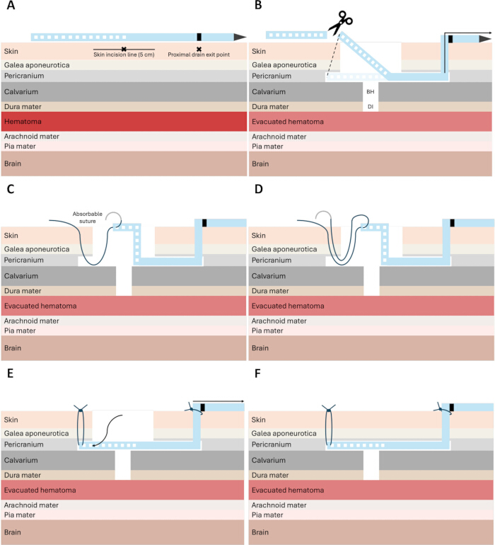
Figure 1: Subperiosteal drain insertion and anchorage following evacuation of chronic subdural hematoma through a single burr hole. (A) A 5 cm-long intended scalp incision is marked over the planned burr hole center. The drain is positioned in a straight line along the incision, with the most proximal fenestrae overlying the marked burr hole center. The intended proximal exit point of the drain is identified using the black skin-exit mark on the drain. (B) The proximal end of the drain is introduced through the skin incision, and the scalp is punctured from below using the drain trocar at the marked exit point. The proximal end of the drain is pulled until the most proximal fenestrae align with the border of the burr hole. The distal fenestrated end of the drain is trimmed so that the distal tip extends just beyond the end of the skin incision. (C) A 2-0 absorbable suture is passed through the skin just beyond the end of the incision into the subperiosteal space and then through the distal hole of the drain and the most distal fenestra. (D) The absorbable suture is passed back from the subperiosteal space to the skin. (E) The distal end of the drain is positioned across and beyond the burr hole while the absorbable suture is tightened and tied. Gentle traction is applied to the proximal end of the drain to ensure that the drain is taut and positioned straight across the burr hole. The proximal end of the drain is anchored to the skin at the proximal exit site using a simple interrupted 2-0 absorbable suture. (F) The skin incision is closed. Please click here to view a larger version of this figure.
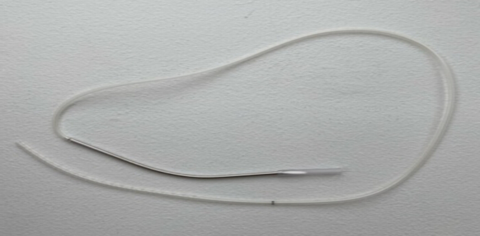
Figure 2: The entire drain, including the distal fenestrated end, the black skin-exit marking, and the proximal end with the trocar. Please click here to view a larger version of this figure.
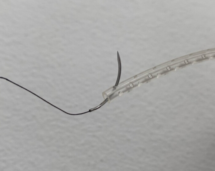
Figure 3: Insertion of a 2-0 absorbable suture through the distal hole of the drain and the most distal fenestra. Please click here to view a larger version of this figure.
Discussion
The goal of this subperiosteal drain insertion and anchorage technique (Figure 1A-F) is to achieve reproducible subperiosteal drain insertion, anchored with the greatest effective suction directly above and across the burr hole. There are four particularly critical steps in this protocol to achieve this goal.
Firstly, accurately marking the burr hole center, the skin incision, and the proximal exit point of the drain along the same axis using the drain itself (as shown in Figure 1A) is necessary to ensure the final position of the drain is directly above and straight across the burr hole.
Secondly, once the skin has been punctured using the trocar, the drain must be pulled until the most proximal fenestrae of the drain align with the proximal border of the burr hole. This is important, as the effective suction in fenestrated drains is typically highest at the most proximal fenestrae24. The distal drain tip is then cut to a length just further than the end of the skin incision (as shown in Figure 1B) in preparation for distal drain tip anchorage.
Thirdly, the entry and exit of the absorbable suture anchoring the distal drain tip (as shown in Figure 1C-E) must be along the same axis as the skin incision, and the proximal drain exit to prevent displacement of the drain away from the burr hole. After distal drain tip anchorage, the drain is gently pulled proximally to ensure it is tight and located straight across the burr hole, prior to proximal drain exit anchorage, to prevent displacement from the burr hole.
Finally, the integrity of both drain anchorage points must be assessed by gently pulling the drain away from each anchorage point. If the drain is not held in place at both points or if the most proximal fenestrae do not align with the proximal border of the burr hole, the protocol clearly states how to correct this by returning to a previous step.
If the final position of the drain is not above and straight across the center of the burr hole, it is likely that the proximal drain exit point is not on the same axis as the distal drain anchorage point and the center of the burr hole. To correct this, keep the distal drain tip anchoring suture in place, cut the proximal anchoring suture, retract the drain back through the proximal drain skin exit, and create a new proximal exit on the correct axis using the trocar. Continue the protocol from step 4.7.
We believe the technique is reproducible for other surgeons and institutions because it is supported by a detailed step-by-step protocol accompanied by illustrations and troubleshooting guidance. Furthermore, the technique has already been applied to more than 20 patients by multiple surgeons across all four neurosurgical departments in Denmark without requiring any modifications.
A limitation of this study is that the position of the drain is only confirmed by visual inspection and manual palpation, as a routine post-operative CT-scan to evaluate the drain position is not performed in Denmark. CT scans are only done in the case of clinical deterioration27. Additionally, the technique is not directly applicable to multiple burr hole evacuation of CSDH. However, the drain may possibly be sufficiently anchored straight above and across both burr holes by including an additional suture between the burr hole on the same axis in addition to the distal and proximal sutures.
Future studies may investigate whether different drain and fenestra diameters have a clinically meaningful impact on outcomes. The drain used in this study has an outer diameter of 10 F and a fenestra diameter of 1 mm, and was selected based on the recommendations of colleagues. We found no studies comparing different drain and fenestra diameters. Additionally, the drain anchorage can be further improved by adding a suture at the most proximal fenestrae. It is uncertain whether this would affect clinically meaningful outcomes, and it was also not done due to a fear of intracranial infection once the suture is removed, as the needle hole would be located on the border of the burr hole and the underlying intracranial space. However, this risk is theoretical and not reported in the literature.
Disclosures
The authors declare no conflicts of interest.
Acknowledgements
The authors would like to thank Dr. Jiri Jr. Bartek for sharing his experiences with subperiosteal drain insertion.
Materials
| Name | Company | Catalog Number | Comments |
|---|---|---|---|
| 2-0 Vicryl suture | Ethicon Inc. | D6242 | Any similar suture is usable |
| Anatomical artery clamp curved Halstead-Mosquito, 125 mm | Scan-Med A/S | BH111R | Any similar instrument is usable |
| Anatomical artery clamp straight, 125 mm | BH110R | No specific manufacturer, any similar instrument is usable | |
| Anatomical forceps straight, 115 mm | No specific manufacturer, any similar instrument is usable | ||
| Bipolar Coagulation forceps, 200 mm | BRAUN SCANDINAVIA A/S | GK644R | Any similar instrument is usable |
| Bone hook angled 90 degrees blunt Crile, 200 mm | Scan-Med A/S | BT080R | Any similar instrument is usable |
| DeBakey atraumatic forceps straight, 200 mm | B. BRAUN MEDICAL A/S | FB402R | Any similar instrument is usable |
| Dissector double curved Olivecrona, 2.5/3 x 240 mm | Scan-Med A/S | R0178 | Any similar instrument is usable |
| Exudrain with trocar 10 FG | Mediplast | 68409 | Any similar instrument is usable |
| Gillies surgical forceps straight, 155 mm | B. BRAUN MEDICAL A/S | BD660R | Any similar instrument is usable |
| Gross-Maier straight tampon forceps, 200 mm | No specific manufacturer, any similar instrument is usable | ||
| Gruenwald anatomical bayonet forceps, 200 mm | B. BRAUN MEDICAL A/S | BD883R | Any similar instrument is usable |
| Local anesthetic | To be chosen from available standard surgical supplies | ||
| Maestro Chuck Perforator 125 mm | Stryker | 5400-210-060 | Any similar instrument is usable with the same size |
| Materials for disinfection | To be chosen from available standard surgical supplies | ||
| Mayo TC dissecting scissor curved blunt, 170 mm | KEBOMED A/S | BC253R | Any similar instrument is usable |
| Metzenbaum dissecting scissor blunt, 145 mm | Karl Storz Endoskopi DANMARK A/S | BC605R | Any similar instrument is usable |
| Metzenbaum dissecting scissor blunt, 180 mm | Scan-Med A/S | BC606R | Any similar instrument is usable |
| Metzenbaum dissecting scissor blunt, Wavecut TC, 180 mm | B. BRAUN MEDICAL A/S | BC602R | Any similar instrument is usable |
| Needle Holder straight TC Halsey, 130 mm | KEBOMED A/S | BM012R | Any similar instrument is usable |
| Officer surgical forceps straight, 150 mm | B. BRAUN MEDICAL A/S | BD598R | Any similar instrument is usable |
| Pean artery clamp straight, 140 mm | Scan-Med A/S | BH304R | Any similar instrument is usable |
| pi drive Signature drill | Stryker | 5407-100-000 | Any similar instrument is usable |
| Raspatorium curved Olivecrona, 11 x 170 mm | Scan-Med A/S | 111-22150 | Any similar instrument is usable |
| Retractor 10 x 30 x 205 mm | No specific manufacturer, any similar instrument is usable | ||
| Scalpel blade No. 11 | No specific manufacturer, any similar instrument is usable | ||
| Scalpel blade No. 24 | No specific manufacturer, any similar instrument is usable | ||
| Scalpel handle straight No. 3, 125 mm | Scan-Med A/S | BB073R | Any similar instrument that fits the scalpel blade is usable |
| Scalpel handle straight No. 4, 135 mm | Scan-Med A/S | BB084R | Any similar instrument that fits the scalpel blade is usable |
| Self-retaining retractor 2 x 2 blunt Mayo-Adams, 40 x 165 mm | B. BRAUN MEDICAL A/S | BV112R | Any similar instrument is usable |
| Self-retaining retractor 3 x 4 Weitlaner, 135 mm | No specific manufacturer, any similar instrument is usable | ||
| Spatula double 15/20 x 185 mm | No specific manufacturer, any similar instrument is usable | ||
| Sterile dressing | To be chosen from available standard surgical supplies | ||
| Syringe + needle | To be chosen from available standard surgical supplies | ||
| Tissue marking pen | To be chosen from available standard surgical supplies |
References
- Santarius, T., et al. Use of drains versus no drains after burr-hole evacuation of chronic subdural haematoma: a randomised controlled trial. Lancet. 374 (9695), 1067-1073 (2009).
- Soleman, J., et al. Drain insertion in chronic subdural hematoma: an international survey of practice. World Neurosurg. 104, 528-536 (2017).
- Jensen, T. S. R., et al. National randomized clinical trial on subdural drainage time after chronic subdural hematoma evacuation. J Neurosurg. 137 (3), 799-806 (2022).
- Hjortdal Grønhøj, M., et al. Optimal drainage time after evacuation of chronic subdural haematoma (DRAIN TIME 2): A multicentre, randomised, multiarm and multistage non-inferiority trial in Denmark. Lancet Neurol. 23 (8), 787-796 (2024).
- Jensen, T. S. R., Poulsen, F. R., Bergholt, B., Hundsholt, T., Fugleholm, K. Drain type and technique for subdural insertion after burr hole evacuation of chronic subdural hematoma. Acta Neurochir. 162 (9), 2015-2017 (2020).
- Greuter, L., Hejrati, N., Soleman, J. Type of drain in chronic subdural hematoma: A systematic review and meta-analysis. Front Neurol. 11, 312(2020).
- Chew, Z. H., Cheong, T. M., Ling, J. M., Saffari, S. E., Lee, L. Use of active low suction pressure (subgaleal) drains in chronic subdural hematoma surgery. World Neurosurg. 194, 123457(2025).
- Soleman, J., et al. Subperiosteal vs. subdural drain after burr-hole drainage of chronic subdural hematoma: a randomized clinical trial (cSDH-Drain-Trial). Neurosurgery. 85 (5), E825-E834 (2019).
- Hwang, Y., et al. Comparative analysis of safety and efficacy in subperiosteal versus subdural drainage after burr-hole trephination for chronic subdural hematoma. Clin Neurol Neurosurg. 212, 107068(2022).
- Akhtar, M. S., et al. Outcomes of subgaleal drain placement after two burr-holes craniectomy for chronic subdural hematoma. J Coll Physicians Surg Pak. 33 (4), 460-464 (2023).
- Bartley, A., Hallén, T., Tisell, M. Is a drainage time of less than 24 h sufficient after chronic subdural hematoma evacuation. Acta Neurochir. 165 (3), 711-715 (2023).
- Kaliaperumal, C., et al. A prospective randomised study to compare the utility and outcomes of subdural and subperiosteal drains for the treatment of chronic subdural haematoma. Acta Neurochir. 154 (11), 2083-2089 (2012).
- Zolfaghari, S., et al. Burr hole craniostomy versus minicraniotomy in chronic subdural hematoma: a comparative cohort study. Acta Neurochir. 163 (11), 3217-3223 (2021).
- Zhang, J. J. Y., et al. Outcomes of subdural versus subperiosteal drain after burr-hole evacuation of chronic subdural hematoma: A multicenter cohort study. World Neurosurg. 131, e392-e401 (2019).
- Zumofen, D., Regli, L., Levivier, M., Krayenbühl, N. Chronic subdural hematomas treated by burr hole trepanation and a subperiosteal drainage system. Neurosurgery. 64 (6), 1112-1116 (2009).
- Zolfaghari, S., et al. Risk factors for need of reoperation in bilateral chronic subdural haematomas. Acta Neurochir. 163 (7), 1849-1856 (2021).
- Ebel, F., Guzman, R., Soleman, J. Burr hole trepanation and insertion of a subperiosteal drain for chronic subdural haematoma: how I do it. Acta Neurochir. 162 (11), 2707-2710 (2020).
- Gazzeri, R., et al. Clinical investigation of chronic subdural hematoma: relationship between surgical approach, drainage location, use of antithrombotic drugs and post-operative recurrence. Clin Neurol Neurosurg. 191, 105705(2020).
- Ishfaq, A. Outcome in chronic subdural hematoma after subdural vs. subgaleal drain. J Coll Physicians Surg Pak. 27 (7), 419-422 (2017).
- Gelabert-González, M., Garcia-Allut, A. Continuous subgaleal suction drainage for the treatment of chronic subdural haematoma. Acta Neurochir. 149 (9), 973-974 (2007).
- Carter, S. H., et al. Clinical outcome of subdural versus subgaleal drain after burr-hole drainage for chronic subdural hematoma. Acta Neurochir. 166 (1), 433(2024).
- Baschera, D., Tosic, L., Westermann, L., Oberle, J., Alfieri, A. Treatment standards for chronic subdural hematoma: Results from a survey in Austrian, German, and Swiss neurosurgical units. World Neurosurg. 116, e983-e995 (2018).
- Ozgen, U., et al. A comparison of subgaleal active drainage and subdural passive drainage and an analysis of factors affecting chronic subdural hematoma outcomes. Turk Neurosurg. 32 (4), 688-696 (2022).
- Wickramarachchi, A., Gregory, S. D., Burrell, A. J. C., Khamooshi, M. Flow characterization of Maquet and Bio-Medicus multi-stage drainage cannulae during venoarterial extracorporeal membrane oxygenation. Comput Biol Med. 171, 108135(2024).
- Deng, X., Chen, J., Du, X. Letter to the editor regarding "Use of active low suction pressure (subgaleal) drains in chronic subdural hematoma surgery". World Neurosurg. 197, 123929(2025).
- Májovský, M., Netuka, D., Beneš, V., Kučera, P. Burr-hole evacuation of chronic subdural hematoma: Biophysically and evidence-based technique improvement. J Neurosci Rural Pract. 10 (1), 113-118 (2019).
- Ng, H. Y., Ng, W. H., King, N. K. K. Value of routine early post-operative computed tomography in determining short-term functional outcome after drainage of chronic subdural hematoma: An evaluation of residual volume. Surg Neurol Int. 5, 136(2014).
Reprints and Permissions
This article has been published
Video Coming Soon