Bilateral acupuncture-related digital osteoarticular infection caused by Enterobacteriaceae was successfully managed with staged surgery and sequential antimicrobial therapy, resulting in infection control, wound healing, and short-term functional recovery.
Case Report
Acupuncture-related Digital Joint Infection Managed With Staged Surgery And Oral Antibiotic Therapy
* These authors contributed equally
In This Article
Summary
Abstract
Acupuncture is generally considered safe; however, inadequate aseptic technique may result in infections involving the skin, soft tissue, bone, or joints. Gram-negative bacilli causing hand infections are uncommon and may present diagnostic and therapeutic challenges, particularly when multiple digits are involved. A 73-year-old woman developed progressive pain, swelling, and functional limitation of the right third and left fifth fingers approximately 1 month after acupuncture. Imaging suggested osteomyelitis with involvement of the interphalangeal joint. Separate sterile specimens were obtained from each affected digit before antibiotic initiation. Cultures identified Morganella morganii in the right third finger and Proteus mirabilis in the left fifth finger. Both isolates were susceptible to third-generation cephalosporins, beta-lactam/beta-lactamase inhibitor combinations, carbapenems, and fluoroquinolones, with low minimum inhibitory concentrations supporting levofloxacin use. A staged surgical approach was performed, consisting of initial debridement with placement of antibiotic-loaded bone cement, followed by delayed wound repair after infection control. Systemic antimicrobial therapy was guided by susceptibility testing, transitioning from intravenous ceftriaxone to oral therapy. At discharge, oral levofloxacin (750 mg once daily) was prescribed to complete an approximately 6-week treatment course. At 6-week follow-up, both wounds had completely epithelialized, with resolution of swelling and marked pain reduction. Functional recovery was evident, with substantial improvement in interphalangeal joint range of motion. Inflammatory markers normalized, and follow-up radiographs demonstrated bone consolidation without recurrent infection. At 3 months, no relapse or systemic complications were observed. This case illustrates that, in selected patients with acupuncture-related digital osteoarticular infection, a stepwise approach integrating surgical debridement, microbiological confirmation, and susceptibility-guided antimicrobial therapy may achieve satisfactory short-term outcomes. High-bioavailability oral agents such as levofloxacin may be considered after initial infection control in carefully selected cases; however, this observation is based on a single case and requires validation in larger studies.
Introduction
Despite the generally favorable safety profile of acupuncture, infectious complications, when they occur, are most commonly limited to superficial soft tissue infections at needle insertion sites. These infections are typically caused by conventional pathogens such as Staphylococcus aureus, although atypical organisms, including nontuberculous mycobacteria (NTM), have also been increasingly recognized1. Epidemiologic data suggest that serious adverse events associated with acupuncture are rare; however, infection remains one of the most frequently reported complications among documented cases2. Most reported infections involve localized cutaneous or soft tissue disease, whereas more complex presentations, such as deep musculoskeletal involvement, disseminated infection, or atypical pathogen-related disease, are relatively uncommon and remain insufficiently characterized in the literature2.
This case is clinically relevant for several reasons. First, bilateral multi-digit involvement following acupuncture is exceptionally rare; most reported cases are limited to localized, single-site infection3. Such an atypical distribution may delay recognition and complicate management. Second, infections caused by Gram-negative bacilli such as Morganella morganii and Proteus mirabilis are uncommon in hand osteoarticular infections, which are more often caused by Gram-positive organisms4. Third, although staged surgical management is an established strategy for complex hand infections, its combination with oral step-down antimicrobial therapy has been less frequently described in acupuncture-related osteoarticular infection, particularly in elderly patients in whom treatment tolerability and adherence are important considerations5,6.
The overall goal of this report is to describe the diagnosis and management of an unusual case of acupuncture-related bilateral digital osteoarticular infection and to illustrate a practical treatment approach integrating microbiological confirmation, staged surgery, and sequential antimicrobial therapy. This case may help readers recognize when a structured, multidisciplinary approach is appropriate for similar atypical presentations, while underscoring that conclusions regarding broader applicability remain limited by the single-case design.
Case Presentation:
A 73-year-old woman presented with progressive pain, swelling, and limited movement of the right third and left fifth fingers for approximately 1 month. She reported undergoing multiple acupuncture sessions, approximately 3 to 4 sessions over a 2-week period, at a local outpatient clinic for chronic hand stiffness 4 weeks before symptom onset. According to the patient, needles were inserted directly into both the right third and left fifth digits, corresponding to the sites that subsequently developed infection. The procedures were performed by a non-hospital practitioner in a community setting using silver needles. Although disposable needles were reportedly used, the details of sterilization procedures and skin disinfection could not be fully verified.
The patient had a medical history of coronary artery disease and hypertension, but no diabetes or immunosuppressive therapy. She denied recent trauma, fever, or other systemic symptoms. On examination, she demonstrated marked limitation in both active and passive range of motion. The right third finger showed proximal interphalangeal (PIP) joint flexion limited to approximately 30°, accompanied by pain and incomplete extension. The left fifth finger exhibited severe restriction at the distal interphalangeal (DIP) joint, with flexion limited to less than 20° and near-complete loss of extension. Grip strength was reduced, and fine motor tasks such as pinching and grasping were significantly impaired. The overlying skin on the right third finger showed a small puncture mark consistent with an acupuncture entry site, with mild purulent discharge.
Laboratory testing demonstrated elevated inflammatory markers, including an erythrocyte sedimentation rate (ESR) of 33 mm/h and a high-sensitivity C-reactive protein (hs-CRP) level of 24.96 mg/L. The white blood cell count was within the normal range at 7.6 × 10⁹/L. Radiographic findings suggested involvement of the middle phalanx and PIP joint in the right third finger and the distal phalanx and DIP joint in the left fifth finger, consistent with osteomyelitis with associated septic arthritis.
Pus specimens were collected separately from each affected digit under strict aseptic conditions before antibiotic initiation. Cultures identified Morganella morganii in the right third finger and Proteus mirabilis in the left fifth finger. No pooling of specimens was performed. Both isolates were susceptible to third-generation cephalosporins, beta-lactam/beta-lactamase inhibitor combinations, carbapenems, and fluoroquinolones, but resistant to ampicillin and first-generation cephalosporins (Table 1). Based on the clinical, radiographic, and microbiological findings, a diagnosis of acupuncture-related bilateral digital osteomyelitis with interphalangeal joint involvement was established.
| Site | Pathogen | Ceftriaxone | Piperacillin–tazobactam | Carbapenems | Fluoroquinolones | Ampicillin | First-generation cephalosporins |
| Right third finger | Morganella morganii | Susceptible | Susceptible | Susceptible | Susceptible | Resistant | Resistant |
| Left fifth finger | Proteus mirabilis | Susceptible | Susceptible | Susceptible | Susceptible | Resistant | Resistant |
Table 1: Microbiological findings and antimicrobial susceptibility profile of isolates obtained from the affected digits. Cultures from separately collected specimens identified Morganella morganii in the right third finger and Proteus mirabilis in the left fifth finger. Antimicrobial susceptibility testing showed that both isolates were susceptible to ceftriaxone, piperacillin-tazobactam, carbapenems, and fluoroquinolones, but resistant to ampicillin and first-generation cephalosporins.
The patient underwent staged surgical management. During Stage I on June 18, 2025, thorough debridement of necrotic tissue and infected bone was performed under regional anesthesia, followed by placement of gentamicin-loaded bone cement to fill the defect and provide local antibacterial coverage. Postoperatively, empirical intravenous antibiotics were initiated and subsequently adjusted according to culture results. During Stage II on July 23, 2025, after clinical and laboratory improvement, secondary wound repair and soft tissue closure were performed.
During hospitalization, the patient received microbiologically guided systemic antimicrobial therapy, transitioning from intravenous ceftriaxone to oral faropenem. At discharge, oral levofloxacin 750 mg once daily was prescribed to complete a total treatment course of 6 weeks, including the inpatient course. Post-discharge care included dressing changes every 2 to 3 days, serial monitoring of CRP and ESR, and gradual initiation of supervised functional exercises. At 6-week follow-up, both surgical sites had healed completely, with resolution of pain and restoration of finger flexion-extension function. Follow-up radiographs demonstrated bone consolidation without recurrent infection. At 3 months, no local relapse or systemic complications were observed.
Diagnosis, Assessment, and Plan:
Based on the clinical presentation, imaging findings, and microbiological results, the patient was diagnosed with acupuncture-related bilateral digital osteomyelitis with interphalangeal joint involvement. Specifically, the right third finger involved the middle phalanx and PIP joint, whereas the left fifth finger involved the distal phalanx and DIP joint.
Several alternative diagnoses were considered. Septic arthritis unrelated to acupuncture was considered but was less likely because of the clear temporal association with needling and the presence of puncture-related skin findings. Crystal arthropathy was excluded because of purulence and positive bacterial cultures. Mycobacterial infection was also considered, but routine microbiological findings did not support this diagnosis. Degenerative osteoarthritis was excluded on the basis of the acute progression and inflammatory profile. The diagnosis of osteomyelitis with joint involvement was established through the combined interpretation of clinical findings, radiographic evidence of cortical erosion and joint-space narrowing, and microbiological confirmation, rather than reliance on a single modality.
This case was assessed as a complex bilateral infection involving both bone and joint structures, with risk of progression and functional impairment. Because necrotic tissue and radiographic bone involvement were present, conservative treatment alone was considered insufficient. A staged surgical approach was therefore selected to allow initial debridement and infection control, followed by delayed wound repair after clinical improvement.
Pathogen-directed antimicrobial therapy was implemented in parallel. Based on susceptibility testing, the availability of effective oral agents, and the need to support adherence in an elderly patient, sequential antimicrobial therapy was used after initial intravenous treatment. This approach was selected to balance microbiological control, surgical management, and treatment feasibility in a complex osteoarticular infection.
Protocol
This study was conducted in accordance with the Declaration of Helsinki and was approved by the Ethics Committee of the First Affiliated Hospital, Zhejiang University School of Medicine (No. 2024-0844). Written informed consent was obtained from the patient for participation and publication. The reagents and the equipment used are listed in the Table of Materials.
1. Initial clinical assessment
- A detailed clinical history was obtained, including the timing, sites, and setting of acupuncture exposure, relevant comorbidities, and symptom progression.
- A focused physical examination of the affected digits was performed, documenting swelling, erythema, tenderness, purulent discharge, and limitation of motion.
- Functional impairment was assessed on the basis of active and passive joint mobility and hand function.
2. Imaging studies
- Plain radiographs of the affected digits were obtained at baseline to assess cortical integrity, joint involvement, and bone destruction.
- Radiographic evaluation was repeated during follow-up to assess progression or resolution of osseous changes.
- Magnetic resonance imaging and ultrasonography were not performed in this case.
3. Laboratory and microbiological tests
- Routine laboratory studies, including complete blood count, erythrocyte sedimentation rate (ESR), and C-reactive protein (CRP), were obtained to evaluate systemic inflammation.
- Pus specimens were collected separately from each affected digit under sterile conditions before antibiotic administration.
- The specimens were processed using standard aerobic culture methods, and antimicrobial susceptibility testing was performed according to established clinical laboratory procedures.
4. Surgical intervention
- Stage I: Debridement and infection control
Thorough surgical debridement was performed to remove necrotic soft tissue and infected bone. The wound cavity was irrigated, and antibiotic-loaded bone cement was placed to provide local antimicrobial activity and structural support. - Stage II: Delayed wound repair
After achieving clinical and laboratory evidence of infection control, a second-stage procedure was performed for wound closure and soft-tissue repair.
5. Antimicrobial therapy
- Empirical intravenous antibiotic therapy was initiated after specimen collection.
- Antimicrobial treatment was adjusted according to microbiological identification and susceptibility testing, with intravenous ceftriaxone used as directed therapy.
- During hospitalization, the regimen was transitioned to oral faropenem on the basis of clinical improvement and pathogen susceptibility.
- At discharge, oral levofloxacin (750 mg once daily) was prescribed to complete a total treatment duration of approximately 6 weeks.
6. Follow-up and functional rehabilitation
- Regular postoperative wound care was provided with dressing changes under sterile conditions.
- Inflammatory markers, including ESR and CRP, were monitored at follow-up visits.
- Functional rehabilitation was initiated after wound stabilization, with gradual progression from passive to active range-of-motion exercises to restore finger mobility.
- Follow-up assessments were performed at regular intervals to evaluate wound healing, pain resolution, and functional recovery.
- No additional monitoring, such as electrocardiography or advanced imaging, and no adjunctive physical therapies, such as paraffin bath treatment, were performed in this case.
Results
The overall clinical course, including staged surgery, antimicrobial therapy, and follow-up assessments, is summarized in Figure 1. At the 2-week follow-up, both surgical wounds were clean, with no evidence of persistent infection. Local swelling and erythema had significantly subsided, and purulent discharge was no longer observed. Inflammatory markers showed a clear downward trend, with C-reactive protein (CRP) markedly reduced from baseline and erythrocyte sedimentation rate (ESR) progressively decreasing. By 6 weeks postoperatively, complete wound healing had been achieved in both digits, with intact skin coverage and no signs of sinus formation or recurrence. Pain was largely resolved, with only minimal discomfort during forceful use. Functional recovery was evident: the right third finger regained near-functional mobility at the proximal interphalangeal (PIP) joint, while the left fifth finger demonstrated substantial improvement in distal interphalangeal (DIP) joint motion, allowing effective grasping and pinching. Radiographic evaluation at this stage demonstrated progressive bone consolidation and restoration of joint alignment, without evidence of ongoing osteolysis or recurrent infection. At the 3-month follow-up, the patient remained free of local recurrence or systemic complications. Inflammatory markers had returned to within normal limits. Functional recovery was stable, with satisfactory flexion-extension of both affected digits and the ability to perform daily activities independently. No additional surgical intervention was required. Overall, the combined staged surgical approach and pathogen-directed sequential antimicrobial therapy resulted in effective infection control, structural healing, and meaningful functional recovery during the short-term follow-up period. Representative intraoperative findings and the key steps of staged surgical management are shown in Figure 2.
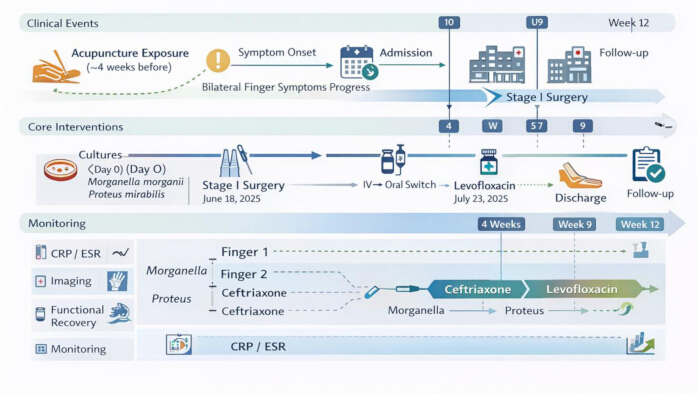
Figure 1: Timeline of case management. Schematic overview of the clinical course from acupuncture exposure to follow-up. The timeline shows key events, including symptom onset, hospital admission, microbiological sampling, staged surgical procedures, and systemic antimicrobial therapy. Stage I debridement with antibiotic-loaded bone cement implantation was performed on June 18, 2025, followed by Stage II wound repair on July 23, 2025. Separate cultures obtained from each affected digit identified Morganella morganii and Proteus mirabilis. Monitoring parameters, including C-reactive protein (CRP), erythrocyte sedimentation rate (ESR), imaging findings, and functional recovery, are shown across follow-up time points. Please click here to view a larger version of this figure.
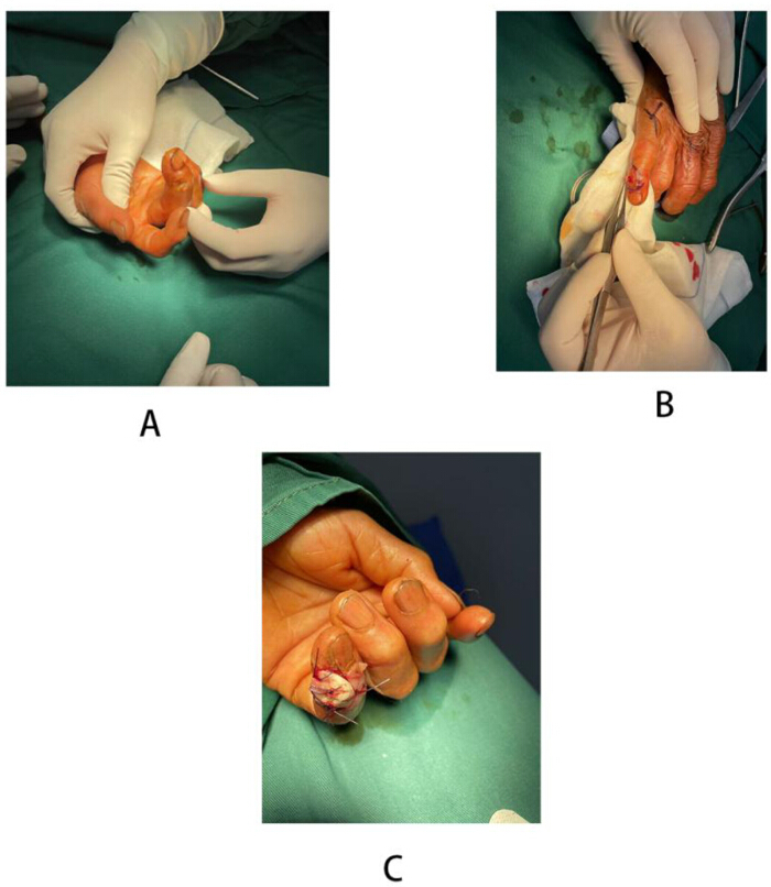
Figure 2: Key intraoperative steps of staged surgical management. (A) Preoperative appearance of the affected digit, showing swelling, deformity, and limited mobility. (B) Stage I procedure, showing thorough debridement of infected and necrotic tissue under sterile conditions. (C) Postoperative appearance after Stage II reconstruction, showing soft tissue closure and restoration of anatomical contour. Please click here to view a larger version of this figure.
Discussion
This case should be interpreted in the context of the existing literature on acupuncture-related infections, while recognizing that its primary value is illustrative rather than broadly generalizable. Most reported acupuncture-related infections involve superficial soft tissue disease or single-site musculoskeletal involvement and are typically caused by Staphylococcus aureus or nontuberculous mycobacteria7,8,9. In contrast, the present case represents a less typical clinical scenario, characterized by bilateral multi-digit osteoarticular infection caused by Gram-negative Enterobacteriaceae. Its value lies less in novelty alone than in the combination of features that may complicate recognition and management, including bilateral involvement, isolation of Morganella morganii and Proteus mirabilis, and the use of staged surgical management combined with sequential antimicrobial therapy in an elderly patient.
From a diagnostic perspective, this case highlights the importance of integrating clinical, radiographic, and microbiological findings rather than relying on any single modality. The diagnosis of osteomyelitis with interphalangeal joint involvement was supported by progressive local symptoms, radiographic evidence of cortical erosion and joint-space narrowing, and pathogen identification from separately collected sterile specimens obtained before antibiotic exposure10,11,12,13. Differential diagnoses, including septic arthritis unrelated to acupuncture, crystal arthropathy, mycobacterial infection, and isolated soft-tissue infection, were considered and excluded based on the overall clinical picture. This structured diagnostic approach is particularly important in atypical or multisite infections following minimally invasive procedures.
The microbiological findings also warrant emphasis. Separate sampling from each affected digit reduced the risk of specimen cross-contamination and allowed site-specific pathogen identification. The recovery of M. morganii from the right third finger and P. mirabilis from the left fifth finger suggests localized inoculation or spread rather than a single homogeneous infectious source. As members of the Enterobacteriaceae family, these organisms may contribute to chronic or treatment-refractory infection through biofilm formation and persistence on necrotic tissue surfaces14,15,16,17,18. This microbiological context provides a mechanistic rationale for aggressive surgical debridement, which remains essential for reducing bacterial burden, disrupting biofilm structure, and improving the likelihood of infection control.
The treatment strategy used in this case was based on both surgical and pharmacologic considerations. Staged debridement was selected because the presence of necrotic bone and uncertainty regarding infection control made immediate definitive closure less favorable. This approach is consistent with established principles in the management of complex hand and osteoarticular infections, in which infection control and adequate debridement take priority over early reconstruction13,16,19. The use of antibiotic-loaded bone cement further supported local antimicrobial delivery and dead-space management20,21. Antimicrobial selection was guided by susceptibility results and by the pharmacologic properties of available oral agents. Because both isolates were susceptible to fluoroquinolones, levofloxacin was considered a reasonable step-down option given its high oral bioavailability and favorable bone penetration22,23,24,25,26. These features make it a practical option in selected patients after initial infection control, although this conclusion should be approached cautiously in a single-case report.
The outcome assessment indicated that the combined strategy was effective in the short term. Clinical improvement was accompanied by objective findings, including declining inflammatory markers, improved range of motion, pain reduction, and follow-up radiographs showing bone consolidation without recurrent infection. At the same time, the favorable outcome likely reflected the combined contribution of several interventions, including timely diagnosis, adequate debridement, susceptibility-guided antimicrobial therapy, and structured postoperative care. Accordingly, this case should not be interpreted as establishing a standard treatment model. Rather, it illustrates a structured and individualized approach that may be informative in similarly atypical presentations, particularly when an acupuncture-related infection is suspected, and reliable microbiological sampling can be obtained before antibiotic exposure.
Limitations:
This report describes a single clinical case, and the generalizability of its findings is therefore limited. Although staged surgical management combined with sequential antimicrobial therapy resulted in favorable short-term outcomes, this observation should be interpreted cautiously. A single case cannot establish the comparative effectiveness of different treatment strategies, including levofloxacin-based oral sequential therapy versus prolonged intravenous therapy. In addition, the relative contribution of each treatment component could not be determined because surgical debridement, local infection control, pathogen-directed antibiotic selection, and structured follow-up were implemented concurrently. The 3-month follow-up period was sufficient to document wound healing and early functional recovery, but it was not long enough to assess long-term recurrence, chronic osteomyelitis, tendon adhesion, or joint stiffness. Microbiological assessment was limited to conventional culture and susceptibility testing; advanced analyses such as molecular resistance profiling or biofilm characterization were not performed. Pharmacokinetic monitoring of oral levofloxacin was also not performed, so the relationship between drug exposure and clinical efficacy could not be directly assessed. Taken together, these limitations indicate that the findings should be regarded as hypothesis-generating rather than practice-defining.
Disclosures
The authors have nothing to disclose.
Acknowledgements
The authors have no acknowledgments.
Materials
| Name | Company | Catalog Number | Comments |
|---|---|---|---|
| Antibiotic-loaded bone cement (PMMA + gentamicin) | Heraeus / equivalent | 40 g PMMA with 2 g gentamicin | Local antimicrobial delivery and dead space management |
| Antibiotics (ceftriaxone) | Roche / equivalent | Injectable, 2 g/vial | Intravenous antimicrobial therapy |
| Antibiotics (levofloxacin) | Daiichi Sankyo / equivalent | Oral tablet, 500–750 mg | Oral sequential antimicrobial therapy |
| Antibiotics (piperacillin–tazobactam) | Pfizer / equivalent | 4.5 g/vial | Empirical broad-spectrum coverage |
| Blood agar plates | Oxoid / Thermo Fisher | Standard culture medium | Aerobic bacterial culture |
| Chlorhexidine–ethanol solution | 3M / equivalent | 0.5% chlorhexidine in 70% ethanol | Skin disinfection |
| Chocolate agar plates | Oxoid / Thermo Fisher | Enriched medium | Fastidious organism culture |
| E. coli ATCC 25922 | ATCC | Quality control strain | Antimicrobial susceptibility testing validation |
| Hydrogen peroxide solution | Local supplier | 3% solution | Wound irrigation |
| Iodophor solution | Betadine / equivalent | 1 g/L iodine solution | Wound irrigation and antisepsis |
| MacConkey agar plates | Oxoid / Thermo Fisher | Selective medium for Gram-negative bacteria | Enterobacteriaceae isolation |
| MALDI-TOF MS system | Bruker | Biotyper system | Bacterial identification |
| Normal saline | Baxter / equivalent | 0.9% NaCl solution | Irrigation and dilution |
| Sterile transport tubes | Copan / equivalent | With transport medium | Specimen collection and transport |
| VITEK 2 Compact system | bioMérieux | Automated system | Antimicrobial susceptibility testing |
| X-ray imaging system | Siemens | Multix Fusion | Radiographic evaluation |