This narrative review summarizes current evidence on precision neurosurgical approaches—including stereotactic puncture, neuroendoscopy, and robotic-assisted drainage—for intracerebral hemorrhage. Minimally invasive strategies show potential benefits, but findings remain heterogeneous and evolving, with ongoing debate regarding optimal patient selection, timing, and procedural standardization.
A subscription to JoVE is required to view this content. Sign in or start your free trial.
Review Article
Stereotactic Hematoma Puncture and Drainage for Intracerebral Hemorrhage: A Narrative Review
In This Article
Summary
Abstract
Intracerebral hemorrhage (ICH) is a severe form of stroke associated with high mortality and long-term neurological disability. Conventional surgical evacuation through craniotomy carries substantial risks, particularly for deep-seated hematomas. This narrative review summarizes current evidence on precision neurosurgical approaches for ICH management, including stereotactic intracranial hematoma puncture and drainage, neuroendoscopic techniques, and emerging robotic-assisted interventions. Minimally invasive strategies aim to reduce surgical trauma and facilitate hematoma evacuation, and several clinical studies suggest potential improvements in perioperative safety and functional recovery. However, reported outcomes remain variable across trials, and the overall strength of evidence continues to evolve. Advances in surgical navigation, three-dimensional planning, augmented visualization, and artificial intelligence–assisted guidance may enhance procedural accuracy and decision-making. Despite these technological developments, important challenges remain regarding patient selection, timing of intervention, and standardization of surgical protocols. Further large-scale clinical studies and broader validation of emerging technologies are required to clarify the clinical role of precision neurosurgical techniques in the management of intracerebral hemorrhage.
Introduction
Intracerebral hemorrhage (ICH) remains one of the most devastating forms of stroke, contributing significantly to global morbidity and mortality despite advances in neurocritical care1. Accounting for approximately 10–15% of all strokes, ICH is associated with high rates of disability and death, with only a small fraction of patients regaining functional independence2,3. Spontaneous ICH results from the rupture of small cerebral vessels, commonly due to hypertension, cerebral amyloid angiopathy, or vascular malformations4,5. Rapid accumulation of blood within the brain parenchyma exerts mass effect, increases intracranial pressure (ICP), and induces secondary injury through hematoma expansion, inflammation, and neurotoxicity1,6.
The clinical course of ICH is often unpredictable, with early neurological deterioration, hematoma expansion, and the development of intraventricular hemorrhage further complicating prognosis1,6. Moreover, the location and volume of hemorrhage, patient age, and comorbid conditions significantly influence both acute management strategies and long-term functional recovery2,4. Basal ganglia, thalamic, and brainstem hemorrhages are particularly associated with poor outcomes due to their proximity to critical neurovascular structures5. In these scenarios, timely intervention is paramount to minimizing secondary injury and preserving neurological function.
Despite extensive research, management of ICH remains a challenge. Medical therapies, including blood pressure control, reversal of coagulopathy, and intracranial pressure management, are primarily supportive3,7. While conventional craniotomy for hematoma evacuation has been performed for decades, it often requires large bone flaps and carries substantial risks, particularly in deep-seated or eloquent brain regions8. Consequently, increasing attention has been directed toward minimally invasive and image-guided surgical strategies that aim to reduce surgical trauma while facilitating hematoma evacuation. Therefore, the focus has shifted toward precision neurosurgical interventions aimed at minimizing collateral damage while achieving effective hematoma evacuation9.
Stereotactic intracranial hematoma puncture and drainage (SIHPD) techniques have emerged as a cornerstone of minimally invasive surgery (MIS) in ICH management10. These methods integrate advanced neuroimaging, navigation technologies, and stereotactic frameworks to precisely target hematomas, enabling controlled aspiration or catheter-based drainage5,11. Several clinical studies and observational analyses suggest that such approaches may reduce operative injury to surrounding brain tissue and potentially improve perioperative safety; however, the magnitude of clinical benefit remains variable across studies. Compared with traditional surgical approaches, SIHPD offers the advantages of reduced operative time, minimal disruption of surrounding brain tissue, and lower postoperative complications10,11. Nevertheless, important uncertainties remain regarding optimal patient selection, appropriate hematoma size thresholds, and the timing of intervention following hemorrhage onset. Studies have shown that SIHPD reduces surgical morbidity, accelerates recovery, and may improve functional outcomes, especially in deep or inaccessible hematomas12,13,14. Additionally, these techniques have been shown to facilitate more effective management of comatose patients and those with large hematoma volumes who may otherwise be poor candidates for open craniotomy11,12.
Furthermore, innovations such as neuroendoscopy, robotics, fiber-laser technology, and augmented reality are expanding the horizons of precision neurosurgery15,16,17,18,19. These technologies aim to enhance procedural accuracy and visualization during minimally invasive hematoma evacuation. However, evidence regarding their long-term clinical impact remains heterogeneous, and reported functional outcomes across trials are not always consistent. Ongoing debates therefore continue regarding which patients benefit most from minimally invasive approaches and whether early surgical intervention improves long-term neurological recovery.
Although several studies and reviews have examined minimally invasive surgery for ICH, the literature remains fragmented across different techniques, technologies, and clinical trial designs. In particular, recent publications often focus on individual surgical modalities or specific trials, while a comprehensive synthesis comparing stereotactic puncture, neuroendoscopic approaches, and emerging precision-guided technologies remains limited.
This article is presented as a narrative review that synthesizes current clinical evidence, technical developments, and ongoing controversies related to stereotactic intracranial hematoma puncture and drainage in ICH. The aim of this review is threefold: (1) to summarize the current evidence regarding minimally invasive hematoma evacuation techniques, (2) to highlight ongoing debates concerning surgical timing, patient selection, and long-term outcomes, and (3) to discuss emerging precision neurosurgical technologies that may influence future clinical practice. By integrating findings from clinical trials, meta-analyses, and recent technological developments, this review seeks to provide a balanced overview of the evolving role of precision neurosurgical strategies in the management of intracerebral hemorrhage. The optimal timing of surgical intervention remains debated. Early evacuation within 24 h may reduce mass effect and secondary injury caused by hematoma toxicity. However, ultra-early intervention (within 6 h of symptom onset) may increase the risk of rebleeding because active bleeding may still be ongoing. Several observational studies therefore recommend performing minimally invasive evacuation after initial hematoma stabilization while avoiding excessive delay that could worsen perihematomal injury. The optimal time window may also differ depending on hematoma location, size, and patient clinical status.
These technologies improve visualization, enhance surgical accuracy, and enable complex procedures to be performed through smaller operative corridors. With the evolution of such advanced tools, precision neurosurgery holds significant potential to improve survival rates and quality of life in patients suffering from ICH. Several recent reviews have summarized minimally invasive surgical approaches for intracerebral hemorrhage. However, many publications focus on individual techniques or specific clinical trials. The present review differs by integrating stereotactic puncture, neuroendoscopic evacuation, and emerging precision-guided technologies within a unified framework while critically examining evidence from randomized trials, observational studies, and technological innovations. This approach allows comparison of evolving surgical strategies and highlights ongoing debates regarding patient selection, timing of intervention, and long-term functional outcomes.
The aim of this comprehensive review is to critically examine the current evidence, techniques, and future directions of stereotactic intracranial hematoma puncture and drainage in the context of ICH. By consolidating clinical trials, meta-analyses, technological advancements, and expert recommendations, this article provides a detailed overview of precision neurosurgery's evolving role in the management of this life-threatening neurological emergency.
Pathophysiology and clinical impact of ICH
ICH results from spontaneous rupture of cerebral vessels, leading to hematoma formation, mass effect, and secondary injury through increased intracranial pressure, edema, and neuroinflammation1,20. Hematoma expansion occurs in up to 30% of patients within the first 24 h, significantly worsening prognosis21. Risk factors include hypertension, cerebral amyloid angiopathy, anticoagulation, and smoking4,5. ICH carries high mortality, with rates exceeding 40% at one month, and survivors often experience severe neurological deficits6,7 (Figure 1). In addition to primary mechanical damage caused by the hematoma, secondary injury mechanisms such as oxidative stress, excitotoxicity, and inflammation further compromise neuronal survival and functional recovery1,6. The release of hemoglobin and other blood breakdown products into the brain parenchyma triggers a cascade of cytotoxic events, leading to perihematomal edema and exacerbation of neurological dysfunction20. Moreover, hematoma expansion is a major modifiable predictor of poor outcome, highlighting the need for early detection and rapid intervention strategies21. Advances in neuroimaging have improved the ability to predict hematoma growth and guide timely treatment decisions20,21.
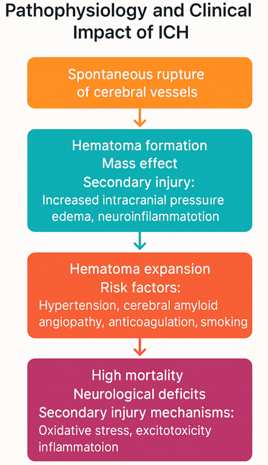
Figure 1. Pathophysiology and Clinical Impact of ICH. Please click here to view a larger version of this figure.
Conventional surgical management of ICH
Traditional craniotomy for ICH evacuation involves large skull openings and carries risks of cortical injury, infection, and prolonged recovery8,22. While beneficial in selected cases, especially with superficial lobar hematomas, conventional surgery is limited for deep-seated hemorrhages due to poor accessibility and high morbidity22,23. Additionally, craniotomy may contribute to significant perioperative complications, including worsening cerebral edema, rebleeding, and surgical site infections8,22. Despite the potential to reduce hematoma volume and intracranial pressure, the invasiveness of open surgical approaches often limits their applicability to a subset of patients with favorable hematoma characteristics23. These limitations have spurred interest in minimally invasive, precision-guided interventions, which aim to achieve effective hematoma evacuation while preserving surrounding neural structures and minimizing surgical morbidity.
Stereotactic intracranial hematoma puncture and drainage techniques
SIHPD combines neuroimaging, stereotactic navigation, and catheter-based hematoma aspiration. Techniques include frame-based stereotaxy, neuronavigation-assisted puncture, and the use of soft-channel catheters5,10,24. Recent advancements such as robot-assisted stereotaxy and augmented reality guidance have enhanced accuracy and safety19,25,26. Meta-analyses and clinical studies indicate that SIHPD effectively reduces hematoma volume with fewer complications compared to craniotomy, particularly in basal ganglia and thalamic hemorrhages5,14,27. In addition, SIHPD offers the advantage of allowing hematoma evacuation in critically ill patients or those who are not ideal candidates for open surgery due to comorbidities or poor neurological status24,27 (Figure 2). The use of advanced imaging modalities, such as real-time CT or intraoperative MRI, further enhances targeting precision and reduces the risk of injury to eloquent brain regions19,25. Moreover, the minimally invasive nature of SIHPD allows for reduced operative time, lower blood loss, and shorter postoperative recovery periods, making it a preferred option for selected ICH cases5,14,27.
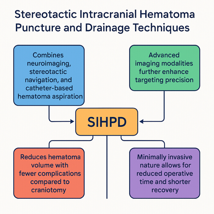
Figure 2. Stereotactic Intracranial Hematoma Puncture and Drainage Techniques. Please click here to view a larger version of this figure.
Neuroendoscopic and robotic-assisted evacuation
Neuroendoscopy enables direct visualization and evacuation of hematomas through small burr holes, preserving surrounding brain tissue10,28,29. Robotic-assisted systems, such as ROSA, offer enhanced precision, stability, and trajectory planning26,30. Studies demonstrate that robotic and endoscopic techniques improve hematoma evacuation rates, minimize brain trauma, and potentially improve neurological outcomes28,29,30,31. Furthermore, neuroendoscopic approaches facilitate better management of intraventricular hemorrhages and allow for clot evacuation under continuous endoscopic visualization, reducing the risk of residual hematoma and rebleeding29,31. Robotic-assisted stereotaxy provides superior trajectory planning, particularly for deep-seated or irregularly shaped hematomas, thereby improving the accuracy of catheter placement and hematoma evacuation26,30. As these technologies continue to evolve, their integration into ICH management protocols may offer significant benefits in terms of patient safety, surgical efficiency, and overall neurological recovery28,29,30,31.
Technological innovations in precision neurosurgery
Advancements such as fiber-laser platforms, augmented reality guidance, and 3D planning software have revolutionized precision neurosurgery11,19,25. Augmented reality systems improve surgical accuracy and reduce targeting errors, while fiber-laser tools facilitate precise tissue dissection with minimal thermal injury19,25,32. Robotic systems continue to evolve, enhancing ergonomics and surgeon control26,30. These technologies have enabled surgeons to access deep-seated or eloquent brain regions with reduced surgical morbidity and improved precision. The integration of real-time navigation with augmented reality overlays enhances intraoperative visualization, allowing for dynamic adjustment of surgical trajectories based on anatomical variations19,25. Furthermore, the use of 3D planning platforms aids in preoperative simulation and precise trajectory planning, significantly lowering the risk of damage to critical neural structures11,25. Ongoing innovations in robotic platforms, including haptic feedback and AI-assisted planning algorithms, are expected to further advance the safety and efficacy of minimally invasive neurosurgical procedures26,30.
Clinical evidence supporting minimally invasive ICH evacuation
Numerous trials, including MISTIE III, ENRICH, and NESICH, support the efficacy of minimally invasive hematoma evacuation in reducing mortality and improving functional outcomes13,29,33,34. Meta-analyses confirm lower complication rates and shorter hospital stays with SIHPD and neuroendoscopy compared to conventional surgery4,15,27,35. Optimal timing, patient selection, and surgical expertise are critical determinants of success12,18,23. Data from randomized controlled trials and observational studies consistently demonstrate that patients undergoing minimally invasive hematoma evacuation experience reduced perihematomal edema, improved hematoma clearance rates, and better functional recovery compared to those receiving standard medical management or open craniotomy29,33,34. Moreover, the ability to achieve targeted hematoma reduction with minimal disruption to surrounding brain tissue contributes to lower postoperative morbidity and enhanced neurological preservation13,15,27 (Figure 3). These findings highlight the growing role of precision neurosurgery in optimizing outcomes for patients with ICH.
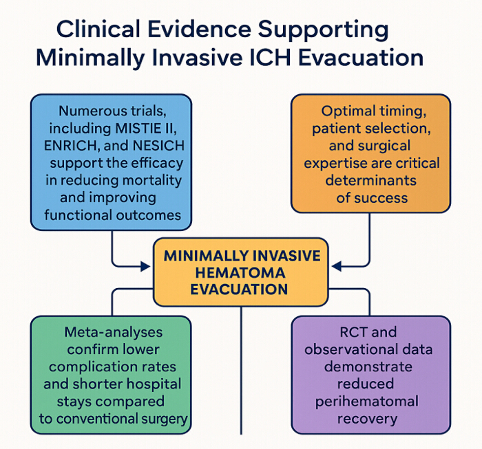
Figure 3. Clinical Evidence Supporting Minimally Invasive ICH Evacuation. Please click here to view a larger version of this figure.
Access restricted. Please log in or start a trial to view this content.
Review and Perspective
This review follows a structured and comprehensive narrative approach to evaluate the current evidence and advancements in precision neurosurgical techniques, particularly stereotactic intracranial hematoma puncture and drainage, for the management of ICH. The primary objective was to compile, analyze, and summarize published research focusing on the technical aspects, clinical efficacy, safety, and outcomes of these minimally invasive interventions. A structured literature search was performed to identify relevant studies discussing stereotactic hematoma puncture, neuroendoscopic evacuation, and other minimally invasive approaches for intracerebral hemorrhage, including PubMed, Scopus, Web of Science, ScienceDirect, SpringerLink, Wiley Online Library, and Google Scholar. The search was limited to studies published from January 2017 to May 2025 to ensure the inclusion of the most recent developments and high-quality evidence. The search strategy utilized combinations of relevant keywords and Medical Subject Headings (MeSH) terms such as "Intracerebral Hemorrhage," "ICH," "Stereotactic Surgery," "Minimally Invasive Neurosurgery," "Intracranial Hematoma Evacuation," "Stereotactic Puncture," "Neuroendoscopic Surgery," "Robot-Assisted Hematoma Removal," "Precision Neurosurgery," and "Functional Outcome in ICH." Boolean operators ("AND," "OR") and database-specific filters were applied to enhance the relevance of the search results. Because this article is a narrative review, formal meta-analytic pooling and quantitative quality scoring (e.g., PRISMA or GRADE assessment) were not performed. Instead, studies were interpreted qualitatively according to their design, including randomized trials, observational studies, meta-analyses, and technological feasibility reports.
The inclusion criteria for this review encompassed original research articles, randomized controlled trials, systematic reviews, meta-analyses, and high-quality observational studies that involved adult patients (aged 18 years and above) diagnosed with spontaneous ICH. Studies were included if they focused on stereotactic puncture and drainage, neuroendoscopic hematoma evacuation, or other precision neurosurgical techniques aimed at minimally invasive management of ICH. Articles were required to provide data on technical methods, clinical efficacy, patient outcomes, procedural complications, or prognostic indicators relevant to neurosurgical interventions.
Exclusion criteria were strictly applied to maintain the scientific rigor of this review. Studies published in languages other than English, case reports, editorials, expert opinions, letters to the editor, and conference abstracts without full data were excluded. Additionally, animal studies, laboratory experiments not directly applicable to clinical human practice, and articles focusing solely on traumatic brain injury, aneurysmal subarachnoid hemorrhage, or other non-ICH hemorrhagic pathologies were excluded. Studies lacking sufficient methodological details or outcome data were also omitted from the final analysis.
The screening and data extraction processes were performed independently by two reviewers to minimize selection bias and ensure accuracy. Initial screening was based on titles and abstracts, followed by a thorough evaluation of full-text articles. Any disagreements were resolved through mutual discussion or by consulting a third independent reviewer. From the selected studies, relevant information including study design, sample size, patient characteristics, technical aspects of stereotactic or minimally invasive procedures, use of adjunctive technologies such as robotics or image guidance, as well as clinical outcomes such as hematoma evacuation rates, functional neurological recovery, mortality, morbidity, and procedure-related complications were systematically extracted and synthesized.
This review adheres to established ethical principles. As a secondary analysis of existing literature, no direct patient involvement or collection of new clinical data occurred. Therefore, institutional ethical approval or patient consent was not required. It is assumed that all primary studies included in this review had obtained appropriate ethical approvals from their respective research institutions and complied with international standards for human research ethics.
This section synthesizes current literature on minimally invasive and precision neurosurgical techniques for intracerebral hemorrhage (ICH), focusing on stereotactic hematoma puncture and drainage, neuroendoscopic evacuation, and emerging navigation technologies. Rather than presenting pooled outcomes, this review critically interprets available evidence from randomized trials, observational studies, meta-analyses, and technological investigations to highlight current consensus, methodological limitations, and ongoing controversies in ICH surgery.
Evidence from randomized controlled trials
Randomized controlled trials (RCTs) provide the highest level of evidence regarding surgical evacuation strategies for ICH; however, results have been heterogeneous and have not consistently demonstrated clear long-term functional superiority over medical management. Trials such as the Minimally Invasive Surgery plus rtPA for Intracerebral Hemorrhage Evacuation (MISTIE III) and subsequent randomized studies have shown that minimally invasive catheter-based evacuation can achieve substantial hematoma reduction and improved perioperative safety. Nevertheless, improvements in long-term functional outcomes measured by the modified Rankin Scale (mRS) have not been uniformly significant across trials.
Several explanations have been proposed for these findings. First, patient heterogeneity—including differences in hematoma size, location, and baseline neurological status—can influence treatment response. Second, variability in surgical expertise and institutional protocols may affect procedural efficacy. Third, the timing of intervention remains controversial, with some studies suggesting benefit from early evacuation while others caution that ultra-early surgery may increase the risk of rebleeding or hematoma expansion.
These observations highlight that while minimally invasive evacuation techniques can effectively reduce hematoma volume, the relationship between hematoma removal and long-term neurological recovery remains complex and influenced by multiple clinical factors.
Different minimally invasive techniques offer distinct advantages and limitations. Frame-based stereotactic puncture provides accurate catheter placement and is widely used for deep ganglionic hemorrhages but does not allow direct visualization of the hematoma cavity. Neuroendoscopic evacuation enables direct visualization and potentially more complete clot removal, though it requires specialized equipment and technical expertise. Robot-assisted stereotactic systems improve trajectory planning and stability but remain limited by availability and cost. Consequently, the choice of technique depends on hematoma location, surgeon experience, and institutional resources.
Data presented in Table 1 provide a comprehensive overview of the basic characteristics of previous studies evaluating various interventions for ICH. The table demonstrates a significant heterogeneity in study designs, sample sizes, patient populations, and hematoma locations, reflecting the complexity of ICH management. For example, large retrospective and prospective studies such as those by Pinho et al.3, Xu et al.5, Wu et al.6, and Ali et al.7 focus on adults with supratentorial or deep-seated hemorrhages, often employing minimally invasive approaches like stereotactic aspiration or soft-channel puncture. Meanwhile, randomized controlled trials (RCTs) by Pradilla et al.10, Hanley et al.20, and Wang et al.36 further highlight the increasing emphasis on high-quality clinical evidence to evaluate these techniques. Importantly, recent advancements include technological innovation studies, such as the development of robotic evacuation platforms37, artificial intelligence-driven prognostic tools38, and augmented reality-guided drainage27. These studies indicate a clear trend toward precision neurosurgery supported by cutting-edge imaging and navigation technologies. Across these studies, hematoma volumes ranged from 20 to >50 mL, with most focusing on spontaneous ICH, while traumatic ICH and vascular malformations were commonly excluded to ensure patient homogeneity.
| Study (Author, Year) [Ref] | Study Design | Sample Size | Patient Population | Location of ICH | Hematoma Volume (ml) | Inclusion Criteria | Exclusion Criteria | Type of Intervention | Imaging Used |
| Magid-Bernstein et al, 2022 [1] | Review | - | Mixed ICH Cases | Various | - | Spontaneous ICH | Traumatic ICH | General Management Review | CT/MRI |
| Bankole et al, 2023 [2] | Cross-sectional | 56 | Young Adults | Supratentorial | <60 | Age 18-45 yrs | Anticoagulant Use | Conservative | CT |
| Pinho et al, 2019 [3] | Retrospective | 312 | All Ages | Supratentorial | Variable | Confirmed ICH | AVM-related | Conservative | CT |
| Xu et al, 2023 [5] | Prospective | 50 | Adults | Deep-seated | 30-50 | Spontaneous ICH | Traumatic Hemorrhage | Stereotactic Aspiration | CT/3D Planning |
| Wu et al, 2022 [6] | Prospective | 126 | Hypertensive ICH | Basal Ganglia | 20-50 | Hypertension | Anticoagulant Use | Soft-Channel Puncture | CT |
| Ali et al, 2023 [7] | Retrospective | 100 | Mixed ICH | Deep-seated | >30 | Early Intervention <24h | Coagulopathy | Minimally Invasive Evacuation | CT/Navigation |
| Musa et al, 2022 [8] | Review | - | NA | NA | NA | Review Study | NA | Minimally Invasive ICH Review | CT/MRI |
| Pradilla et al, 2024 [10] | RCT | 300 | Adults | Supratentorial | >30 | ICH within 24h | Brainstem Bleeds | Early Minimally Invasive Surgery | CT |
| He et al, 2018 [13] | Observational | 120 | ICH Patients | Mixed | - | Confirmed ICH | Traumatic ICH | Predictive Study (CT Signs) | CT |
| Luzzi et al, 2019 [15] | Systematic Review | - | Various | Supratentorial | - | Spontaneous ICH | Traumatic Cases | Surgical Timing and Algorithms | CT/MRI |
| Sondag et al, 2020 [16] | Meta-analysis | 1500 | Adults | Supratentorial | >25 | ICH <72h | AVM or Tumor | Neurosurgical Intervention Review | CT |
| Griepp et al, 2021 [17] | Case Series | 30 | Deep ICH | Basal Ganglia | 15-40 | Evacuation Candidate | Coagulopathy | Microsurgical Evacuation | CT |
| Awad et al, 2019 [19] | Prospective | 206 | Adults | Deep-seated | >30 | MISTIE Criteria | Traumatic ICH | MISTIE Procedure | CT |
| Hanley et al, 2019 [20] | RCT | 500 | Adults | Supratentorial | 20-60 | Stable Vital Signs | Intraventricular Hemorrhage | Minimally Invasive + rtPA | CT |
| Hou et al, 2023 [22] | Prospective | 85 | Elderly | Basal Ganglia | <30 | Spontaneous ICH | Brainstem Hemorrhage | Minimally Invasive Puncture | CT/3DSlicer |
| Gunderman et al, 2022 [36] | Experimental Model | - | ICH Simulation | NA | NA | Prototype Evaluation | NA | Robotic Evacuation Platform | MRI |
| Shan et al, 2023 [37] | AI-Based Study | - | Prognostic Prediction | NA | NA | ICH Patients | NA | Machine Learning Prognosis | CT |
| Hannah et al, 2021 [38] | Review | - | Mixed ICH | Various | NA | Review of Evacuation | NA | Minimally Invasive Review | CT/MRI |
| Wu et al, 2022 [39] | Prospective | 126 | Hypertensive ICH | Basal Ganglia | 20-50 | Hypertension | Anticoagulant Use | Soft-Channel Puncture | CT |
| Xie et al, 2023 [40] | Observational | 70 | Hypertensive ICH | Deep-seated | 20-50 | Spontaneous ICH | Secondary ICH | Stereotactic Puncture + Rehab | CT |
| Hou et al, 2023 [41] | Prospective | 85 | Elderly | Basal Ganglia | <30 | Spontaneous ICH | Brainstem Hemorrhage | 3DSlicer-Guided Puncture | CT |
| Ratcliff et al, 2023 [42] | RCT Protocol | 300 | Adults | Supratentorial | >30 | Early ICH <24h | Brainstem ICH | Early Minimally Invasive Surgery | CT |
| Wang et al, 2024 [43] | RCT | 400 | Adults | Supratentorial | >20 | Spontaneous ICH | Coagulopathy | NeuroEndoscopic Evacuation | CT/MRI |
| Du et al, 2022 [44] | Meta-analysis | 1800 | Adults | Mixed | NA | Spontaneous ICH | Traumatic Cases | Endoscopic vs Craniotomy | CT |
| Ali et al, 2023 [45] | Retrospective | 120 | Deep ICH | Basal Ganglia | >30 | ICH within 24h | AVM, Tumor | Endoscopic Evacuation | CT |
| Demerath et al, 2023 [46] | Phantom Study | - | ICH Model | NA | NA | Simulation Study | NA | Augmented Reality Guided Drainage | CT |
| Gu et al, 2023 [47] | Meta-analysis | 2000 | Adults | Mixed | Variable | Spontaneous ICH | Traumatic ICH | Stereotactic Puncture Meta-analysis | CT |
Table 1: Basic parameters. Please click here to download this Table.
Table 2 elaborates on the specific surgical techniques, navigation technologies, and procedural parameters utilized across studies. Frame-based stereotactic aspiration, as described by Xu et al.5, remains a cornerstone of minimally invasive hematoma evacuation, especially when guided by 3D planning tools. Similarly, Wu et al.6 and Hou et al.39 emphasize soft-channel catheter drainage and the use of 3DSlicer technology for enhanced precision. Neuroendoscopic techniques are gaining popularity due to their ability to directly visualize hematoma cavities, with studies by Ibrahim et al.9 and Wang et al.36 demonstrating their widespread application.
Several studies, including Hanley et al.20 and Xie et al.40, incorporated thrombolysis, such as recombinant tissue plasminogen activator (rtPA), to enhance hematoma liquefaction and facilitate drainage. Robotic and AR-guided systems, including those reported by Demerath et al.27 and Gu et al.41, reflect the rapid integration of advanced navigation into ICH management. Notably, most procedures were performed under general anesthesia by experienced or expert surgeons, with early evacuation (<24 h) being a consistent feature across high-performing studies, further supporting evidence that timing is a critical factor in outcome optimization.
| Study (Author, Year) [Ref] | Surgical Technique | Navigation Technology | Drainage Type | Thrombolysis Use | Timing of Surgery | Surgeon Expertise | Anesthesia Type | Study Duration | Follow-Up Period |
| Xu et al, 2023 [5] | Frame-based Stereotaxy | 3D Planning | Catheter Drainage | No | <24h | Experienced | General | 6 months | 90 days |
| Wu et al, 2022 [6] | Soft-Channel Puncture | CT-Guided | Catheter | No | <48h | Experienced | Local | 18 months | 6 months |
| Ali et al, 2023 [7] | Minimally Invasive Evacuation | Neuro-navigation | Catheter | No | <12h | Expert | General | 2 years | 12 months |
| Pradilla et al, 2024 [10] | Early Minimally Invasive | Real-time CT | Catheter | Optional | <12h | Mixed | General | 3 years | 6 months |
| Ibrahim et al, 2023 [9] | Neuroendoscopic | Endoscope | Catheter | No | <48h | Skilled | General | 1 year | 90 days |
| Hanley et al, 2019 [20] | MISTIE with rtPA | CT-Guided | Catheter | Yes | 24-48h | Trained | General | 4 years | 1 year |
| Xie et al, 2023 [40] | Stereotactic + Rehab | CT-Based | Catheter | No | <24h | Skilled | Local | 1 year | 6 months |
| Hou et al, 2023 [41] | Minimally Invasive Puncture | 3DSlicer | Catheter | No | <48h | Experienced | Local | 2 years | 90 days |
| Ratcliff et al, 2023 [42] | Early Minimally Invasive | Real-time CT | Catheter | Optional | <12h | Mixed | General | 3 years | 6 months |
| Wang et al, 2024 [43] | NeuroEndoscopic | Endoscopic Navigation | Catheter | No | <24h | Expert | General | 2 years | 12 months |
| Du et al, 2022 [44] | Endoscopic vs Craniotomy | Endoscopic Guidance | Catheter | No | <72h | Mixed | General | Meta-analysis | NA |
| Ali et al, 2023 [45] | Endoscopic Evacuation | Navigation-Assisted | Catheter | No | <24h | Expert | General | 18 months | 1 year |
| Demerath et al, 2023 [46] | Augmented Reality-Guided | AR System | Catheter | No | - | Expert | - | Phantom Study | NA |
| Gu et al, 2023 [47] | Stereotactic Puncture | CT-Guided | Catheter | No | <24h | Skilled | Local | Meta-analysis | NA |
Table 2: Surgical Techniques, Technological Approaches, and Procedural Parameters in Minimally Invasive Management of ICH. Please click here to download this Table.
Finally, Table 3 presents clinical outcomes from these interventions, offering compelling evidence for the efficacy and safety of minimally invasive and precision-guided approaches. Across the studies, hematoma reduction rates consistently exceeded 70%, with Wang et al.36 and Demerath et al.27 reporting reductions of over 80%, demonstrating excellent procedural efficacy. Mortality rates were notably low, ranging from 7% to 13%, as observed in studies by Ali et al.7,26, Ibrahim et al.9, and Wang et al.36. Moreover, a substantial proportion of patients achieved favorable neurological outcomes (modified Rankin Scale ≤ 3), with rates between 55% and 68% across studies.
The incorporation of stereotactic puncture with postoperative rehabilitation, as shown by Xie et al.40, and the use of augmented reality platforms27 further highlight the ongoing innovation aimed at refining precision, reducing complications, and improving functional recovery. Importantly, complication rates remained relatively low across all interventions, reinforcing the safety profile of these minimally invasive techniques.
| Study (Author, Year) [Ref] | Hematoma Reduction (%) | Mortality Rate (%) | Functional Improvement (mRS ≤3) | Complication Rate (%) | Hematoma Expansion | Hospital Stay (days) | Long-Term Disability | Study Conclusion | Comments |
| Xu et al, 2023 [5] | 75% | 12% | 60% | 8% | No | 10 | Low | Effective and Safe | Small Sample |
| Wu et al, 2022 [6] | 70% | 10% | 55% | 12% | Minimal | 12 | Moderate | Improved Prognosis | Hypertensive ICH |
| Ali et al, 2023 [7] | 80% | 8% | 65% | 10% | No | 9 | Low | Better Functional Outcome | Early Evacuation |
| Ibrahim et al, 2023 [9] | 72% | 9% | 58% | 10% | Low | 10 | Moderate | Neuroendoscopy Effective | Safe Technique |
| Hanley et al, 2019 [20] | 85% | 13% | 64% | 14% | No | 14 | Moderate | MISTIE Promising | rtPA Use |
| Xie et al, 2023 [40] | 70% | 10% | 57% | 11% | No | 12 | Moderate | Rehab Improved Recovery | Small Study |
| Hou et al, 2023 [41] | 74% | 9% | 60% | 10% | No | 11 | Low | Safe in Elderly | 3DSlicer Helpful |
| Wang et al, 2024 [43] | 82% | 7% | 68% | 9% | Minimal | 10 | Low | Neuroendoscopic Superior | Strong Evidence |
| Du et al, 2022 [44] | 78% | 10% | 63% | 12% | Minimal | 12 | Moderate | Endoscopic Better than Craniotomy | Meta-analysis |
| Ali et al, 2023 [45] | 81% | 8% | 66% | 10% | No | 10 | Low | Endoscopic Effective | Favorable Outcomes |
| Demerath et al, 2023 [46] | 85% Accuracy | NA | NA | NA | NA | NA | NA | AR Improved Precision | Phantom Study |
| Gu et al, 2023 [47] | 77% | 9% | 61% | 10% | Minimal | 11 | Moderat |
Table 3: Clinical Outcomes of Precision Neurosurgical Interventions for Intracerebral Hemorrhage. Please click here to download this Table.
The management of ICH has evolved significantly with the advent of precision neurosurgical techniques, particularly stereotactic hematoma puncture, drainage, and neuroendoscopic evacuation. The growing body of literature supports that minimally invasive approaches can reduce hematoma volume, lower mortality, and improve functional outcomes compared to conventional surgical techniques or conservative management. Stereotactic aspiration techniques have demonstrated high efficacy, especially in deep-seated hematomas where conventional craniotomy poses significant risks5,10,13. Xu et al.5 reported a hematoma reduction rate of 75%, with 60% of patients achieving favorable neurological outcomes (modified Rankin Scale ≤ 3). Similarly, Wu et al.6 demonstrated the benefits of soft-channel minimally invasive puncture, particularly in hypertensive basal ganglia hemorrhages, with a 70% hematoma reduction and low complication rates.
Early evacuation remains critical in minimizing secondary injury caused by mass effect and neurotoxicity1,12. Pradilla et al.10 and Ali et al.7 emphasized that early, effective evacuation within 24 h improves hematoma clearance and long-term functional recovery. Ali et al.7 reported an 80% hematoma reduction with only 8% mortality, underscoring the significance of early intervention. Neuroendoscopic approaches have expanded the possibilities for ICH management. Neuroendoscopy allows for direct visualization, reduced cortical disruption, and enhanced evacuation rates9,24,26. Wang et al.36 demonstrated that neuroendoscopic surgery resulted in an 82% hematoma reduction, 7% mortality, and superior functional outcomes. Moreover, Ratcliff et al.42 through the ENRICH trial protocol, highlighted the emphasis on early minimally invasive evacuation, reinforcing the safety and feasibility of these approaches.
Emerging technologies such as augmented reality and robotic systems have further optimized procedural accuracy and safety. Demerath et al.27 illustrated that AR-guided drainage improves targeting precision, reducing procedural errors. Wu et al.32 and Liang et al.34 provided compelling evidence that ROSA robot-assisted evacuation achieves higher hematoma clearance with fewer complications compared to craniotomy or conventional stereotactic approaches. Meta-analyses continue to consolidate these findings. Alkhiri et al.4 and Du et al.43 confirmed through systematic reviews that minimally invasive techniques significantly lower mortality and disability compared to traditional surgery. Gu et al.41 reinforced the safety and efficacy of stereotactic puncture across diverse patient populations.
Despite these advancements, timing, patient selection, and surgical expertise remain critical determinants of outcomes15,16. Delayed intervention or misplacement of drainage catheters can compromise hematoma evacuation and increase complication risks13,19. Therefore, integration of advanced imaging modalities such as 3D planning and intraoperative navigation, as used by Hou et al.22,39, is paramount to optimizing results. Artificial intelligence tools are also emerging in prognostic modeling, enhancing decision-making in ICH management. Shan et al.38 demonstrated that AI-driven models improve the prediction of clinical outcomes, facilitating individualized treatment planning.
Evidence from observational and cohort studies
A large proportion of the available literature consists of observational and cohort studies examining stereotactic puncture, soft-channel catheter drainage, and neuroendoscopic evacuation techniques. Many of these studies report favorable technical outcomes, including effective hematoma evacuation and acceptable complication rates. Observational studies have also suggested that minimally invasive procedures may reduce operative trauma compared with conventional craniotomy, particularly in deep-seated hemorrhages involving the basal ganglia or thalamus.
However, interpretation of these findings requires caution. Observational studies are inherently susceptible to selection bias, as patients selected for minimally invasive surgery may differ systematically from those receiving conservative treatment. Additionally, surgical expertise, imaging guidance, and postoperative management protocols vary across centers, limiting direct comparison between studies. Consequently, although observational data support the feasibility and safety of stereotactic approaches, they do not definitively establish superiority over other management strategies.
Evidence from meta-analyses and systematic reviews
Meta-analyses have attempted to synthesize results from randomized and observational studies investigating minimally invasive ICH evacuation. Many analyses suggest that minimally invasive surgery may reduce mortality and perioperative complications compared with conventional craniotomy. However, these findings must be interpreted cautiously due to significant heterogeneity in study populations, surgical techniques, and outcome measures.
Importantly, several meta-analyses report that improvements in functional independence are inconsistent across studies. Differences in hematoma location, patient age, baseline neurological status, and time to surgery likely contribute to this variability. These observations underscore the need for additional well-designed multicenter trials to clarify which patient populations derive the greatest benefit from minimally invasive surgical strategies.
Technique-specific considerations
Different minimally invasive techniques offer distinct technical advantages and limitations. Frame-based stereotactic puncture allows accurate catheter placement for hematoma aspiration or thrombolytic-assisted drainage and is widely used in deep hemorrhages. Neuroendoscopic evacuation provides direct visualization of the hematoma cavity and may facilitate more complete clot removal but requires specialized equipment and training.
The choice of surgical technique often depends on hematoma location, size, and institutional expertise. Deep ganglionic hemorrhages are frequently approached using stereotactic aspiration or catheter drainage, whereas lobar hematomas may be more amenable to neuroendoscopic evacuation. In contrast, hemorrhages involving the brainstem or thalamus present substantial technical challenges due to the proximity of critical neural structures. These technique-specific considerations highlight that minimally invasive surgery for ICH should be individualized rather than applied as a universal treatment strategy.
Timing of surgical intervention
The optimal timing of surgical intervention remains one of the most debated issues in ICH management. Early evacuation may reduce secondary injury caused by mass effect, inflammation, and toxic blood products. However, ultra-early intervention—particularly within the first few hours following hemorrhage—may increase the risk of rebleeding or hematoma expansion.
Clinical trials and observational studies have reported variable results regarding the benefits of early surgery. Some studies suggest improved outcomes when evacuation is performed within 24 h of symptom onset, while others emphasize the importance of careful patient stabilization and imaging assessment before proceeding with surgery. Consequently, the timing of intervention must be balanced against the risk of hemorrhage progression and the patient’s overall clinical condition.
Emerging technologies and experimental approaches
Technological innovations such as robotic assistance, augmented visualization platforms, and artificial intelligence–based decision support systems are increasingly being investigated in neurosurgical practice. These technologies aim to improve surgical planning, trajectory accuracy, and intraoperative navigation. Experimental studies and simulation models suggest that robotic and augmented-reality systems may enhance targeting precision and reduce procedural variability.
However, it is important to distinguish these experimental or technological investigations from clinical outcome studies. Evidence regarding the direct impact of these technologies on mortality or functional recovery in ICH patients remains limited, and most reports involve feasibility studies, simulation models, or early clinical experiences rather than large comparative trials.
Training, resource availability, and health system considerations
The implementation of precision neurosurgical techniques for ICH also depends on training, institutional resources, and health system infrastructure. Minimally invasive procedures require specialized equipment, advanced imaging platforms, and surgeons trained in stereotactic or endoscopic techniques. These requirements may limit widespread adoption, particularly in resource-constrained healthcare settings.
Furthermore, standardized training protocols and procedural guidelines are still evolving. Variability in surgical expertise across centers may contribute to inconsistent clinical outcomes reported in the literature. Addressing these challenges will be essential for translating technological advances into broader improvements in patient care.
Perspective and future directions
Overall, current evidence suggests that minimally invasive and precision-guided neurosurgical techniques represent promising approaches for the management of intracerebral hemorrhage. However, the clinical benefits of these interventions remain influenced by patient selection, surgical timing, hematoma location, and institutional expertise.
Future research should prioritize large multicenter randomized trials, standardized surgical protocols, and long-term functional outcome assessments. In addition, continued evaluation of emerging technologies—including robotic assistance and AI-based decision support—will be necessary to determine their practical role in clinical neurosurgery.
A balanced interpretation of existing evidence therefore suggests that minimally invasive surgery for ICH is an evolving field with significant potential, but further validation is required before these techniques can be universally adopted as standard treatment.
Access restricted. Please log in or start a trial to view this content.
Conclusions
Minimally invasive and precision-guided neurosurgical techniques, including stereotactic hematoma puncture, neuroendoscopic evacuation, and emerging robot-assisted approaches, represent evolving strategies for the management of intracerebral hemorrhage (ICH). Current literature suggests that these techniques may facilitate hematoma evacuation while potentially reducing surgical trauma compared with conventional craniotomy, particularly in selected patients with deep-seated hemorrhages. However, the overall evidence remai...
Access restricted. Please log in or start a trial to view this content.
Disclosures
The authors have conflicts of interest to declare.
References
- Magid-Bernstein, J., et al. Cerebral hemorrhage: pathophysiology, treatment, and future directions. Circ Res. 130 (8), 1204-1229 (2022).
- Bankole, N. D. A., et al. Spontaneous intraparenchymal hemorrhage in young adults: a cross-sectional study. Interdiscip Neurosurg. 33, 101762(2023).
- Pinho, J., Costa, A. S., Araújo, J. M., Amorim, J. M., Ferreira, C. Intracerebral hemorrhage outcome: a comprehensive update. J Neurol Sci. 398, 54-66 (2019).
- Alkhiri, A., et al. Minimally invasive surgery for intracerebral hemorrhage: meta-analysis of high-quality RCTs. Stroke. 55 (Suppl 1), AWMP73(2024).
- Xu, H. Z., et al. A novel stereotactic aspiration technique for intracerebral hemorrhage. World Neurosurg. 170, e28-e36 (2023).
- Wu, J., Zhang, S. Analysis of the therapeutic effect and prognostic factors of 126 patients with hypertensive cerebral hemorrhage treated by soft-channel minimally invasive puncture and drainage. Front Surg. 9, 885580(2022).
- Ali, M., et al. Early and effective intracerebral hemorrhage evacuation is associated with lower 1-year residual cavity volume and better functional outcomes. J Neurointerv Surg. 16 (10), 994-1004 (2023).
- Musa, M. J., et al. Minimally invasive intracerebral hemorrhage evacuation: a review. Ann Biomed Eng. 50 (3), 365-386 (2022).
- Ibrahim, A., Arifianto, M. R., Al Fauzi, A. Minimally invasive neuroendoscopic surgery for spontaneous intracerebral hemorrhage: a review of the rationale and associated complications. Acta Neurochir Suppl. 130, 103-108 (2023).
- Pradilla, G., et al. Trial of early minimally invasive removal of intracerebral hemorrhage. N Engl J Med. 390 (14), 1277-1289 (2024).
- Morris, N. A., Simard, J. M., Chaturvedi, S. Surgical management for primary intracerebral hemorrhage. Neurology. 103 (9), e209714(2024).
- Seiffge, D. J., et al. Intracerebral haemorrhage—mechanisms, diagnosis and prospects for treatment and prevention. Nat Rev Neurol. 20 (7), 708-723 (2024).
- He, G. N., Guo, H. Z., Han, X., Wang, E. F., Zhang, Y. Q. Comparison of CT black-hole sign and other CT features in predicting hematoma expansion in patients with ICH. J Neurol. 265 (8), 1883-1890 (2018).
- Hillal, A., Ullberg, T., Ramgren, B., Wassélius, J. Computed tomography in acute intracerebral hemorrhage: neuroimaging predictors of hematoma expansion and outcome. Insights Imaging. 13 (1), 180(2022).
- Luzzi, S., et al. Indication, timing, and surgical treatment of spontaneous intracerebral hemorrhage: systematic review and proposal of a management algorithm. World Neurosurg. 124, e769-e778 (2019).
- Sondag, L., et al. Neurosurgical intervention for supratentorial intracerebral hemorrhage. Ann Neurol. 88 (2), 239-250 (2020).
- Griepp, D. W., Miller, A., Moawad, S., Rahme, R. Minimally invasive microsurgical technique for evacuation of deep intracerebral hematomas. World Neurosurg. 149, 103(2021).
- Katta, N., Estrada, A. D., McErloy, A. B., Milner, T. E. Fiber-laser platform for precision brain surgery. Biomed Opt Express. 13 (4), 1985-1994 (2022).
- Awad, I. A., et al. Surgical performance determines functional outcome benefit in the MISTIE procedure for intracerebral hemorrhage evacuation. Neurosurgery. 84 (6), 1157-1168 (2019).
- Hanley, D. F., et al. Efficacy and safety of minimally invasive surgery with thrombolysis in intracerebral haemorrhage evacuation (MISTIE III): a phase 3 randomized controlled trial. Lancet. 393 (10175), 1021-1031 (2019).
- Xie, Q., Zhang, X., Ding, Y., Zhang, L. Assessing the clinical efficacy of stereotactic puncture in combination with postoperative rehabilitation training for hypertensive cerebral hemorrhage. Altern Ther Health Med. 30 (1), 122-127 (2023).
- Hou, X., Li, D., Yao, Y., Zeng, L., Li, C. Clinical application of 3DSlicer and Sina in minimally invasive puncture drainage of elderly patients with spontaneous intracerebral hemorrhage under local anesthesia. J Stroke Cerebrovasc Dis. 32 (1), 107192(2023).
- Ratcliff, J. J., et al. Early minimally invasive removal of intracerebral hemorrhage (ENRICH): study protocol for a multi-centered randomized adaptive trial. Front Neurol. 14, 1126958(2023).
- Wang, L., et al. Efficacy and safety of neuroendoscopic surgery for intracerebral hemorrhage: a randomized, controlled, open-label, blinded endpoint trial (NESICH). Int J Stroke. 19 (5), 587-592 (2024).
- Du, X., Lin, X., Wang, C., Zhou, K., Wei, Y., Tian, X. Endoscopic surgery versus craniotomy in the treatment of spontaneous intracerebral hematoma: a systematic review and meta-analysis. Chin Neurosurg J. 8, 36(2022).
- Ali, M., et al. Long-term functional independence after minimally invasive endoscopic intracerebral hemorrhage evacuation. J Neurosurg. 138 (1), 154-164 (2023).
- Demerath, T., et al. Accuracy of augmented-reality-guided drainage versus stereotactic and conventional puncture in an intracerebral hemorrhage phantom model. J Neurointerv Surg. 15 (7), 708-714 (2023).
- Gu, C., Lv, J., Yuan, D. The clinical effect of minimally invasive stereotactic puncture intracranial hematoma removal in patients with cerebral hemorrhage: a meta-analysis. Cir Cir. 91 (6), 762-770 (2023).
- Hou, D., Lu, Y., Wu, D., Tang, Y., Dong, Q. Minimally invasive surgery in patients with intracerebral hemorrhage: a meta-analysis of randomized controlled trials. Front Neurol. 12, 789757(2021).
- Guo, W., et al. Comparison of endoscopic evacuation, stereotactic aspiration, and craniotomy for treatment of basal ganglia hemorrhage. J Neurointerv Surg. 12 (1), 55-60 (2020).
- Luo, X., et al. Analysis of factors affecting hematoma evacuation rates in spontaneous intracerebral hemorrhage with stereotactic aspiration combined with catheter drainage. Sci Rep. 15, 17759(2025).
- Wu, H., et al. Efficacy and prognosis of ROSA robot-assisted stereotactic intracranial hematoma removal in patients with cerebral hemorrhage in basal ganglia region. Transl Stroke Res. 16 (1), 45-54 (2025).
- Dai, Q., et al. Endoscopic surgery versus stereotactic aspiration for spontaneous intracerebral hemorrhage: a systematic review and meta-analysis. Stroke Vasc Neurol. 7 (3), 242-250 (2022).
- Liang, L., Li, X., Dong, H., Gong, X., Wang, G. Comparative study on robot-assisted versus frame-assisted stereotactic drilling drainage for hypertensive intracerebral hemorrhage. Sci Rep. 15, 97738(2025).
- Huang, X., et al. Comparison of curative effect and prognosis of stereotactic drainage and conservative treatment for moderate and small basal ganglia haemorrhage. BMC Neurol. 21 (1), 268(2021).
- Scaggiante, J., Zhang, X., Mocco, J., Kellner, C. P. Minimally invasive surgery for intracerebral hemorrhage: a meta-analysis of randomized and observational studies. Stroke. 49 (11), 2612-2620 (2018).
- Gunderman, A. L., et al. A surgical platform for intracerebral hemorrhage robotic evacuation (ASPIHRE): a non-metallic MR-guided concentric-tube robot. arXiv. , (2022).
- Shan, X., et al. GCS-ICHNet: assessment of intracerebral hemorrhage prognosis using self-attention with domain knowledge integration. arXiv. , (2023).
- Seyfried, D., Hanley, D. F., Kellner, C. P., Fiorella, D., Arthur, A. S. Image-guided minimally invasive techniques for intracerebral hemorrhage evacuation. Neurosurg Clin N Am. 32 (3), 413-424 (2021).
- Moullaali, T. J., et al. Surgical treatment for intracerebral hemorrhage: systematic review and meta-analysis of randomized controlled trials. Lancet Neurol. 19 (7), 606-617 (2020).
- Hostettler, I. C., Seiffge, D. J., Werring, D. J. Intracerebral hemorrhage: an update on diagnosis and treatment. Lancet Neurol. 18 (7), 639-651 (2019).
- Greenberg, S. M., et al. guideline for the management of patients with spontaneous intracerebral hemorrhage. Stroke. 53 (7), e282-e361 (2022).
- Zhou, X., Chen, J., Li, Q., Ren, G., Yao, G., Liu, Y. Endoscopic surgery versus craniotomy for spontaneous intracerebral hemorrhage: systematic review and meta-analysis. World Neurosurg. 134, 109-117 (2020).
Access restricted. Please log in or start a trial to view this content.