This study evaluates biofilm formation on 3D-printed polylactic acid (PLA) and polyethylene terephthalate glycol (PETG) using static, semi-static, and dynamic in vitro models. The methodology supports safer medical device development and advances biofilm research in 3D-printed biomaterials.
A subscription to JoVE is required to view this content. Sign in or start your free trial.
Research Article
Comparative In Vitro Staphylococcus aureus Biofilm Evaluation on 3D-Printed Polylactic Acid and Polyethylene Terephthalate Glycol-modified Surfaces
In This Article
Summary
Abstract
Three-dimensional (3D) printing has accelerated the development of customized medical devices, but biofilm formation on printed materials remains a major threat to implant safety and performance. Because material chemistry and print-dependent surface features can influence bacterial attachment and antibiotic tolerance, standardized in vitro approaches that enable meaningful comparisons across commonly used 3D-printing polymers are needed. Here, we compare biofilm development on polylactic acid (PLA) and polyethylene terephthalate glycol (PETG) using a multi-model in vitro evaluation framework that captures complementary aspects of biofilm biology, including early adhesion, maturation, and antimicrobial tolerance, under both static and flow conditions. S. aureus biofilms were established and assessed using quantitative (viable bacterial load and minimal biofilm eradication concentration [MBEC]) and qualitative (scanning electron microscopy) endpoints. Both PLA and PETG supported biofilm formation across models; however, PLA tended to show higher early adhesion and greater biofilm density. In static assays, PLA demonstrated higher CFU values than PETG, whereas vancomycin MBEC values were similar between materials. Assay-dependent differences in MBEC were observed across platforms, underscoring how model structure can influence apparent antimicrobial susceptibility. Under dynamic flow, biofilm burden increased relative to static conditions, with minimal material-dependent differences. Collectively, these results highlight the susceptibility of both polymers to biofilm formation and demonstrate the value of a multi-model framework for evaluating material-associated biofilm behavior and benchmarking antimicrobial performance on 3D-printed device-relevant substrates.
Introduction
Recent advances in 3D printing have led to the growing adoption of 3D-printed medical devices due to their cost-effectiveness, high precision, complex structural design, efficient material utilization, and patient-specific design1,2. These advancements are driving significant clinical progress in the development of customized orthopedic3,4,5,6, craniofacial7,8, dental1,9, and spinal10,11 implants.
Numerous techniques and materials now allow precise control over design parameters, making it possible to optimize surface characteristics and enhance overall device functionality2,12,13. The implant’s surface is critically important, as it serves as the main interface with the host environment and influences immune responses, bacterial adhesion, and tissue integration—factors essential for the device’s long-term performance14,15. However, this same interface also presents a major challenge, as infections associated with implantable medical devices can lead to severe complications and are often difficult to treat due to the ability of pathogens to form surface-attached biofilms16,17,18,19.
In this context, in vitro testing of biofilm models on materials intended for the manufacture of implantable medical devices represents a fundamental step in the risk assessment and product development process20. These assays enable the investigation of the material’s susceptibility to microbial adhesion, biofilm formation, and maturation, as well as the evaluation of the effectiveness of antimicrobial strategies or surface modifications aimed at preventing microbial growth.
A further limitation of many in vitro biofilm assays is that they are frequently performed on a single, standardized substrate (often one polymer or a generic laboratory material), implicitly assuming that results are transferable across device materials21. This is problematic for 3D-printed medical devices, where polymer chemistry and manufacturing-dependent surface features can vary substantially and may directly influence bacterial attachment, biofilm maturation, and antimicrobial tolerance22. Consequently, evaluating biofilm behavior on the actual polymers used in fabrication, rather than extrapolating from a single reference substrate, addresses an important gap in current testing practice.
Because the implant surface is the key interface with the host, it strongly influences bacterial adhesion and biofilm formation, which underpin many device-associated infections and treatment failures. We hypothesized that commonly used 3D-printed polymers differ in their ability to support S. aureus biofilm development and in their susceptibility to vancomycin-mediated biofilm eradication. Accordingly, we performed a qualitative and quantitative evaluation of biofilm formation on 3D-printed material models. In the present study, a qualitative and quantitative evaluation of biofilm formation on 3D-printed material models was performed.
Access restricted. Please log in or start a trial to view this content.
Protocol
All hazardous chemicals (e.g., glutaraldehyde, sodium cacodylate buffer, Hexamethyldisilazane [HMDS], and ethanol) were handled in accordance with institutional Environmental Health and Safety (EHS) policies. Preparation and use of volatile/toxic reagents were performed in a certified chemical fume hood with appropriate personal protective equipment (lab coat, eye protection, and chemically resistant gloves), and flammable solvents were kept away from ignition sources. Chemical wastes were segregated and collected in properly labeled containers (including dedicated arsenic-containing waste for cacodylate and solvent waste for HMDS/ethanol) for disposal through the institutional hazardous-waste program. Biohazardous materials (bacterial cultures, contaminated plates, and disposables) were decontaminated according to biosafety procedures (e.g., autoclaving or approved disinfectants) prior to disposal, and any ethylene oxide sterilization was conducted in accordance with certified facility protocols and required aeration practices.
3D printed models
The models were designed using free computer-aided design software and printed using a 3D printer equipped with fused deposition modeling technology, with 1.75 mm (diameter) filaments of polylactic acid (PLA) and polyethylene terephthalate glycol (PETG) (Table of materials). The printing parameters used for fabricating the specimens were as follows: nozzle (0.4 mm) movement speed of 1800 mm/min; first layer speed of 300 mm/min; nozzle temperature of extrusion of 220 °C (PLA) and 255 °C (PETG); bed temperature of 60 °C; layer height of 0.08 mm; extrusion width of 0.48 mm and; infill density of 100% for all samples. None of the samples required the use of support structures. 3D models consisted of discs, flat-bottom plates, the Calgary biofilm device (CBD), and the modified Robbins device (MRD). The discs were designed as a cylinder with a 6 mm (diameter) x 6 mm (height).
For the CBD, a model was created (Table of materials). The CBD consisted of two parts: a base plate, a standard 96-well microtiter plate (ANSI/SLAS format), and a lid (peg plate): a polymer (PLA or PETG) lid with 96 cylindrical pegs with 2 mm (diameter) and 5 mm (height). The MRD was developed with 4 mm (diameter and height) pins fixed in a screw-thread model (Table of materials). After completing the printing process, the models were carefully removed from the build plate, visually inspected for any deformations, and subsequently sterilized with ethylene oxide.
Disc model of static biofilm
This method was used to induce biofilm formation on PLA using the previously described 6 mm × 6 mm cylindrical discs23 (Figure 1).
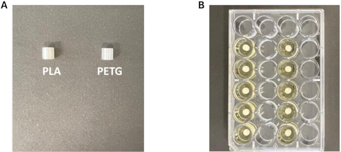
Figure 1: PLA and PETG discs and a 24-well plate for biofilm formation on these discs’ models. (A) Representative 3D-printed PLA and PETG discs prior to bacterial inoculation and biofilm formation. (B) PLA and PETG discs positioned in a 24-well plate during biofilm induction under in vitro culture conditions. Abbreviations; PLA = polylactic acid; PETG = polyethylene terephthalate glycol modified. Please click here to view a larger version of this figure.
S. aureus was stored at −80 °C in tryptic soy broth (TSB) supplemented with 15% glycerol and, prior to each experiment, was cultured on blood agar and incubated for 24 h at 37 °C. An isolated colony was resuspended in 10 mL of TSB supplemented with 50 mM glucose, added to a 50 mL conical tube, and incubated for 24 h at 37 °C in an orbital shaker at 120 rpm. The resulting culture was centrifuged for 5 min at 200 x g, and the supernatant was discarded. The pellet was washed three times with 10 mL of sterile 0.9% NaCl. In the final washing step, the pellet was resuspended in 10 mL of fresh TSB to obtain a bacterial suspension with a turbidity equivalent to the 0.5 McFarland standard (approximately 1.5 × 10⁸ CFU/mL), as determined by nephelometry. The suspension was then diluted 1:10 in 10 mL of TSB in a 15 mL conical tube to achieve a final bacterial concentration of 107 CFU/mL. For experiments with discs, 2 mL of the previous bacterial suspension was added to each well of sterile 24-well plates to cover each group of discs (PLA and PETG) for 2 h to allow cell adhesion. The discs were then aseptically transferred using sterile tweezers to a new sterile 24-well plate containing 2 mL of 0.9% NaCl to remove planktonic cells. Fresh 10 mL of TSB was added to the wells, and the plates were incubated at 37 °C for 24 h. During this step, cells adhering to the disc surfaces formed a biofilm. Following incubation, the culture medium was aspirated, and the discs were washed with 2 mL of 0.9% NaCl to remove residual medium and non-adherent cells. A 24-well plate was prepared with 1 mL of vancomycin solution at serial dilutions (2–4,096 mg/L), and the discs were submerged for 24 h. A control group without vancomycin (NaCl 0.9%) was included. After 24 h, the discs were washed with 2 mL NaCl 0.9%.
To quantify adhered bacteria on the discs, each disc was transferred using sterile tweezers to a microtube containing 1 mL of sterile 0.9% NaCl and sonicated in an ultrasonic bath for 15 min at 40 kHz and 37 °C to detach the adhered cells. The microtubes were vortexed for 60 s to ensure complete disaggregation of cell clusters. Aliquots of 100 µL of the undiluted sample (10°) and 10⁻2 and 10⁻4 dilutions in sterile 0.9% NaCl were plated on TSA and incubated for 24 h at 37 °C. Bacterial growth was quantified by counting CFU on a plate and expressed as CFU/mL.
24-well flat-bottom plates for minimal biofilm eradication concentration
For experiments in 24-well plates, 3 mL of bacterial suspension (prepared as described above) was added to each well for 2 h to allow adequate cell adhesion. Afterwards, the bacterial suspension was aspirated, and 3 mL of sterile 0.9% NaCl was added to remove planktonic cells. Fresh 3 mL of TSB was then added, and the plates were incubated at 37 °C for 24 h. During this step, cells adhering to the well surfaces formed a biofilm. Following incubation, the TSB was aspirated, and the wells were washed with 3 mL of 0.9% NaCl to remove residual medium and non-adherent cells. 3 mL of vancomycin solution at serial dilutions (2–4,096 mg/L) was added to each well. A control group without vancomycin (NaCl 0.9%) was included.
After 24 h, the solution was removed, and each well was gently washed twice with 3 mL of 0.9% NaCl. Three milliliters of 0.9% NaCl were added to each well, the plate was capped and sealed with parafilm, and then sonicated in an ultrasonic bath for 15 min at 40 kHz and 37 °C to detach the adhered biofilm. Aliquots of 100 µL were collected from the wells, and the undiluted sample (10°) and 10⁻2 and 10⁻4 dilutions prepared in sterile 0.9% NaCl were plated on TSA and incubated for 24 h at 37 °C. Bacterial growth was then quantified by counting CFU/mL. All experiments were performed in triplicate. Minimal biofilm eradication concentration (MBEC) was defined to be a reduction of at least 3 log10 in comparison with the control group (Figure 2).
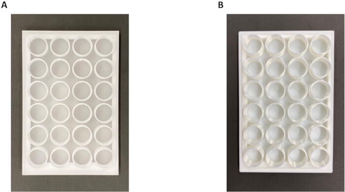
Figure 2: Printed 24-well plates. (A) 24-well plates printed with PLA. (B) 24-well plates printed with PETG. Abbreviations; PLA = polylactic acid; PETG = Polyethylene terephthalate glycol. Please click here to view a larger version of this figure.
The Calgary biofilm device
The CBD consists of a 96-well microtiter plate and a lid with 96 pegs, each designed to fit precisely into a well when the lid is placed on the plate. To induce biofilm formation, each well of a sterile 96-well plate was filled with 200 µL of the bacterial suspension, and sterile peg lids were then placed onto the plate. The assembled plates were incubated at 37 °C for 24 h under static conditions to allow biofilm development on the pegs. Following incubation, the peg lids were gently rinsed in a 96-well plate containing 200 µL of 0.9% NaCl to remove planktonic cells. The plate with the pegs was submerged in another 96-well plate containing vancomycin at progressive concentrations (2–4,096 mg/L) and incubated at 37 °C for 24 h under static conditions.
The plate with pegs was rinsed in a 96-well plate containing 200 µL of 0.9% NaCl, and the pegs were carefully stamped in a 15 cm TSA plate and incubated at 37 °C for 24 h. Bacterial growth was observed, and MBEC was defined as the lowest concentration at which no growth occurred. All experiments were performed in triplicate (Figure 3).
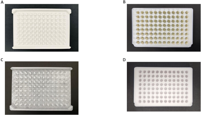
Figure 3: Calgary device and 96-well flat-bottom plates printed with PLA and PETG. (A) Calgary device printed with PLA. (B) 96-well flat-bottom plates printed with PLA. (C) Calgary device printed with PETG. (D) 96-well flat-bottom plates printed with PETG. Abbreviations; PLA = polylactic acid; PETG = polyethylene terephthalate glycol. Please click here to view a larger version of this figure.
A dynamic system under continuous conditions of flow
Each MRD contains five individual pegs fixed in a screw-thread model and arranged linearly along a channel with a rectangular cross-section. The dynamic system consisted of a flask containing 1 L of solution with the antibiotic to be tested connected to a peristaltic pump via a silicone tube coupled to the MRD. Assembly was done in a laminar air flow cabinet to prevent contamination. The medium flow rate was maintained at 1 mL/min and circulated through the system for 24 h. The fluid passing through the MRD is discarded into another glass flask (without recirculation).
S. aureus biofilms were induced on the pegs of an MRD, dropping the pegs on 24-well plates with TSB with 3 mL at a concentration of 107 CFU/mL. The pegs were gently rinsed by immersion in a 24-well plate filled with 3 mL of 0.9% NaCl solution. The pegs were carefully screwed into the MRD (n = 5). Vancomycin solution (40 mg/L) diluted in 0.9% NaCl was infused through the MDR using the pump as described above for 24 h. A control group without antibiotics (0.9% NaCl) was included. This concentration was chosen based on the serum concentration obtained during high doses of intravenous vancomycin, as previously described24. The pins were aseptically removed from the MRD using sterile tweezers, placed in microtubes containing 1 mL of sterile 0.9% NaCl after washing with 0.9% NaCl, and sonicated in an ultrasonic bath for 15 min at 40 kHz and 37 °C to ensure complete detachment of the biofilm. The microtubes were vortexed for 60 s, and the procedure was repeated three times to disrupt residual biofilm aggregates. Aliquots of 100 µL were plated on TSA, and the plates were incubated for 24 h at 37 °C. Bacterial growth was quantified and expressed as CFU/mL (Figure 4).
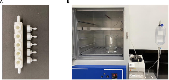
Figure 4: Modified Robbins device with PETG and PLA and a photo of the dynamic system. (A) modified Robbins device printed with PETG or PLA. (B) Photo of the dynamic system. Abbreviations; PLA = polylactic acid; PETG = polyethylene terephthalate glycol. Please click here to view a larger version of this figure.
Scanning electron microscopy
To visualize the morphology and structure of the biofilms, scanning electron microscopy (SEM) was performed on the discs. Biofilm was induced on discs as described in the protocols. The discs were carefully removed from the plate using sterile tweezers and were fixed with a solution (0.68 g sucrose, 0.42 g sodium cacodylate, 0.6 mL 30% glutaraldehyde), with 19.4 mL of deionized water, for 45 min. Subsequently, the specimens were transferred to a buffer solution (0.68 g sucrose and 0.42 g sodium cacodylate) for 10 min. The samples were then dehydrated in a graded ethanol series (35%, 50%, 70%, and 100%) for 10 min each. Afterwards, the specimens were transferred to another Petri dish and covered with 100% hexamethyl disilazane for 10 min. Finally, the discs were sputter-coated with gold using a rotary pump and mounted on a metal stub for observation under different magnifications on a scanning electron microscope (Figure 5).
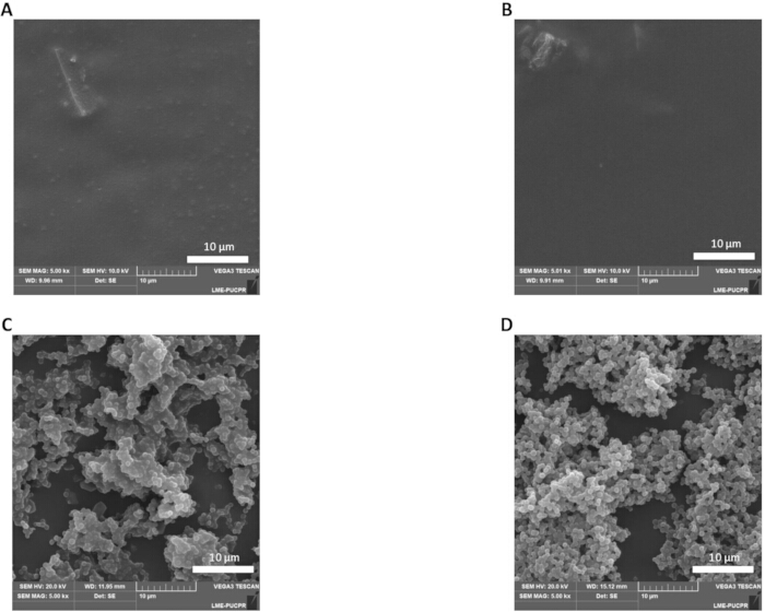
Figure 5: Scanning electron microscopy of PLA and PETG 3D printed models with and without biofilm induction. (A) Scanning electron microscopy photograph of PLA without biofilm. (B) Scanning electron microscopy photograph of PETG without biofilm. (C) Scanning electron microscopy photograph of biofilm-forming bacteria on PLA discs. (D) Scanning electron microscopy photograph of biofilm-forming bacteria on PETG discs. C and D show typical features of biofilms: bacterial organization into a 3D structure, adherence to surfaces, and the presence of an extracellular matrix. White scalebar represents 10 µm. Abbreviations; PLA = polylactic acid; PETG = Polyethylene Terephthalate Glycol. Please click here to view a larger version of this figure.
Statistical analysis
Some analyses were descriptive. For quantitative data, the sample size justified the use of the median with the interquartile range, with the Mann-Whitney test for statistical comparison. A p-value < 0.05 was considered statistically significant.
Access restricted. Please log in or start a trial to view this content.
Results
Disc model of static biofilm
This assay is used to determine the MBEC on individual specimens. Progressive two-fold dilutions of vancomycin demonstrated baseline biofilm formation of 7.48 × 10⁵ CFU/mL [IQR 6.34–8.72] for PLA and 3.60 × 10⁵ CFU/mL [IQR 3.08–5.52] for PETG. Although both materials showed biofilm densities within the 105 range, a statistically significant difference in biofilm production was observed between them (p = 0.015). When exposed to vancomycin, ...
Access restricted. Please log in or start a trial to view this content.
Discussion
This study proposes a comprehensive in vitro evaluation of biofilm formation on 3D-printed medical-grade polymers, specifically PLA and PETG, using four complementary experimental platforms: static disc assays, 24-well plate models, the CBD, and a dynamic flow system based on an MRD. By integrating these models, the study aims to characterize how distinct printing materials behave when exposed to S. aureus biofilm induction, and to determine the MBEC of vancomycin across different test conditions. This ...
Access restricted. Please log in or start a trial to view this content.
Disclosures
Authors have no conflicts of interest. ChatGPT was used for grammar review.
Acknowledgements
We would like to thank Renee Fleeman for the invitation.
Access restricted. Please log in or start a trial to view this content.
Materials
| Name | Company | Catalog Number | Comments |
|---|---|---|---|
| 0.9% NaCl | Fresenius Kabi Brasil, Ceara, Brazil | 17811 | |
| 3D printer with Fused Deposition Modeling technology (Bambu X1-Carbon) | Bambu Lab, Shenzhen, China | PF001-P | |
| Blood agar | NewProv, Paraná, Brazil | PA31 | |
| Calgary device for biofilm downloadable | No company | See link (without catalogue numbers) | https://www.printables.com/model/1464751-calgary-model-biofilm |
| Cell culture multiwell plate, 24 well | Greiner Bio-One, Kremsmünster Österreich, Austria | 662160 | |
| Centrifuge (5910 Ri) | Eppendorf, Hamburg, Germany | 5943000511 | |
| Centrifugue tube sterile 15 mL | Corning, Missouri, USA | 430790 | |
| Centrifugue tube sterile 50 mL | Corning, Missouri, USA | 430291 | |
| Electronic Pipette Controller (Easypet® 3) | Eppendorf, Hamburg, Germany | 4430000018 | |
| Ethanol (≥99.5%, ACS reagent) | Sigma-Aldrich, Missouri, USA | 459844 | |
| Glutaraldehyde | Merck, Darmstadt, Germany | 8143931000 | |
| Hexamethyldisilazane | Merck, Darmstadt, Germany | 440191 | |
| Incubator (502/4-C) | Fanem, São Paulo, Brazil | 502007700 | |
| Laminar Air Flow cabinet (BioSEG 9) | Veco, São Paulo, Brazil | 1EAC3770 | |
| Mechanical pipette (Eppendorf Reference® 2) | Eppendorf, Hamburg, Germany | 4924000061 | |
| Modified Robbins Device downloadable | No company | See link (without catalogue numbers) | https://www.printables.com/model/1481700-robbins-device-modified |
| Nephelometer | Alfakit, Florianópolis, Brazil | 7718 | |
| Orbital shaker (INNOVA 44R) | Eppendorf, Hamburg, Germany | M1282-0314 | |
| Parafilm M | Amcor, Illinois, USA | PM-996 | |
| Peristaltic pump (Minipuls® 3) | Gilson, Villiers-le-Bel, France | F117604 | |
| Petri dish (90 x 15 mm) | Greiner Bio-One, São Paulo, Brazil | 630164 | |
| Pipette tip 1–200μL | Corning, Missouri, USA | CLS4142 | |
| Polyethylene terephthalate glycol (PETG) | Creality, China | 44759 | |
| Polylactic acid (PLA) Ultrafuse® PLA PRO1 | BASF, Maarssen, Netherlands | PR1-7501A075 | |
| Rotary pump (Q150R ES Plus) | Quorum Technologies, Lewes, UK | 13034 | |
| Scanning electron microscope (Vega3 LMU) | Tescan, Brno, Czech Republic | See link (without catalogue numbers) | https://tescan.com/product-portfolio |
| Serological pipette 10 mL | Corning, Missouri, USA | CLS4488 | |
| Sodium cacodylate | Sigma-Aldrich, Missouri, USA | C0250 | |
| Staphylococcus aureus ATCC® 25923 strain | Laborclin, Pinhais, Brazil | 660674 | |
| Sucrose | Sigma-Aldrich, Missouri, USA | 59378 | |
| The 24-well flat-bottom downloadable | No company | See link (without catalogue numbers) | www.printables.com/model/104339-24-well-plates |
| Tryptic soy agar | Kasvi, Madrid, Spain | K25-1068 | |
| Tryptic soy broth | Sigma-Aldrich, Missouri, USA | 22092 | |
| Ultra Low Freezer ( IULT 335 D) | Indrel, Paraná, Brazil | 12038 | |
| Ultrasonic bath (Soniclean 15) | Sanders Medical, Minas Gerais, Brazil | See link (without catalogue numbers) | https://sandersdobrasil.com.br/produto/lavadora-ultrassonica-soniclean-15d/ |
| Vancomycin | ABL, São Paulo, Brazil | 15,56,20,041 | |
| Vortex (FLEXVORTEX) | Loccus, São Paulo, Brazil | 4500 |
References
- Kouhi, M., et al. Recent advances in additive manufacturing of patient-specific devices for dental and maxillofacial rehabilitation. Dent Mater. 40 (4), 700-715 (2024).
- Mamo, H. B., Adamiak, M., Kunwar, A. 3D printed biomedical devices and their applications: A review on state-of-the-art technologies, existing challenges, and future perspectives. J Mech Behav Biomed Mater. 143, 105930(2023).
- Przadka, M., et al. Advances in 3D printing applications for personalized orthopedic surgery: From anatomical modeling to patient-specific implants. J Clin Med. 14 (11), (2025).
- Ling, K., Wang, W., Liu, J. Current developments in 3D printing technology for orthopedic trauma: A review. Medicine (Baltimore). 104 (12), e41946(2025).
- Long, T., Tan, L., Liu, X. Three-dimensional printing in modern orthopedic trauma surgery: A comprehensive analysis of technical evolution and clinical translation. Front Med (Lausanne). 12, 1560909(2025).
- Cong, B., Zhang, H. Innovative 3D printing technologies and advanced materials revolutionizing orthopedic surgery: Current applications and future directions. Front Bioeng Biotechnol. 13, 1542179(2025).
- Slavin, B. V., et al. 3D printing applications for craniomaxillofacial reconstruction: A sweeping review. ACS Biomater Sci Eng. 9 (12), 6586-6609 (2023).
- Moncayo-Matute, F. P., et al. 3D printing and virtual surgical planning in craniofacial and thoracic surgery: Applications to personalised medicine. J Pers Med. 15 (9), (2025).
- Dimitrova, M., et al. Recent advances in 3D printing of polymers for application in prosthodontics. Polymers (Basel). 15 (23), (2023).
- Iqbal, J., et al. Recent advances of 3D-printing in spine surgery. Surg Neurol Int. 15, 297(2024).
- Hajnal, B., et al. Clinical applications of 3D printing in spine surgery: A systematic review. Eur Spine J. 34 (2), 454-471 (2025).
- Paul, G. M., et al. Medical applications for 3D printing: Recent developments. Mo Med. 115 (1), 75-81 (2018).
- Liaw, C. Y., Guvendiren, M. Current and emerging applications of 3D printing in medicine. Biofabrication. 9 (2), 024102(2017).
- Mendonça, C. J. aD., Dantas, L. R., Soni, J. F., Tuon, F. F. Antimicrobial action of a biodegradable thermoplastic impregnated with vancomycin for use in 3D printing technology. Braz Arch Biol Tech. 67, 13(2024).
- Dantas, L. R., Witt, M. A., Carneiro, E., Tuon, F. F. Nanoarchitectonics for advancing bone graft technology: Integration of silver nanoparticles against bacteria and fungi. Microorganisms. 12 (12), 2616(2024).
- Stewart, P. S., Bjarnsholt, T. Risk factors for chronic biofilm-related infection associated with implanted medical devices. Clin Microbiol Infect. 26 (8), 1034-1038 (2020).
- Amod, A., Anand, A. A., Sahoo, A. K., Samanta, S. K. Diagnostic and therapeutic strategies in combating implanted medical device-associated bacterial biofilm infections. Folia Microbiol (Praha). 70 (2), 321-342 (2025).
- Oliva, A., et al. Challenges in the microbiological diagnosis of implant-associated infections: A summary of the current knowledge. Front Microbiol. 12, 750460(2021).
- Veerachamy, S., Yarlagadda, T., Manivasagam, G., Yarlagadda, P. K. Bacterial adherence and biofilm formation on medical implants: A review. Proc Inst Mech Eng H. 228 (10), 1083-1099 (2014).
- Ortis, G. B., et al. Additive manufacturing, thermoplastics, CAD technology, and reverse engineering in orthopedics and neurosurgery applications to preventions and treatment of infections. Antibiotics (Basel). 14 (6), 561(2025).
- Coenye, T., Nelis, H. In vitro and in vivo model systems to study microbial biofilm formation. J Microbiol Methods. 83 (2), 89-105 (2010).
- Riga, E. K., Vohringer, M., Widyaya, V. T., Lienkamp, K. Polymer-based surfaces designed to reduce biofilm formation: From antimicrobial polymers to strategies for long-term applications. Macromol Rapid Commun. 38 (20), 1700216(2017).
- Soni, J. F., et al. Evaluation of silver nanoparticle-impregnated PMMA loaded with vancomycin or gentamicin against bacterial biofilm formation. Injury. 54 (6), 110649(2023).
- Borges, N. H., Suss, P. H., Ortis, G. B., Dantas, L. R., Tuon, F. F. Synergistic activity of vancomycin and gentamicin against Staphylococcus aureus biofilms on polyurethane surface. Microorganisms. 13 (5), 1119(2025).
- Pedroni, M. A., et al. Different concentrations of vancomycin with gentamicin-loaded PMMA to inhibit biofilm formation of Staphylococcus aureus and their implications. J Orthop Sci. 29 (1), 334-340 (2024).
- Tuon, F. F., et al. Antimicrobial treatment of Staphylococcus aureus biofilms. Antibiotics (Basel). 12 (1), 87(2023).
- Tuon, F. F., Dantas, L. R., Suss, P. H., Tasca Ribeiro, V. S. Pathogenesis of the Pseudomonas aeruginosa biofilm: A review. Pathogens. 11 (3), 300(2022).
- Carstensen, S., et al. Transcriptional evaluation of functional genes of resistance, biofilm, and quorum sensing in ctx-m15 ESBL-producing Klebsiella pneumoniae after meropenem concentration based on serum level. Res Microbiol. , (2025).
- Kraft, L., Ribeiro, V. S. T., Goncalves, G. A., Suss, P. H., Tuon, F. F. Comparison of amphotericin B lipid complex, deoxycholate amphotericin B, fluconazole, and anidulafungin activity against Candida albicans biofilm isolated from breakthrough candidemia. Enferm Infecc Microbiol Clin (Engl Ed). 41 (10), 596-603 (2023).
- Chaiben, V., et al. A carbapenem-resistant Acinetobacter baumannii outbreak associated with a polymyxin shortage during the COVID pandemic: An in vitro and biofilm analysis of synergy between meropenem, gentamicin, and sulbactam. J Antimicrob Chemother. 77 (6), 1676-1684 (2022).
- Da Rocha, L., et al. Evaluation of Staphylococcus aureus and Candida albicans biofilm adherence to PEEK and titanium-alloy prosthetic spine devices. Eur J Orthop Surg Traumatol. 32 (5), 981-989 (2022).
- Mendonca, J. R., Dantas, L. R., Tuon, F. F. Activity of multipurpose contact lens solutions against Staphylococcus aureus, Pseudomonas aeruginosa, Serratia marcescens, and Candida albicans biofilms. Ophthalmic Physiol Opt. 43 (5), 1092-1099 (2023).
- Koch, J. A., et al. Staphylococcus epidermidis biofilms have a high tolerance to antibiotics in periprosthetic joint infection. Life (Basel). 10 (11), 314(2020).
- Dunne, W. M. Jr Bacterial adhesion: Seen any good biofilms lately. Clin Microbiol Rev. 15 (2), 155-166 (2002).
- Beckman, R. L. T., Cella, E., Azarian, T., Rendueles, O., Fleeman, R. M. Diverse polysaccharide production and biofilm formation abilities of clinical Klebsiella pneumoniae. NPJ Biofilms Microbiomes. 10 (1), 151(2024).
- Dantas, L. R., Ortis, G. B., Suss, P. H., Tuon, F. F. Advances in regenerative and reconstructive medicine in the prevention and treatment of bone infections. Biology (Basel). 13 (8), 582(2024).
- Molina-Manso, D., et al. In vitro susceptibility to antibiotics of staphylococci in biofilms isolated from orthopaedic infections. Int J Antimicrob Agents. 41 (6), 521-523 (2013).
- Cieslinski, J., et al. Direct detection of microorganisms in sonicated orthopedic devices after in vitro biofilm production and different processing conditions. Eur J Orthop Surg Traumatol. 31 (6), 1113-1120 (2021).
- Fernandez-Grajera, M., Pacha-Olivenza, M. A., Fernandez-Calderon, M. C., Gonzalez-Martin, M. L., Gallardo-Moreno, A. M. Dynamic adhesive behavior and biofilm formation of Staphylococcus aureus on polylactic acid surfaces in diabetic environments. Materials (Basel). 17 (13), 3184(2024).
- Franklin, M. J., Chang, C., Akiyama, T., Bothner, B. New technologies for studying biofilms. Microbiol Spectr. 3 (4), (2015).
- Sikdar, R., Elias, M. H. Evidence for complex interplay between quorum sensing and antibiotic resistance in Pseudomonas aeruginosa. Microbiol Spectr. 10 (6), e0126922(2022).
- Kruger, H. C., et al. Antimicrobial action, cytotoxicity, calcium ion release, and pH variation of a calcium hydroxide-based paste associated with Myracrodruon urundeuva Allemao extract. Aust Endod J. 48 (1), 170-178 (2022).
Access restricted. Please log in or start a trial to view this content.