Here, we present a protocol for dissecting and extracting whole brain and gut components of surface and cave-adapted morphotypes of Astyanax mexicanus. This protocol can be adapted for use in downstream applications, including transcriptomics, metabolomics, and gut microbiota analysis.
A subscription to JoVE is required to view this content. Sign in or start your free trial.
Method Article
Brain and Gut Dissection in Surface Fish and Cavefish Populations of Astyanax mexicanus
In This Article
Summary
Abstract
Teleost fish are used to answer a wide range of research questions spanning from development to genetics and neuroscience, and more recently, to understand the complexities of the gut-brain axis, and even in a wider context, the gut-brain-liver axis. The Mexican tetra, Astyanax mexicanus, has been increasingly used in comparative and developmental evolution frameworks due to the availability of morphologically and physiologically distinct morphotypes, including river-dwelling surface fish and cave-adapted, blind cavefish. Adaptation to cave habitats is associated with changes in morphological traits, behavioral traits, and gut microbiota diversity that are also found in laboratory conditions. This has opened up the possibility to ask questions about the interactions between the microbiome, metabolic activity, neural adaptation, and complex behavioral outputs. These interactions can now be addressed through whole-brain mapping studies with cleared brains and gut microbiota sequencing to detect bacterial diversity associated with surface and cave-adapted phenotypes. However, most of these studies would benefit from intact, carefully dissected, and preserved whole organs and tissues, which allow for post-processing in downstream applications. This protocol describes a step-by-step method for brain and gut dissection, where the whole brain and abdominal organs are removed and isolated for both surface fish and cavefish morphotypes.
Introduction
Animal models, including vertebrate fish, are critical tools for modeling biological phenomena and complex pathologies in relatively simpler, accessible systems. For instance, the zebrafish, Danio rerio, is one of the most widely used teleost fish models, with a plethora of methods applied to study their development, genetics, physiology, and behavior, with significant impact in the biomedical and neurobehavioral sciences1,2,3,4,5,6,7,8. A huge advantage to using zebrafish in the present is the availability of a number of methods to study organ and tissue histology and pathology in loss-of-function mutant lines, for which step-by-step dissection protocols exist3,9,10.
However, a number of other teleost fish models have been on the rise over the past decades that are suitable to ask other types of questions, including those within the fields of evolutionary biology11,12,13,14,15,16,17,18,19,20,21, biological mechanisms underlying aging2,22, toxicology23,24, and others25,26. An example of this is the case of the Mexican tetra, Astyanax mexicanus, a teleost fish originating from Northeastern Mexico, with ancestral populations of river-dwelling surface fish and over 30 populations of blind, cave-dwelling cavefish (Figure 1)12.
Cavefish have adapted a suite of morphological characteristics, including eye loss12,27, loss of pigmentation28, expansion of sensory systems17,29,30, and increased fat storage12,31, that allowed them to survive in hostile cave habitats. These fish also adapted a suite of behavioral traits, including reductions in sleep19,20, anxiety32, schooling33, and aggression13,18,19,20,29,34,35,36,37, compared to their ancestral surface fish. Adaptation of most of these traits most likely resulted from a combination of genetic variation and environmental pressures35,36,28, which include habitat differences between the rivers and the caves. An increasingly useful model to ask questions about the impact of genetic variation on morphological and behavioral trait adaptation16,18,19,20,35,38,39 and more recently, on gut microbiota diversity14,15, questions that may require the collection of organs like the brain, guts, liver, and others, there is a need for a standardized gross anatomy dissection protocol that may be used by the community at large. While whole brain dissections and organ dissections in A. mexicanus have been done before, there is a lack of a one-stop, comprehensive method that allows understanding how to perform these types of dissections in a time-efficient manner, which is critical for many types of projects, including those that require collecting samples for metabolomics40, metagenomics41, or whole-organ imaging16,38. Our goal is to remove the troubleshooting barrier towards accessing organs in this model for post-processing, allowing users in the community to seamlessly focus on their research.
In this protocol, we outline a step-by-step method for brain and gastrointestinal dissection in adult surface fish and Pachón cavefish of the species Astyanax mexicanus. This methodology can be adapted to a variety of downstream protocols, including those associated with the gut microbiome, tissue metabolomics, and brain clearing for whole-brain imaging on a light-sheet microscope. This technique is also adaptable for simultaneous application to the same specimen.
Access restricted. Please log in or start a trial to view this content.
Protocol
This protocol is conducted in accordance with IACUC-approved protocols 2024-0004 and A790125 at the Molecular Sciences Center and Medical Sciences Campus of the University of Puerto Rico.
1. Pre-dissection set-up
- If using a Sylgard Petri (SP) dish to dissect, make sure they are prepared prior to the procedure, taking into account that it may take a few days to set.
- To prepare a SP dish, use Sylgard 184 and Elastomer 184 in a 10:1 ratio. Mix 200 g of Sylgard 184 base with 20 g of elastomer in a disposable cup. Pour into Petri dishes to about one-third to one-half depth. Tap the dish to remove bubbles and cap. Ensure dishes are filled and left on a level surface. Allow to cure for at least 1 week42.
NOTE: If collecting samples for 16s rRNA sequencing or metagenomics, refrain from using SP dishes. Instead, use sterilized, single-use Petri plates and follow aseptic technique under a laminar flow hood. If following aseptic technique, cleaning the laminar flow hood with 70% ethanol, a germicidal detergent, and ideally exposing the hood to a germicidal ultraviolet C-lamp for 15 min is recommended. This should be done prior to placing disinfected and autoclaved materials/tools within the laminar flow hood. - Set up a dissection microscope with paper towels on both sides. Designate one side for waste storage (e.g., excess organ tissue) and the other side for placing sterilized dissection tools. Cut small pieces of paper towel to clean tools in between dissections.
- Prepare 1 PBS and keep it in a cold ice slurry and on hand in case it is needed to clean areas or specimens for better visibility. Depending on the protocol, choose not to use 1PBS.
NOTE: Not using it is recommended, for instance, while collecting samples for metagenomic sequencing43. If using 1 PBS on this type of approach, collecting a 1PBS sample for sequencing is recommended to screen for possible contamination. - Place the SP (or a single-use, sterilized Petri dish) under the microscope and make sure to have all the tools at hand (dissecting kit, microcentrifuge tubes for tissue collection on ice, etc.).
- Place 1 PBS and labeled microcentrifuge tubes in an ice bucket nearby. Have a plastic dropper in hand for 1 PBS.
- Set up a fish euthanasia station close by. Prepare an ice bath by placing ice slurry in a tank with a plastic grate inside, then fill it with fish system water (from their home system or home tank).
NOTE: MS-222 may be used as well, depending on downstream protocols (sample post-processing needs).
2. Brain dissection in adult surface fish and cavefish
- Euthanize fish in an ice slurry. Make sure there is no movement, and the fish sinks to the bottom, with no opercular movement. Time depends on size. Larger fish will take longer to cease opercular movement.
NOTE: Juvenile fish take approximately 2-5 min for cessation of opercular movement (euthanasia), while adult and large fish may take about 10-15 min. Morphotype-based differences exist. Some cavefish populations may take longer than surface fish for anesthesia/euthanasia. Husbandry and experimental protocols recommend validation of anesthesia and euthanasia procedures across populations, as responses may vary between morphs and populations11. - Transfer fish to a SP (or single-use, sterilized Petri dish inside the hood) and, with a dropper, bathe with ice-cold 1x PBS (Figure 1).
- Detach the head from the body by cutting with a scalpel about 2 mm behind the gills (Figure 2). This will protect part of the midbrain and allow for the manipulation of the brain once dissected, without causing damage. Remove the body toward the waste area using a paper towel, then place it in a biohazard bag in the biohazard waste container for disposal.
- With fine forceps or a hemostat, get a good grip of the posterior part of the fish head. With a micro-scissor, cut through the edges of the mouth towards the back of the head and gills (Figure 3).
- Repeat on the other side. Remove the mandible and then clean the underside well with a second pair of fine or ultra-fine forceps.
NOTE: Follow the inner contour of the gills. - By this point, keep holding firmly to the head of the fish with a hemostat or pin it with a T pin to the SP (if using), carefully by the tip of the mouth.
- Detach both eyes using ultra-fine forceps or scissors. Insert the tip into the edge of the eye and then gently pull or cut the eye out (Figure 4).
NOTE: Not applicable to Cavefish (scrape skin off eye area). - Scratch a sagittal line at the top of the head that runs between the eyes from front to back and divides the head into left and right. Afterward, use the micro scissors to cut through the line shallowly (Figure 5).
- With fine forceps, carefully begin to pull the two sides of the head, taking extra care not to break the brain beneath, embedded under connective tissue. Once the brain is visible, with the tip of superfine forceps, detach the brain from the cranium (Figure 6).
- Avoid crushing the brain by pulling out with forceps. Instead, hold it by the hindbrain or spinal cord if a remnant of it is still present to preserve integrity.
- If the brain is still on one side of the head, use the ultra-fine forceps to carefully detach it from the bed skull by gently inserting the tips behind the brain and gently lifting. Avoid pulling at all times, as this might break it.
- Once the brain is extracted, clean gently with ultra-fine forceps, eliminating any fat or surrounding tissues. Place in a microcentrifuge tube or appropriate storage for the downstream protocol. At this point, preserve the whole brain or collect sub-regions of the brain (e.g., telencephalon, optic tectum, olfactory bulbs) (Figure 7).
- Telencephalon isolation: To extract the telencephalon, pinch at the base between the telencephalon and the optic tectum with ultra-fine forceps and separate the telencephalon from the rest of the brain. Transfer to the appropriate storage tube.
- If using for metagenomics, flash-freeze collected samples in liquid nitrogen and store at -80 °C. Do this by dipping the microcentrifuge tube in liquid nitrogen for 5 s.
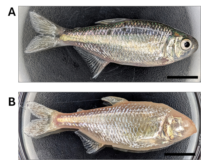
Figure 1: Astyanax mexicanus morphotypes. (A) Surface Fish. (B) Pachón cavefish. Scale bar: 10 mm Please click here to view a larger version of this figure.
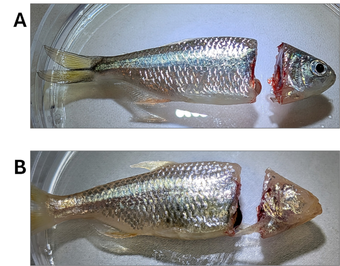
Figure 2: Detachment of the head. (A) Surface Fish. (B) Pachón cavefish Please click here to view a larger version of this figure.
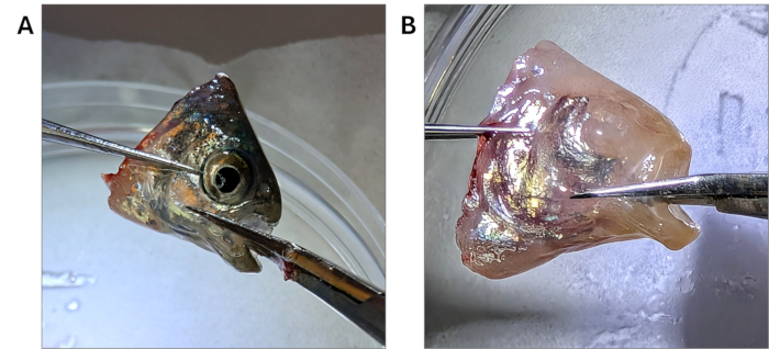
Figure 3: Removal of the jaw from a detached fish head. (A) Surface Fish. (B) Pachón cavefish Please click here to view a larger version of this figure.
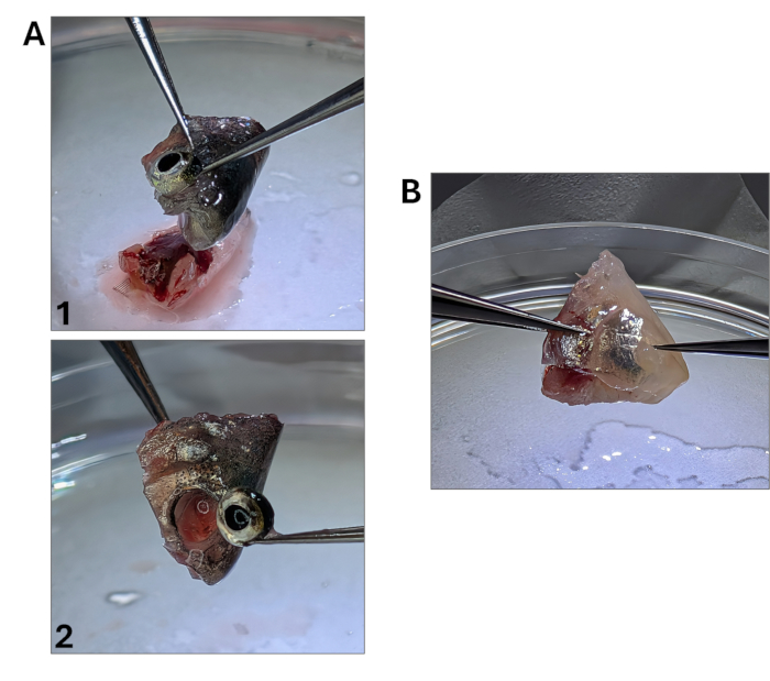
Figure 4: Removal of the eye from a detached fish head. In cavefish, scraping of the area can be performed to thin the tissue. (A) Surface Fish. (B) Pachón cavefish Please click here to view a larger version of this figure.
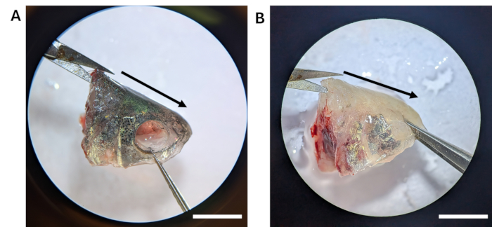
Figure 5: Sagittal cut through the superior aspect of the fish head. (A) Surface Fish. (B) Pachón cavefish. Scale bar: 5 mm Please click here to view a larger version of this figure.
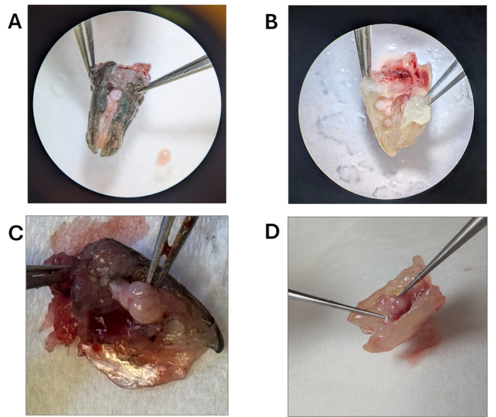
Figure 6: Separation of the lateral portions of the head and exposure of the brain within the cranial cavity. (A) Surface Fish. (B) Pachón cavefish. (C,D) Removal of the brain from the skullbed. Scale bar: 5 mm Please click here to view a larger version of this figure.
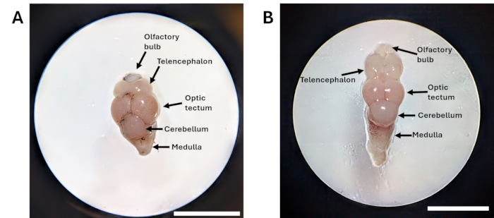
Figure 7: A. mexicanus regional brain anatomy. (A) Surface Fish. (B) Pachón cavefish. Scale bar: 5 mm Please click here to view a larger version of this figure.
3. Gut dissection in adult surface fish and cavefish
- Follow the method for euthanasia as described above.
- Pat the fish dry with a lint-free wipe before placing it on a clean SP and covering it with ice-cold 1 PBS (or system water, depending on downstream application) with the dropper to keep tissue moist.
- Secure the fish on the SP (if using) with T pins.
- If not using a SP, a hemostat to strongly grip anteriorly from the mouth while making cuts will do. Place the pin anteriorly within the eye socket (also true for cavefish), ensuring it reaches the SP.
- Posteriorly pin through the tail to the plate. Ensure the fish remains still during the procedure.
NOTE: If performing gastrointestinal dissection on the same specimen as brain dissection, quickly decapitate with a scalpel before initiating dissection and pinning the fish to the surface of the plate. Make an anteriorly diagonal cut behind the operculum that preserves the top of the head and avoids the gut. Ideally, split the head and body and have two experienced dissectors work on each for time efficiency. The anterior pin can be placed at the uppermost anterior part of the body post-decapitation, while the posterior pin remains in the same location (Figure 8).
- Using dissection micro-scissors, cut the skin and underlying muscle along the ventral midline from the anal fin/anus toward the operculum (the hard covering over the gills) in a semicircular line following the fish's underside curve (Figure 9).
- Remove the operculum and the pectoral fin with the micro-scissors and fine forceps.
NOTE: This step helps make the removal of the skin more efficient, but it may slow down dissection and may be skipped depending on the needs. - Cut the skin and underlying muscle superiorly from the anal fin and then cut anteriorly towards the gills, extending the ventral incision as needed (Figure 10). This ultimately creates a window.
- Open the incision with the help of fine forceps to expose the body cavity. Use the fine forceps to grasp the skin and lift it gently to remove (Figure 11).
- Cut both anterior and posterior attachments of the gut and carefully take out all internal organs - remove the gastrointestinal system from the body and place it on a clean area of the SP (or single-use, sterilized Petri dish) (Figure 12 and Figure 13).
- First, separate the liver from the rest of the abdominal organs with the ultra-fine forceps. If isolating the liver, place it in a microcentrifuge tube or use an appropriate storage method according to the downstream protocol or need. Carefully stretch out the gut and identify the stomach and midgut/hindgut portions (Figure 14).
- Detach the stomach from the midgut/hindgut.
- Hold the anterior and posterior ends of the midgut/hindgut with fine forceps and gently stretch it out to extend it to its total length.
- Collect tissue in a microcentrifuge tube and store appropriately.
- If using for metagenomics, flash-freeze collected samples in liquid nitrogen and store at -80 °C. This is done by dipping the microcentrifuge tube in liquid nitrogen for 5 s.
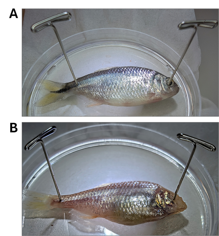
Figure 8: Pin placement for gastrointestinal dissection. (A) Surface Fish. (B) Pachón cavefish Please click here to view a larger version of this figure.
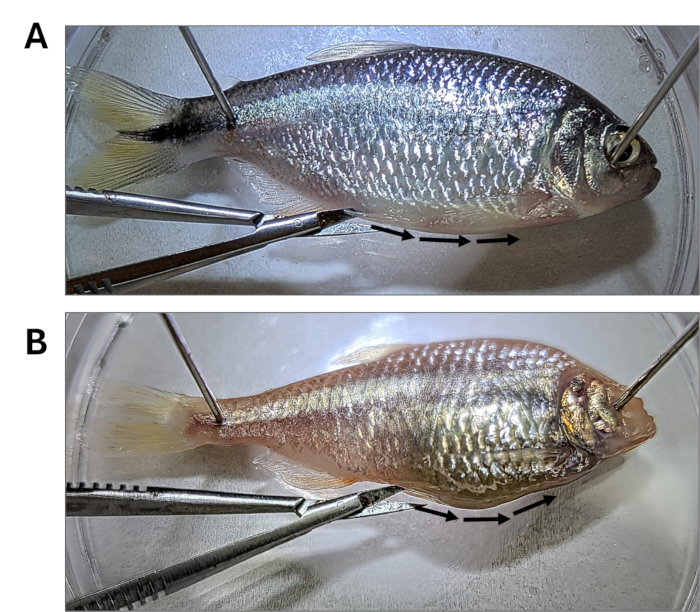
Figure 9: Underside cut to pre-dissection. (A) Surface Fish. (B) Pachón cavefish Please click here to view a larger version of this figure.
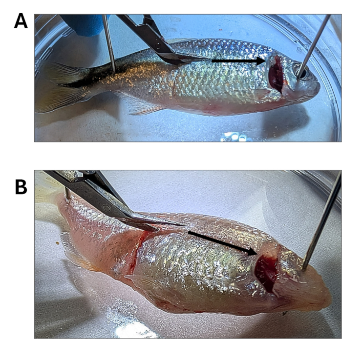
Figure 10: Removal of operculum and creation of a window. (A) Surface Fish. (B) Pachón cavefish Please click here to view a larger version of this figure.
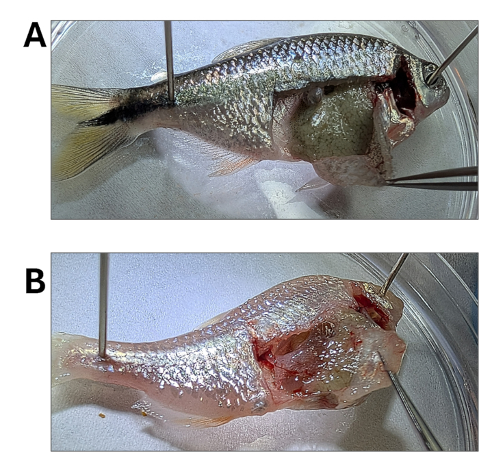
Figure 11: Removal of superficial lateral skin and muscle. (A) Surface Fish. (B) Pachón cavefish Please click here to view a larger version of this figure.
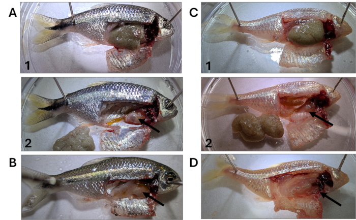
Figure 12: Organs in situ in the adult fish body cavity after removal of muscle and skin. (A) Female Surface Fish with abdominal cavity exposed and eggs removed to expose organs. (B) Male Surface Fish with abdominal cavity and organs exposed. (C) Female Pachón Cavefish with abdominal cavity exposed and eggs removed to expose organs. (D) Male Pachón Cavefish with abdominal cavity and organs. Black arrows point to the region where the gastrointestinal bundle is found. Please click here to view a larger version of this figure.
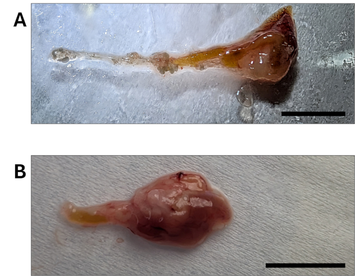
Figure 13: Gastrointestinal bundle removed from the fish's body cavity. (A) Surface Fish. (B) Pachón cavefish. Scale bar: 10 mm Please click here to view a larger version of this figure.
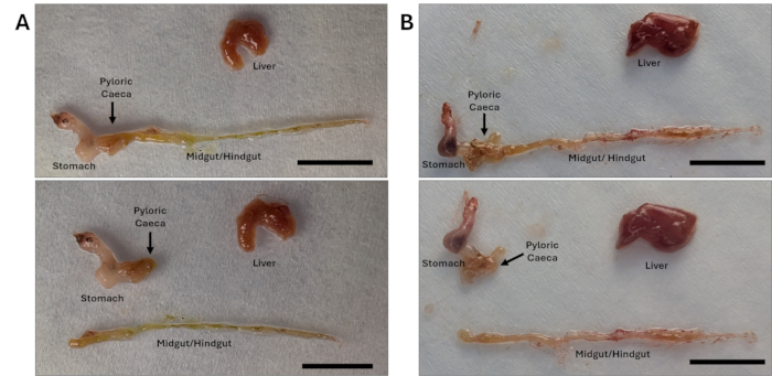
Figure 14: Isolated gastrointestinal organs of Surface fish and Cavefish. (A) Surface Fish. (B) Pachón cavefish. Scale bar: 10 mm Please click here to view a larger version of this figure.
Access restricted. Please log in or start a trial to view this content.
Results
Following this protocol, brain and gut contents were extracted, and measurements of the length of the complete gut, telencephalon, and optic tectum were taken and compared between surface fish and Pachón cavefish morphotypes (Figure 15). We used independent-sample Welch's t-tests for statistical comparisons, with a sample size of n = 6 surface fish and n = 6 Pachón cavefish. Surface fish had significantly larger optic tectum dimensions than pachón cavefish, in line with previously p...
Access restricted. Please log in or start a trial to view this content.
Discussion
The increasing relevance of A. mexicanus as a research model in a number of fields, ranging from evolution to biomedical neuroscience and microbiology, calls for a standardized organ dissection and collection method. While isolated structures have been studied and documented previously, including the optic tectum16,38, adult heart11, and others11,44, a systematic guideline...
Access restricted. Please log in or start a trial to view this content.
Disclosures
There is no conflict of interest. All procedures follow IACUC-approved protocol 2024-0004.
Acknowledgements
This work is funded by NIH grant 5R16EY037336-02, RCMI grant U54 MD007600, and NIH grant 1P20GM156713-01 (COBRE Puerto Rico Center for Microbiome Sciences).
Access restricted. Please log in or start a trial to view this content.
Materials
| Name | Company | Catalog Number | Comments |
|---|---|---|---|
| 1.0 Liter Zebrafish Breeding Tank | Tecniplast | https://shop.tecniplastusa.com/products/1-0-liter-zebrafish-breeding-tank | To prepare the euthanasia ice slurry. The tank is half-filled with ice and then the perforated internal tank is placed on top. Then it is filled with water to the top. |
| 4L Polyurethane Ice Pan | USA Scientific | Item #2328-7242 | |
| Biohazard Waste Bags | Fisherbrand | 14-828-244 | |
| Calbiochem OmniPur PBS 10x Liquid Concentrate, Molecular Biology Grade, Sterile | MilliporeSigma | 65074L | |
| Dissecting Microscope Olympus SZ51 | Olympus | OLSZ51NPS-LED/R | |
| Dissecting Tissue Forceps | Fisherbrand | 08-953G | |
| Dissection Pins, 100 pack | Home Science tools | https://www.homesciencetools.com/product/dissection-pins-100-pack/?srsltid=AfmBOoofk8r6_hd4nx_7bjjP2 GzkNBlnOJ3vUKor5 rFCJvVxPN9xRlpc | |
| Forceps, Delicate, Extra Fine Sharp Tips | MOPEC | AB132 | |
| Forceps, Tissue, Delicate Super Fine Tip | MOPEC | AB047 | |
| Hemostat, 12.5 cm, straight | World Precision instruments | https://wpiinc.com/products/var-15920-halsted-mosquito-hemostatic-forceps | |
| Jewelers forceps, Dumont No. 5 | MilliporeSigma | https://www.sigmaaldrich.com/US/en/product/sigma/f6521 | |
| Kimwipes EX-L | Kimberly-Clark | 34155 | |
| Liquid Nitrogen | Provided by Institution | ||
| Noyes Micro Scissors, 12 cm | World Precision instruments | 500228 | |
| Paper Towels | Provided by Institution | ||
| Petri Dishes with Clear Lid, 95 mm, 15 mm, Round | Fisherbrand | FB0875714G | |
| Scalpel Blades, #22, 10 pack | Home Science Tools | https://www.homesciencetools.com/product/scalpel-blades-22-10-pack/ | |
| Scalpel handle #4 | Surtex | https://surtex-instruments.com/product/scalpel-handle-no-4/ | |
| Scissors, Iris Dissection | MOPEC | AA146 | |
| Scissors, Superfine Micro | MOPEC | AA152 | |
| Standard Disposable Transfer Pipettes | Thermo Scientific | 13-711-9D | |
| Sterile Eppendorf Microcentrifuge 1.5 µL tubes | Eppendorf | 05-402-25 | |
| Sylgard 184 | Electron Microscopy Sciences | 24236-10 | |
| Thermo-Flask | Lab-Line Instruments Inc. | 2119 | |
| Ultra Fine Scissors | NEWMED Instruments | NM-DI-206023 | |
| Water suitable for electrophoresis, PCR | Sigma-Aldrich | W4502-1L |
References
- Ji, J., Thwaite, R., Roher, N. Oral intubation of adult zebrafish: a model for evaluating intestinal uptake of bioactive compounds. J Vis Exp. (139), e58366(2018).
- Örling, J., Kosonen, K., Villman, J., Reichard, M., Paatero, I. Impaired fin regeneration and angiogenesis in aged zebrafish and turquoise killifish. Biol Open. 12 (4), bio059622(2023).
- Turner, K. J., Bracewell, T. G., Hawkins, T. A. Anatomical dissection of zebrafish brain development. Brain Dev Methods Protoc. 1082, 197-214 (2014).
- Barbosa, A., et al. Western Blotting Assay for egr-1 Immediate Early Gene in Brain Tissue of Zebrafish Applied to Neuroethological Study. Zebrafish Protocols for Neurobehavioral Research. , Humana Press. Totowa, NJ. (2012).
- Corradi, L., Zaupa, M., Sawamiphak, S., Filosa, A. Using perk immunostaining to quantify neuronal activity induced by stress in zebrafish larvae. STAR Protoc. 3 (4), 101731(2022).
- Elipot, Y., Legendre, L., Père, S., Sohm, F., Rétaux, S. Astyanax transgenesis and husbandry: how cavefish enters the laboratory. Zebrafish. 11 (4), 291-299 (2014).
- Leigh, S. C., Nguyen-Phuc, B. -Q., German, D. P. The effects of protein and fiber content on gut structure and function in zebrafish (danio rerio). J Comp Physiol B. 188 (2), 237-253 (2018).
- Sassen, W. A., et al. Embryonic zebrafish primary cell culture for transfection and live cellular and subcellular imaging. Dev Biol. 430 (1), 18-31 (2017).
- Jie, J., et al. Adult zebrafish intestine dissection: a technique to analyze compound absorption. J Vis Exp. (10), e20199(2023).
- Gupta, T., Mullins, M. C. Dissection of organs from the adult zebrafish. J Vis Exp. (37), e1717(2010).
- Baumann, D. P., Ingalls, A. Mexican Tetra (astyanax mexicanus): Biology, Husbandry, and Experimental Protocols. Laboratory Fish in Biomedical Research. , Academic Press. (2022).
- Gross, J. B., Meyer, B., Perkins, M. The rise of Astyanax cavefish. Dev Dyn. 244 (9), 1031-1038 (2015).
- Carlson, B. M., Gross, J. B. Characterization and comparison of activity profiles exhibited by the cave and surface morphotypes of the blind Mexican tetra, Astyanax mexicanus. Comp Biochem Physiol C Toxicol Pharmacol. 208, 114-129 (2018).
- Riddle, M., et al. Evolution of the Mexican cavefish gut microbiome. Curr Biol. 23, 01307(2023).
- Riddle, M. R., et al. Host evolution shapes gut microbiome composition in Astyanax mexicanus. Ecol Evol. 14 (4), e11192(2024).
- Jaggard, J. B., et al. Cavefish brain atlases reveal functional and anatomical convergence across independently evolved populations. Sci Adv. 6 (38), eaba3126(2020).
- Rodríguez-Morales, R. Sensing in the dark: constructive evolution of the lateral line system in blind populations of Astyanax mexicanus. Ecol Evol. 14 (4), e11286(2024).
- Rodriguez-Morales, R., et al. Convergence on reduced aggression through shared behavioral traits in multiple populations of Astyanax mexicanus. BMC Ecol Evol. 22 (1), 116(2022).
- Duboué, E. R., Borowsky, R. L., Keene, A. C. β-adrenergic signaling regulates evolutionarily derived sleep loss in the Mexican cavefish. Brain Behav Evol. 80 (4), 233-243 (2012).
- Yoshizawa, M., et al. Distinct genetic architecture underlies the emergence of sleep loss and prey-seeking behavior in the Mexican cavefish. BMC Biol. 13, 15(2015).
- Forberg, J. Temperature effects on the development of the axial skeleton and body shape in Astyanax mexicanus (teleostei: characidae) [Master's Thesis]. , College of Science and Health Theses and Dissertations. https://via.library.depaul.edu/csh_etd/514 (2022).
- Platzer, M., Englert, C. Nothobranchius furzeri: a model for aging research and more. Trends Genet. 32 (9), 543-552 (2016).
- Blackwell, B. R., et al. Effects of metformin and its metabolite guanylurea on fathead minnow (Pimephales promelas) reproduction. Environ Toxicol Chem. 41 (11), 2708-2720 (2022).
- Ankley, G. T., Villeneuve, D. L. The fathead minnow in aquatic toxicology: past, present and future. Aquat Toxicol. 78 (1), 91-102 (2006).
- Butler, J. M., Whitlow, S. M., Roberts, D. A., Maruska, K. P. Neural and behavioural correlates of repeated social defeat. Sci Rep. 8 (1), 6818(2018).
- Norton, W. H. J., Gutiérrez, H. C. The three-spined stickleback as a model for behavioural neuroscience. PLoS ONE. 14 (3), e0213320(2019).
- Espinasa, L., Jeffery, W. R. Conservation of retinal circadian rhythms during cavefish eye degeneration. Evol Dev. 8 (1), (2006).
- Jeffery, W., Ma, L., Parkhurst, A., Bilandzija, H. Pigment Regression and Albinism in Astyanax Cavefish. Biology and Evolution of the Mexican Cavefish. , Academic Press, Elsevier. (2015).
- Yoshizawa, M., Jeffery, W. R., van Netten, S. M., McHenry, M. J. The sensitivity of lateral line receptors and their role in the behavior of Mexican blind cavefish (Astyanax mexicanus). J Exp Biol. 217 (Pt 6), 886-895 (2014).
- Lloyd, E., et al. Evolutionary shift towards lateral line-dependent prey capture behavior in the blind Mexican cavefish. Dev Biol. 441 (2), 328-337 (2018).
- Xiong, S., Krishnan, J., Peuß, R., Rohner, N. Early adipogenesis contributes to excess fat accumulation in cave populations of Astyanax mexicanus. Dev Biol. 441 (2), 297-304 (2018).
- Chin, J. S. R., et al. Convergence on reduced stress behavior in the Mexican blind cavefish. Dev Biol. 441 (2), 319-327 (2018).
- Kowalko, J. E., et al. Loss of schooling behavior in cavefish through sight-dependent and sight-independent mechanisms. Curr Biol. 23 (19), 1874-1883 (2013).
- Espinasa, L., et al. Divergent evolutionary pathways for aggression and territoriality in Astyanax cavefish. Subterr Biol. 43, 169-183 (2022).
- Kowalko, J. Utilizing the blind cavefish Astyanax mexicanus to understand the genetic basis of behavioral evolution. J Exp Biol. 223 (Suppl_1), jeb208835(2020).
- Sekulovski, B., Miller, N. Mechanisms of social behaviour in the anti-social blind cavefish (Astyanax mexicanus). Proc Biol Sci. 292 (2043), 20250052(2025).
- Chin, J. S. R., et al. Analysis of stress responses in Astyanax larvae reveals heterogeneity among different populations. J Exp Zool B Mol Dev Evol. 334 (7-8), 486-496 (2020).
- Kozol, R. A., et al. A brain-wide analysis maps structural evolution to distinct anatomical module. eLife. 12, e80777(2023).
- Ra, K., et al. Evolution of a central dopamine circuit underlies adaptation of light-evoked sensorimotor response in the blind cavefish, Astyanax mexicanus. bioRxiv. , (2024).
- Medley, J. K., et al. The metabolome of Mexican cavefish shows a convergent signature highlighting sugar, antioxidant, and ageing-related metabolites. eLife. 11, e74539(2022).
- Yen, S., Johnson, J. S. Metagenomics: a path to understanding the gut microbiome. Mamm Genome. 32 (4), 282-296 (2021).
- Marder, E. Marder lab website - Sylgard dishes. , https://sites.google.com/brandeis.edu/marder-lab/experimental-resources/sylgard-dishes (2026).
- Hallmaier-Wacker, L. K., Lueert, S., Roos, C., Knauf, S. The impact of storage buffer, DNA extraction method, and polymerase on microbial analysis. Sci Rep. 8, 6292(2018).
- Riddle, M. R., et al. Evolution of gastrointestinal tract morphology and plasticity in cave-adapted Mexican tetra, Astyanax mexicanus. bioRxiv. , (2020).
- Garduño-Sánchez, M., et al. Phylogeographic relationships and morphological evolution between cave and surface Astyanax mexicanus populations (de Filippi 1853) (actinopterygii, characidae). Mol Ecol. 32 (20), 5626-5644 (2023).
Access restricted. Please log in or start a trial to view this content.
Reprints and Permissions
This article has been published
Video Coming Soon