Polygonal gating (PGG) and cruciform gating (CFG) are flow cytometry methods for detecting sperm DNA integrity. PGG measures the DNA fragmentation index (DFI) and the high DNA staining index (HDS). CFG reports severe DFI, mild DFI, and overall DFI. This study aims to compare the results and correlations between them.
Method Article
Comparison of Sperm DNA Analysis Outcomes Using Different Gating Strategies in Flow Cytometry
Opens in a new tab
In This Article
Summary
Loading...
$$\rightleftharpoonup{xx}$$
$$\longleftharp{xx}$$,
$$\longrightharp{xx}$$,
Abstract
Loading...
$$\rightleftharpoonup{xx}$$
$$\longleftharp{xx}$$,
$$\longrightharp{xx}$$,
Sperm DNA integrity is a key factor in ensuring successful fertilization, embryo development, and ongoing pregnancy. Sperm chromatin structure assessment (SCSA) using flow cytometry after DNA denaturation and acridine orange staining is considered the gold-standard method for evaluating sperm DNA. Two gating strategies for flow cytometry–based assessment of the sperm DNA fragmentation index (DFI) have been reported in the literature: polygonal gating (PGG) and cruciform gating (CFG). The aim of the present study was to compare the relationships of assay results between PGG and CFG, and to analyze their correlations with parameters of basic semen analysis. Data obtained independently by the two methods were compared using statistical analysis, including Bland-Altman plots and regression analysis. A total of 121 male outpatients undergoing fertility assessment at our hospital's reproductive center were selected via a completely random sampling method. Sperm DFI and related indicators were detected by flow cytometry using PGG and CFG, respectively. The results demonstrated that DFI values obtained from the two methods showed good agreement. The correlation coefficients between DFIp from the PGG method and DFIm, DFIs, and DFIc from the CFG method were 0.6497, 0.9404, and 0.9667, respectively (All p < 0.01). High DNA staining index (HDS) showed a slight negative correlation with DFIs (r = -0.3042, p < 0.05). The percentage of progressively motile sperm was negatively correlated with DFIp, DFIs, and DFIc (r = -0.3824, -0.3794, -0.3574, respectively, all p < 0.05). No significant correlation was observed between HDS and semen volume, sperm concentration, total sperm count, or PR (%). These findings indicate that DFIs are more closely associated with male fertility, suggesting that the CFG method, which provides this parameter, may be more suitable for clinical assessment of sperm DNA damage.
Introduction
Loading...
$$\rightleftharpoonup{xx}$$
$$\longleftharp{xx}$$,
$$\longrightharp{xx}$$,
Sperm DNA integrity refers to the integrity and functional stability of the DNA molecular structure in the sperm nucleus. Specifically, it is characterized by normal chromatin conformation, intact base sequences without breakage or oxidative damage, and the capacity to accurately transmit genetic information to offspring. During spermatogenesis, when oxidative stress increases1, chromatin remodeling is abnormal2, or the sperm is affected by environmental toxins3, the integrity of sperm DNA will be damaged, resulting in base mismatch, loss, modification, DNA addition and cross-linking, single-strand and double-strand breaks, etc. Moreover, damaged sperm DNA can impair male fertility and adversely affect pregnancy outcomes in assisted reproductive technology (ART)4, while also increasing the risk of recurrent pregnancy loss5. In 2021, the European Association of Urology (EAU) recommended the inclusion of DFI testing in the male fertility assessment system6.
At present, the integrity of sperm DNA is mainly evaluated by detecting the sperm DNA fragmentation index (DFI). The sperm chromatin structure assay (SCSA) by flow cytometry is the primary method for assessing DFI7. Two flow cytometry gating methods for sperm DFI detection have been reported in the literature. One is the polygonal gating method (PGG), which can simultaneously report two key indicators, such as sperm DFI and HDS8. PGG is a classic SCSA approach characterized by a smaller detection coefficient of variation and superior repeatability9. However, it cannot distinguish the severity of DNA damage. Another type is the cruciform gating method (CFG), which can report severe damage marker of sperm DNA (DFIs), mild damage marker of sperm DNA (DFIm), and the total DFI of the cruciform gating method (DFIc). DFIc is equal to the sum of DFIs and DFIm. Compared with current technologies for assessing sperm DNA integrity, the CFG method can reflect the severity of sperm DNA damage10.
To determine which method is more suitable for assessing sperm quality, this study employed both the PGG method and the CFG method to measure DFI and related parameters in sperm. The correlation between the results of these two methods and conventional semen analysis parameters was compared. To our best knowledge, this is the first study to analyze the correlation of detection results between the two methodologies, PGG and CFG.
Protocol
Loading...
$$\rightleftharpoonup{xx}$$
$$\longleftharp{xx}$$,
$$\longrightharp{xx}$$,
This study was approved by the Medical Ethics Committee of The Affiliated Huai'an First People's Hospital of Nanjing Medical University (Approval number: KY-2024-181-01). Informed consent was obtained from the patients whose samples were used in this study. Overall schematic diagram of the workflow presented in Figure 1. The details of the reagents and equipment utilised are outlined in the Table of Materials.
Settings of CFG for the flow cytometer
After acid treatment, the nuclear chromatin of sperm with damaged DNA shows single - stranded DNA, while the nuclear chromatin of sperm with undamaged DNA retains its intact double - stranded structure. The fluorescent dye acridine orange (AO) binds to single - stranded DNA and emits red fluorescence when excited by a 488 nm laser. When bound to double - stranded DNA, it emits green fluorescence when excited by a 488 nm laser. Based on the number of red and green fluorescent signals captured by the flow cytometer, the ratio of red fluorescence to the total of red and green fluorescence is calculated, which represents the sperm DFI.
The CFG settings were implemented according to the overview provided by Yang et al.10. The flow cytometer should be configured using the lowest green fluorescence and the highest red fluorescence measured in AO-stained normal sperm as boundary references (Figure 2). The following is a list of the cellular characteristics of each quadrant in the CFG:
Quadrant Q1 is indicative of normal sperm. It was observed that the sperm DNA was intact, and the binding amount of AO was minimal, resulting in green fluorescence.
Sperms in quadrant Q2 have partially fragmented DNA and appear orange under fluorescence microscopy. Such DNA may be repairable, these sperms are classified as having mild DNA fragmentation (DFIm)10.
Quadrant Q3 is defined as the non-specific fluorescence that has undergone subtraction.
Sperms in quadrant Q4 have fragmented DNA and appear red under fluorescence microscopy, indicating severe DNA fragmentation (DFIs).
It is proposed that sperms in quadrants Q2 and Q4 both contain fragmented DNA, and their sum is defined as DFIc.
Settings of the PGG for the flow cytometer
The polygonal gate for flow cytometric analysis was established following the method described by Evenson et al.8. As shown in Figure 3, the cellular characteristics associated with each gate are as follows:
P3 is indicative of the percentage of cells with denatured DNA.
P4 represents sperm with high DNA stainability (HDS). This group of sperm lacks the normal histone-to-protamine exchange.
The predominant cell populations located outside the P3 and P4 gates consist of normal spermatozoa, exhibiting reduced red fluorescence and predominantly emitting green fluorescence.
Semen sample collection and analysis
All 121 semen samples were from the men who visited the Reproductive Center of Huai'an First Hospital Affiliated to Nanjing Medical University. Semen samples were collected by masturbating after a period of 2-7 days of abstinence. Routine semen analysis was strictly conducted in accordance with the requirements of the WHO Laboratory Manual for the Examination and Processing of Human Semen11,12. Sperm concentration and motility analyses were carried out using a computer-aided sperm analysis system (CASA). Each sample was analyzed twice. The results were reported as the mean only when the difference between the two results was within the 95% confidence interval; otherwise, resampling and analysis were conducted.
DFI analysis
Calculate the required sample volume for the semen sample so that the final concentration of sperm in reagent A from the flow cytometry kit is 2–3 x 10⁶/mL. Calculation method: Assuming the sperm concentration in the original semen specimen is M x 106/mL, then the required semen volume (µL) = 150/M. Add the calculated semen volume to a flow cytometer tube, then add 50 µL of reagent A and mix gently. Add 100 µL of reagent B, mix gently, and incubate for 30 s at room temperature. Immediately add 300 µL of reagent C and mix gently. Detect the sample using CFG and PGG on the flow cytometer, and analyze at least 5000 spermatozoa.
Statistical analysis
Data were subjected to statistical analysis using GraphPad Prism 6.0. Bland-Altman plots were constructed to assess the level of agreement between PGG and CFG by calculating the mean and differences. The differences between the two methods were plotted against their average values. Furthermore, the Pearson method was utilized to analyze the correlation between PGG and CFG. It was determined that a p-value less than 0.05 was statistically significant.
Results
Loading...
$$\rightleftharpoonup{xx}$$
$$\longleftharp{xx}$$,
$$\longrightharp{xx}$$,
Among the 121 study participants (mean age: 32.5 ± 4.9 years; range: 26–44 years), 28 were diagnosed with asthenozoospermia and 4 with oligozoospermia. Baseline demographic characteristics and semen analysis parameters are summarized in Table 1.
As demonstrated in Figure 4, the Bland-Altman plot revealed that the sperm DFI results obtained by the PGG and CFG methods exhibited excellent consistency (r = 0.9667, p = 0.0000). Only two samples showed discrepancies greater than +10%, and three samples showed discrepancies less than -10%, with the proportions being 1.7% and 2.5%, respectively.
Table 2 shows a strong positive correlation between DFIp from PGG and each of the three CFG-derived parameters: DFIm, DFIs, and DFIc. The correlation coefficients were 0.6497, 0.9404, and 0.9667, respectively, and the p-values were all less than 0.05. The HDS results detected by the PGG method exhibited a significant negative correlation with the DFIs and DFIc detected by the CFG method. The correlation coefficients were -0.3042 and -0.2120, respectively, and the p-values were all less than 0.05. The investigation revealed no correlation between HDS and DFIm (p-value was more than 0.05).
As shown in Table 3, the percentage of progressively motile sperm was significantly negatively correlated with DFIp, DFIm, DFIs, and DFIc (all p values were less than 0.05), but not correlated with HDS (p value was greater than 0.05). Semen volume exhibited a significant positive correlation with DFIp, DFIs, and DFIc (all p values were less than 0.05), but not with HDS (p value was greater than 0.05). Sperm concentration and total sperm count were not found to be correlated with the results of the analysis of PGG and CFG (p-value was more than 0.05).
The CFG method demonstrates excellent consistency with PGG for DFI assessment. CFG enables discrimination of the degree of DNA damage, and its DFI parameters are significantly correlated with sperm motility. The indicators of severe sperm DNA damage, known as DFIs, are more suitable for widespread use in clinical routine practice.
Data availability:
The datasets produced and/or analyzed throughout the course of this research incorporate clinical data at the patient level and are not disclosed to the public owing to institutional privacy and ethical considerations. Upon submitting a reasonable request and securing approval from the Ethics Committee of The Affiliated Huai'an No. 1 People's Hospital of Nanjing Medical University, the corresponding author is authorized to furnish de-identified data and analytical findings. This study did not involve the development of any custom code.
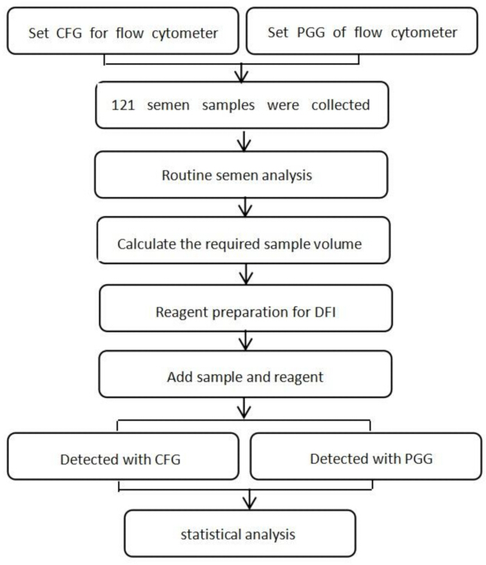
Figure 1: Overall schematic diagram of the workflow. Please click here to view a larger version of this figure.
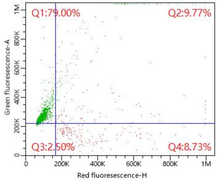
Figure 2: Cruciform gating (CFG) of flow cytometry for detecting DFI. Q1 – Normal sperm. Q2 – Mild DNA fragmentation (DFIm). Q3 – non-specific fluorescence that has been subtracted. Q4 – Severe DNA fragmentation (DFIs). Sperm with DNA damage fall into both Q2 and Q4. Their sum represents the total DNA‑fragmented sperm population (DFIc). Please click here to view a larger version of this figure.
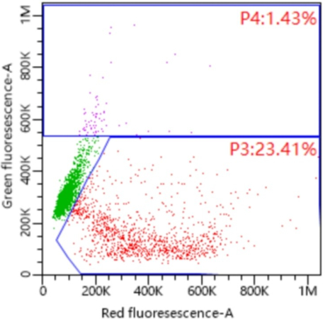
Figure 3: Polygonal gating (PGG) of flow cytometry for detecting DFI. P3: sperms with denatured DNA. P4: High DNA stainability (HDS) Please click here to view a larger version of this figure.
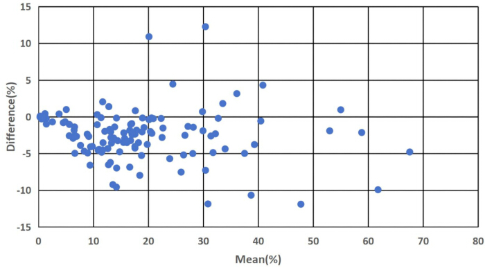
Figure 4: Bland-Altman plots of the results from PGG and CFG assays for DFI. X - axis: Mean of measurement results obtained by the PGG method and the CFG method. Y - axis: Difference in measurement results between the PGG method and the CFG method Please click here to view a larger version of this figure.
| Item | Mean±SD | Range |
| Age(years) | 32.5±4.9 | 26-44 |
| Days of abstinence(Day) | 4.1±1.3 | 3-7 |
| Semen volume(mL) | 3.8±1.6 | 0.5-6.8 |
| Sperm concentration(×106/mL) | 52.0±35.5 | 10.3-185.1 |
| Progressive motility(%) | 42.8±14.9 | 5.2-66.3 |
| Total motility(%) | 52.8±15.9 | 9.6-78.6 |
Table 1: General information and semen analysis data in the study population (n = 121).
| DFIp | HDS | |||||
| r | P | r | P | |||
| DFIm | 0.6497 | 0.0000 | -0.0103 | 0.9109 | ||
| DFIs | 0.9404 | 0.0000 | -0.3042 | 0.0007 | ||
| DFIc | 0.9667 | 0.0000 | -0.212 | 0.0196 | ||
| Note: | DFIp: DFI from polygonal gating method. | |||||
| HDS: high DNA staining index. | ||||||
| DFIm: mild DNA damage index. | ||||||
| DFIs: severe DNA damage index. | ||||||
| DFIc: DFI from cruciform gating method. | ||||||
Table 2: Correlation between the results of different gating methods (n = 121). The Pearson method was utilized to analyze the correlation between PGG and CFG.
| Semen volume | Sperm concentration | Total sperm count | PR (%) | ||||||||
| r | P | r | P | r | P | r | P | ||||
| DFIp | 0.2061 | 0.0233 | -0.0623 | 0.4970 | 0.0652 | 0.4775 | -0.3824 | 0.0000 | |||
| HDS | -0.0184 | 0.8415 | 0.0734 | 0.4234 | -0.0088 | 0.9234 | -0.0314 | 0.7328 | |||
| DFIm | 0.0520 | 0.5713 | -0.0709 | 0.4394 | -0.0384 | 0.6760 | -0.1975 | 0.0299 | |||
| DFIs | 0.2400 | 0.0080 | -0.0590 | 0.5203 | 0.0790 | 0.3890 | -0.3794 | 0.0000 | |||
| DFIc | 0.1893 | 0.0376 | -0.0759 | 0.4082 | 0.0344 | 0.7083 | -0.3574 | 0.0001 | |||
| Note: | PR (%) : percentage of progressively motile sperm. | ||||||||||
Table 3: Correlation between the results of DFI and basic semen examination(n = 121). The Pearson method was utilized to analyze the correlation between PGG and CFG.
Discussion
Loading...
$$\rightleftharpoonup{xx}$$
$$\longleftharp{xx}$$,
$$\longrightharp{xx}$$,
The 6th edition of the WHO Laboratory Manual for the Examination and Processing of Human Semen12 recommends four methods for assessing sperm DNA integrity: terminal deoxynucleotidyl transferase (dUTP) nick end labelling (TUNEL), single cell gel electrophoresis (Comet) assays, SCSA, and sperm chromatin dispersion (SCD) test. Among these, SCSA has been widely adopted in andrology laboratories due to its use of flow cytometry for analysis, which offers high throughput, efficiency, and objectivity, as well as the ability to simultaneously report parameters such as DFI and HDS7. According to a global survey7, nearly one-quarter (24.1%) of reproductive clinicians worldwide selected the SCSA as the method for evaluating sperm DNA integrity.
The most critical step in the SCSA procedure is establishing the appropriate flow cytometric gating. The traditional PGG method can detect both broken fragments (DFI) of DNA strands and abnormally high DNA-staining (HDS) sperm, characterized by the lack of normal histone and protamine exchange13. However, it cannot distinguish between single-strand and double-strand breaks in sperm DNA. In recent years, Yang et al.10 established a CFG method for analyzing the DFI of sperm using flow cytometry, which can distinguish the severity of sperm DNA damage. The key step of this method is to set up a cruciform gate for the flow cytometer. The CFG method exhibits excellent repeatability (coefficient of variation was less than 5%) and a linear range for DFI detection from 8.93% to 53.90%. This method reports three indicators—DFIm, DFIs, and DFIc—but does not provide HDS results10.
This study found that the total DFI of the CFG method (DFIc) was in good agreement with that of the PGG method (DFIp). Correlation analysis revealed that the correlation coefficients of DFIs and DFIp were significantly higher than those of DFIm. Neither sperm concentration nor total sperm count correlated with the CFG or PGG method results. PR (%) and the four types of DFI (DFIm, DFIs, DFIc, DFIp) were all significantly negatively correlated. PR (%) was significantly negatively correlated with all four types of DFI (DFIm, DFIs, DFIc, DFIp). The correlation with DFIm (r = -0.1975) was significantly weaker than that of DFIs, DFIc, and DFIp (r = -0.3794, -0.3574, -0.3824, respectively). These findings are consistent with those reported by Yang et al.10. Furthermore, DFIs and DFIc showed good correlations with semen volume, whereas DFIm did not. Although the clinical utility of an indicator that closely aligns with basic semen parameters may be limited, this finding suggests that the severe sperm DNA damage index (DFIs) may reflect distinct pathological aspects of spermatogenesis. More importantly, as shown in our correlation analysis, DFIs demonstrated a significantly stronger association with percentage of progressively motile sperm compared to DFIm, further supporting its potential as a complementary indicator in male fertility assessments beyond conventional semen analysis.
HDS represents the proportion of immature sperm lacking normal histone and protamine exchange. The protamine 1 precursor in HDS sperm retains terminal amino acids that are typically cleaved8, potentially preventing proper chromatin condensation and resulting in increased acridine orange staining of dsDNA. Previous studies have emphasized the significance of HDS in describing chromatin defects in sperm. Research by Lin et al.14 and Virro et al.15 indicates that the miscarriage rate of in vitro fertilization (IVF) is significantly higher in men with HDS > 15%. However, recent findings suggest that HDS may not reliably indicate nuclear immaturity due to its weak correlation with CMA3, AB, and TB staining results16. Thus, HDS might not be suitable as an indicator of male fertility17. This study also shows no significant correlation between HDS and basic semen parameters, including semen volume, sperm concentration, and motility. Therefore, HDS is not recommended as a marker for sperm DNA integrity18.
During the DFI detection process, inconsistencies in fluorescence boundaries may arise. This phenomenon is primarily attributed to significant variations in sperm concentration and motility among different specimens. To address this issue, it is recommended to use appropriate gating to exclude dead sperm, debris, and other non-specific signals. This approach enhances the accuracy of the data and mitigates the problem of inconsistent fluorescence boundaries.
To the best of our knowledge, this is the first study to compare the analysis results of PGG and CFG methods. A limitation of this study is its single-center, small-sample design, and its conclusions require further validation through multi-center, large-sample studies. Future research directions include ensuring consistency in sperm DFI test results across different flow cytometers and determining whether sperm DFIs can predict assisted reproductive technology outcomes.
In conclusion, the CFG method provides more parameters for sperm DNA integrity. Specifically, DFIm and DFIs reflect the proportions of sperm with mild and severe DNA damage, respectively. DFIc and DFIp results show good consistency. The CFG method is suitable for clinical application and promotion.
Disclosures
Loading...
$$\rightleftharpoonup{xx}$$
$$\longleftharp{xx}$$,
$$\longrightharp{xx}$$,
The authors have nothing to disclose.
Materials
| Name | Company | Catalog Number | Comments |
|---|---|---|---|
| Computer-assisted sperm analysis system | Beijing Suijia Software Co., Ltd | SA-II | |
| Flow cytometer | Shenzhen Mindray Bio-Medical Electronics Co., Ltd | Cyto E6 | |
| Flow cytometry kit | Shenzhen BRED Biotechnology Co., LTD | ||
| Makler counting chamber | Sefi Medical Instruments | ||
| Sperm nucleus DNA integrity Kit | Shenzhen BRED Biotechnology Co., LTD | - |
References
Loading...
$$\rightleftharpoonup{xx}$$
$$\longleftharp{xx}$$,
$$\longrightharp{xx}$$,
- Vaughan, D. A., Tirado, E., Garcia, D., Datta, V., Sakkas, D. DNA fragmentation of sperm: a radical examination of the contribution of oxidative stress and age in 16 945 semen samples. Hum Reprod. 35 (10), 2188-2196 (2020).
- Štiavnická, M., et al. H3K4me2 accompanies chromatin immaturity in human spermatozoa: an epigenetic marker for sperm quality assessment. Syst Biol Reprod Med. 66 (1), 3-11 (2020).
- Jeng, H. A., et al. Mixture analysis on associations between semen quality and sperm DNA integrity and occupational exposure to polycyclic aromatic hydrocarbons. Arch Environ Occup H. 78 (1), 14-27 (2023).
- Li, F., Duan, X., Li, M., Ma, X. Sperm DNA fragmentation index affect pregnancy outcomes and offspring safety in assisted reproductive technology. Sci Rep. 14 (1), 356(2024).
- Zhu, X. B., et al. Sperm DNA fragmentation in Chinese couples with unexplained recurrent pregnancy loss. Asian J Androl. 22 (3), 296-301 (2020).
- Minhas, S., et al. European Association of Urology Guidelines on Male Sexual and Reproductive Health: 2021 Update on Male Infertility. Eur Urol. 80 (5), 603-620 (2021).
- Agarwal, A., et al. Technical Aspects and Clinical Limitations of Sperm DNA Fragmentation Testing in Male Infertility: A Global Survey, Current Guidelines, and Expert Recommendations. World J Mens Health. 42 (1), 202-215 (2023).
- Evenson, D., Jost, L. Sperm chromatin structure assay is useful for fertility assessment. Meth Cell Sci. 22 (2-3), 169-189 (2000).
- B, V. L., et al. Sperm chromatin structure assay versus sperm chromatin dispersion kits: Technical repeatability and choice of assisted reproductive technology procedure. Clin Exp Reprod Med. 47 (4), 277-283 (2020).
- Yang, F., et al. Establishment and evaluation of a flow cytometry technique reflecting the severity of human sperm DNA damage. Zhonghua Nan Ke Xue. 26 (11), 989-995 (2020).
- World Health Organization. WHO laboratory manual for the examination and processing of human semen. , 5th ed., World Health Organization. Geneva. (2010).
- World Health Organization. WHO laboratory manual for the examination and processing of human semen. , 6th ed., World Health Organization. Geneva. (2021).
- Evenson, D. P. The Sperm Chromatin Structure Assay(SCSA) and other sperm DNA fragmentation tests for evaluation of sperm nuclear DNA integrity as related to fertility. Anim Reprod Sci. 169 (6), 56-75 (2016).
- Lin, M. H., et al. Sperm chromatin structure assay parameters are not related to fertilization rates, embryo quality, and pregnancy rates in in vitro fertilization and intracytoplasmic sperm injection, but might be related to spontaneous abortion rates. Fertil Steril. 90 (2), 352-359 (2007).
- Virro, M. R., Larson-Cook, K. L., Evenson, D. P. Sperm chromatin structure assay (SCSA) parameters are related to fertilization, blastocyst development, and ongoing pregnancy in in vitro fertilization and intracytoplasmic sperm injection cycles. Fertil Steril. 81 (5), 1289-1295 (2004).
- Mohammadi, Z., Tavalaee, M., Gharagozloo, P., Drevet, J. R., Nasr-Esfahani, M. H. Could high DNA stainability (HDS) be a valuable indicator of sperm nuclear integrity. Basic Clin Androl. 30, 12(2020).
- Shen, L., et al. High sperm DNA stainability might not be an accurate predictive indicator of male fertility and assisted reproductive technology outcomes. Front Endocrinol (Lausanne). 16, 1510114(2025).
- Lu, J. C. High DNA stainability (HDS) should not be recommended as a marker for the detection of sperm DNA damage. Andrologia. 54 (7), e14442(2022).
Reprints and Permissions
Request permission to reuse the text or figures of this JoVE article
Request Permission