Whole-body magnetic stimulation (WB-MS) was used in a binge-like feeding model involving intermittent access to a palatable diet. WB-MS transiently reduced intake during simulation without altering chow intake. Still, effects were not sustained, as binge-like behavior reemerged after treatment, with no changes in weight, adiposity, or accumbens c-Fos expression.
A subscription to JoVE is required to view this content. Sign in or start your free trial.
Research Article
Whole-Body Magnetic Stimulation Reduces Binge-Like Feeding in a Mouse Model of Disordered Eating
In This Article
Summary
Abstract
Whole-body magnetic stimulation (WB-MS) was used in a binge-like feeding model involving intermittent access to a palatable diet. WB-MS transiently reduced intake during simulation without altering chow intake. Still, effects were not sustained, as binge-like behavior reemerged after treatment, with no changes in weight, adiposity, or accumbens c-Fos expression.
Introduction
Binge eating is a dysregulated feeding pattern characterized by rapid consumption of large quantities of highly palatable food in the absence of physiological hunger. It is a core feature of binge eating disorder (BED), a condition strongly associated with obesity and metabolic dysfunction1,2. Increasing evidence highlights the role of mesolimbic reward circuits in the loss of control over feeding behavior3,4. BED is increasingly conceptualized as a disorder of altered corticostriatal signaling, in which dysregulated communication between reward-related and executive control systems promotes compulsive food intake. At the neurobiological level, dopaminergic signaling within the mesolimbic pathway, together with glutamatergic inputs, contributes to altered incentive salience and excessive motivation for palatable food5,6. In addition, peripheral metabolic signals such as GLP-1, insulin, and ghrelin interact with these circuits to modulate reward valuation and feeding behavior7. Recent evidence further indicates that binge-like behavior is associated with changes in neural dynamics within the NAc, including low-frequency oscillations in the delta and theta ranges (2–8 Hz), which have been linked to reward anticipation and craving states preceding loss of control8.
The NAc acts as an integrative hub linking limbic, cortical, and motor systems, translating motivational signals into goal-directed behavior. Disruption of this system, particularly under repeated exposure to palatable food, promotes compulsive and habit-like patterns of intake9,10. Experimental models using intermittent access to palatable food reliably induce binge-like eating are associated with increased activation of the NAc, supporting its role in compulsive feeding behavior11–14. Noninvasive neuromodulation refers to techniques that alter brain activity without surgical intervention, including magnetic field-based approaches such as transcranial magnetic stimulation15. These strategies have gained clinical relevance for their ability to modulate neural excitability and network activity in neuropsychiatric disorders16,17. In humans, neuromodulation has been shown to reduce binge-eating symptoms and improve behavioral control, as well as to influence metabolic regulation18,19.
Based on this evidence, whole body magnetic stimulation (WB-MS) may represent a noninvasive therapeutic approach capable of modulating circuits involved in compulsive feeding. Unlike focal techniques, low-frequency magnetic fields can influence neuronal excitability and distributed brain networks, suggesting a potential impact on subcortical structures such as NAc8. Experimental studies have shown that such fields modulate dopaminergic and glutamatergic neurotransmission6,7,8,19, providing a plausible mechanism for influencing reward-related circuits. However, its effects on binge-like eating behavior remain unknown. Therefore, the present study evaluated the effects of low-frequency WM-MS in a mouse model of binge-like eating induced by intermittent access to palatable food, assessing food intake, body weight, adiposity, and c-Fos expression in the NAc.
Access restricted. Please log in or start a trial to view this content.
Protocol
All procedures were conducted in compliance with the Mexican Official Standard for the Care and Use of Laboratory Animals (NOM-062-ZOO-199) and approved by the Institutional Committee for the Care and Use of Laboratory Animals (CICUAL) at the Universidad Nacional Autónoma de México (UNAM), under protocol number 035-CIC-2023. All the materials used in the study are mentioned in the Table of Materials.
Population definition and acquisition
A total of 30 C57BL/6 mice (9 females and 21 males) at postnatal day 30 (P30), with an initial body weight of 15–18 g, were used. Mice were housed individually in home cages under controlled environmental conditions: a 12:12 h light-dark cycle (lights on at 6:00 a.m.), an ambient temperature of 23 ± 2 °C, and a relative humidity of 70 ± 10%.
Habituation
At P30, the animals were individually housed in home cages with ad libitum access to water and standard food. All cages were environmentally enriched with cardboard tubes, and wood shavings were used as bedding. During the 7-day habituation period, body weight, food intake, and water consumption were recorded every 24 h.
Binge-like intake
The experimental model used in this study is based on the intermittent binge-intake paradigm described by researchers14. Prior to group allocation, all animals were brought to a comparable mean body weight (17 g) to reduce baseline variability. The animals (N = 30) were then randomly assigned to experimental groups using a digital “spin-the-wheel” tool, in which individual subjects were listed and sequentially allocated to one of the three groups. The standard group included females (standard female group, SFG; n = 3) and males (standard male group, SMG; n = 7), which had continuous access to standard chow and water. The continuous access group included females (continuous female group, CFG; n = 3) and males (continuous male group, CMG; n =7) with continuous ad libitum access to standard chow, water, and palatable food (M&M´s). The intermittent access group included females (intermittent females group, IFG; n =3) and males (intermittent males group, IMG; n = 7), who had ad libitum access to standard chow and water, and were provided access to palatable food three times per week (Monday, Wednesday, and Friday) for 2 h (4:00 to 6:00 p.m.). These feeding conditions were maintained for 35 days.
Whole-body magnetic stimulation (WB-MS)
This stimulation paradigm was selected based on previous studies using low-frequency electromagnetic fields (50 Hz, 1 mT) in C57BL/6 mice under similar experimental conditions20,21. WB-MS was performed using a pulsed magnetotherapy device (Dhan 1000, magnetotherapy unit) that generated a sinusoidal alternating magnetic field of 10 Gauss (1 mT) at 50 Hz. For stimulation sessions, mice remained individually housed in their home cages, which were placed between two electromagnetic plates on opposite sides of each cage. To avoid magnetic-field interference, metallic components, including the wire cage, grid, and water bottle, were temporarily removed during stimulation. This setup allowed animals to move freely throughout the procedure without restraint or additional stress.
To ensure experimental consistency, identical handling and environmental conditions were maintained during both stimulation and non-stimulation sessions. During control sessions (days 15–21), the magnetic plates were positioned identically but remained inactive, thereby controlling for potential effects of handling, cage manipulation, environmental noise, or electromagnetic plate placement.
WB-MS consisted of daily 20 min exposures from 3:40 p.m to 4:00 p.m. over seven consecutive days (days 22–28 of the experimental protocol), immediately before the binge-like eating assessment period (4:00–6:00 p.m). From days 29 to 35, the mice were again subjected to sessions without stimulation, after which their food intake was assessed.
Adipose tissue collection
At the end of the experimental protocol (day 36, 8:00 a.m.), adipose tissue was collected from all experimental groups. The SG consisted of three females and two males (n = 5), the CG three females and three males (n = 6), and the IG three females and four males (n = 7). All animals were anesthetized with ketamine (60 mg/kg) and xylazine (5 mg/kg)22, and the absence of a nociceptive response to toe pinch stimulation confirmed adequate depth of anesthesia. A ventral midline incision was then performed using a No. 3 scalpel blade, extending through the subcostal, inguinal, and midline abdominal regions, followed by gentle lateral retraction of the skin to expose the abdominal cavity.
Gonadal, retroperitoneal, and subcutaneous adipose tissue depots were carefully identified, dissected, and excised under standardized conditions to minimize inter-sample variability. Immediately after dissection, tissues were weighed using a calibrated analytical balance (maximum capacity 220 g, readability 0.1 mg), previously verified according to the manufacturer's specifications23. All samples were processed under identical conditions across groups to ensure experimental consistency.
Brain tissue preparation and immunofluorescence
Mice were euthanized 90 min after the last food consumption session24. Brains were collected following intracardiac perfusion with 4% paraformaldehyde and subsequently processed for histological analysis. Coronal brain sections (30 µm) were obtained using a cryostat maintained at −18 °C and stored in PBS containing sodium azide until processing. A subset of male animals from each experimental group was allocated to brain tissue preparation for immunofluorescence analyses (SG, n = 4; CG, n = 4; IG, n = 4), while the remaining animals were used for adipose tissue collection. This differential allocation was determined a priori based on methodological requirements, as intracardiac perfusion with paraformaldehyde results in tissue fixation that is incompatible with subsequent adipose tissue harvesting due to disruption of adipose tissue integrity. Therefore, sample distribution was driven exclusively by technical constraints and was independent of experimental group assignment or biological outcomes.
For c-Fos immunofluorescence, sections were incubated for 72 h with a mouse monoclonal anti-c-Fos antibody (1:1000, sc-166940) diluted in PBS containing 0.25% porcine skin gelatin and 0.5% Triton X-100, which served as the blocking and permeabilization solution. Primary antibody incubation was performed for 24 h at room temperature, followed by 48 h at 4 °C. After incubation, sections were rinsed thoroughly in PBS and incubated for 2 h with a donkey polyclonal anti-mouse secondary antibody (Alexa Fluor 488-conjugated, 1:500, AB_2338840) diluted in the same solution. Finally, sections were mounted on glass slides using mounting medium.
Image acquisition and quantification
Confocal imaging was performed using a microscope equipped with a 488 nm laser and a 20x objective lens. Z-stack images were acquired using NIS-Elements C software and stored under coded identifiers for blind analysis. Quantification of c-Fos-positive nuclei was performed using ImageJ (ImageJ 2.9.0 / 1.53t). Only fully contained nuclei were included in the counts, and a quadrant-based approach was applied to prevent double-counting of individual cells. For each animal, one image was acquired from each of three independent immunostained sections using identical acquisition parameters across all experimental groups. The number of c-Fos-positive nuclei was quantified individually for every image, and the values obtained from the three sections corresponding to the same animal were summed. This total value was subsequently normalized to the total analyzed area and expressed as c-Fos density (positive nuclei/mm2). The overall schematic of the experimental procedures used is illustrated in Figure 1A–H.
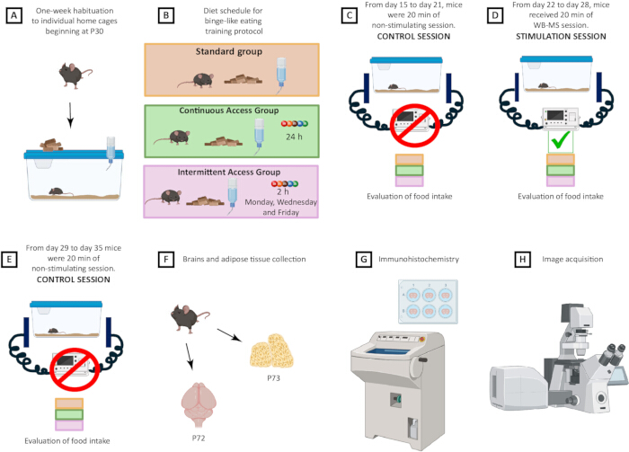
Figure 1: Experimental design and timeline of the binge-like eating protocol and WB-MS intervention. (A) shows a representative image of a mouse indicating the initial housing conditions. A total of 30 mice (9 females and 21 males) were individually housed in home cages starting at P30 and underwent a 1-week habituation period. (B) illustrates the dietary conditions implemented throughout the binge-like eating protocol from experimental day 1 to day 35. The control group had ad libitum access to standard chow and water. Continuous access groups had ad libitum access to standard chow, water, and palatable food. Intermittent access groups had ad libitum access to standard chow and water. At the same time, palatable food was provided only on Mondays, Wednesdays, and Fridays from 4:00 to 6:00 p.m. (C) indicates that from days 15 to 21, animals continued under the same dietary conditions while magnetic stimulation plates were placed beneath the cages without delivering stimulation (sham exposure). (D) shows that from days 22 to 28, WB-MS was administered 20 min before palatable food exposure while maintaining the corresponding dietary conditions for each group. (E) indicates that from days 29 to 35, WB-MS was stopped, although dietary conditions remained unchanged. (F) shows that brains were collected at postnatal day 72 (P72), followed by adipose tissue collection at postnatal day 73 (P73). (G) illustrates brain sectioning using a cryostat and immunohistochemical procedures. Finally, (H) shows confocal microscopy analysis of c-Fos immunoreactivity. The images were created by the authors using Biorender, and a proper license has been obtained. Please click here to view a larger version of this figure.
Statistical analysis
Statistical analyses were performed using SPSS version 25. Since normal distribution could not be assumed, non-parametric tests were used throughout the study. Differences among the SG, CG, and IG were evaluated using the Kruskal–Wallis test. Repeated comparisons within each experimental group were analyzed using the Friedman test, followed by multiple-comparison tests when appropriate. Due to limited sample sizes for certain experimental endpoints, particularly in females (n = 3 per group), data from male and female C57BL/6 mice were pooled for the analyses presented in Figure 2, Figure 3, and Figure 4 to improve statistical robustness and support an exploratory assessment of experimental outcomes. In addition, pooled binge-like intake data from male and female C57BL/6 mice were analyzed using a two-way ANOVA followed by multiple-comparisons testing to evaluate the effects of experimental group and time throughout the feeding protocol, including the WB-MS stimulation week. Statistical significance was defined as p ≤ 0.05.
Access restricted. Please log in or start a trial to view this content.
Results
Throughout the binge, subjects in the intermittent access groups (IMG for males, IFG for females) exhibited significantly higher caloric intake than those in the standard (SFG and SMG) and continuous groups (CFG and CMG) on days 15, 17, and 19 (Kruskal-Wallis test; p < 0.05; Figure 2A–B). WB-MS was administered at an intensity of 1 mT and a frequency of 50 Hz for 20 min per day over seven consecutive days, as indicated by the gray band. During this stimulation ph...
Access restricted. Please log in or start a trial to view this content.
Discussion
The present findings suggest that WB-MS may transiently modulate binge-like eating behavior in a murine model induced by intermittent access to palatable food. Although no significant changes in neuronal activity were detected in the NAc, palatable food intake progressively decreased throughout the intermittent-access sessions during the stimulation period, culminating in a significant decrease during the final binge session. These observations suggest that magnetic stimulation may influence reinforcement or motivational...
Access restricted. Please log in or start a trial to view this content.
Disclosures
The authors declare no competing interests or affiliations that could influence the content of this manuscript.
Acknowledgements
The authors thank Sandra Teresa Flores García, M.Sc. Armando Muñoz-Comonfort, Dr. Francisco Estrada-Rojo, and Dr. Fabiola Hernández-Vázquez for their valuable technical support. They would also like to thank Dr. Gustavo López-Toledo for designing Figure 1. Sonia Ortega-Tinoco is a PhD student in the Programa de Posgrado en Ciencias Biomédicas, UNAM, supported by a CONAHCyT fellowship number 1102522. This work was funded by the UNAM-DGAPA-PAPIIT grant IN214826 awarded to Dr. Julieta Garduño and IA301325 awarded to Dr. Raúl Sampieri-Cabrera.
Access restricted. Please log in or start a trial to view this content.
Materials
| Name | Company | Catalog Number | Comments |
|---|---|---|---|
| ImageJ | imagej.net | Public Domain, BSD-2 | Software. Java-based image processing program. Developed at the National Institutes of Health and the Laboratory for Optical and Computational Instrumentation (LOCI, University of Wisconsin) |
| Coverslips | Corning | 2935 | Cover glass 24 x 50 mm (Thickness 0.13–0.17) |
| Secondary antibody | Jackson ImmunoResearch | AB_2340758 | Donkey polyclonal anti-mouse secondary antibody |
| Laboratory Rodent Diet 5001 | LabDiet | 1320 | Standard food. https://di.facmed.unam.mx/comisiones/Composici%C3%B3n%20del%20alimento%20Laboratory%20Rodent%20Diet%205001.pdf |
| Leica CM1510 S | Leica | CM1510 S | Cryostat for routine histology. |
| M&Ms 43.8g (Brown packaging) | M&M´s | NA | Palatable food. Nutritional content per 100 : 480 kcal, 19 g of fat, 70 g of carbohydrates, 5.1 g of protein, 0.13 g of saltIngredients: sugar, cocoa mass, skimmed milk powder, cocoa butter, lactose, starch, milk fat, palm fat, glucose syrup, shea fat, stabiliser (gum arabic), dextrin, glazing agents (beeswax, carnauba wax), colours (E100, carmine, E132, E133, E150a, E150c, E150d, E153, E160a, E160e, E162, E163, E170), beetroot concentrate, emulsifiers (soya lecithin, E445), salt, flavourings, palm kernel oil, antioxidant (E306). (May contain: peanut, hazelnut, almond). Milk chocolate contains milk solids 14% minimum. Milk chocolate contains vegetable fats in addition to cocoa butter. |
| Dhan 1000 | Magnetoterapia SA de CV | MAG1000 | Pulsed magnetotherapy device |
| PBS | MEYER | Monobasic 2415. Dibasic 2410. NaCl S3014 | Sodium Phsphate Monobasic monohydrate . Sodium Phospate Dibasic heptahydrate. Crystalline Sodium Chloride grade molecular biology. |
| Nikon Microscope | Nikon Instruments, Inc | A1R+ STORM | Confocal Microscope |
| Polycarbonate mice cage | Orchid Scientific | EM01 | Non ferromagnetic policarbonate cage used for WB-MS exposure, fully transparent and autovlave (373 x 190 x 138 mm). |
| Anesket | Pisa | 040.000.0226.00 | Ketamine injectable solution |
| Procin Equus | Pisa | Q-7833-111 | Xylazine injectable solution |
| c-Fos antibody | Santa Cruz Biotechnology | sc-166940 | Mouse monoclonal anti-c-Fos antibody |
| Laboratory balance | Sartorius | Analytical balance | |
| VECTASHIELD Mounting Medium | Vector Laboratories | H-1000-10 | Antifade mounting medium is specially formulated to preserve fluorescence signals in immunofluorescence (IF) microscopy by minimizing photobleaching. |
| Glass slides | VELAB | VE-P30 | Microscope glass slides Frosted on one side 26 x 76 mm (Thickness 0.8–1.1 mm) |
| Sodium azide | K 6048.0100 | 99% Sodium azide |