This review examines the role of ultrasound intervention in diagnosing and treating non-breast superficial and abdominal diseases. Originally a diagnostic tool, ultrasound intervention has expanded into a platform integrating thermal ablation, drainage, and vascular therapy. Advances such as contrast-enhanced imaging and fusion navigation have improved precision, although standardization and long-term efficacy remain challenges.
A subscription to JoVE is required to view this content. Sign in or start your free trial.
Review Article
The Development of Ultrasound Intervention in the Diagnosis and Treatment of Non-breast Superficial and Abdominal Diseases
In This Article
Summary
Abstract
Interventional ultrasound is undergoing a paradigm shift from a traditional diagnostic adjunct to a diversified therapeutic platform, particularly in non-breast superficial and abdominal diseases. This review provides a domain-specific analysis of this clinical transition, focusing on two primary use cases: precise thermal ablation for superficial organ lesions (e.g., thyroid nodules) and image-guided tumor management in solid abdominal organs (e.g., hepatocellular carcinoma). Through a narrative review of recent literature, we describe how the modality has advanced from simple puncture biopsy to a comprehensive diagnostic and therapeutic system integrating complex drainage and vascular interventions. For thyroid diseases, thermal ablation is increasingly recommended by international guidelines (e.g., ATA, ETA) as a viable alternative to surgery for symptomatic benign nodules and strictly selected, low-risk papillary thyroid microcarcinomas. In hepatic malignancies, thermal ablation is endorsed by major guidelines (e.g., EASL, AASLD) as a potentially curative option for early-stage hepatocellular carcinoma in patients with small tumors (typically ≤3 cm) or those ineligible for surgical resection. This transformation is driven by the integration of technologies such as contrast-enhanced ultrasound, fusion imaging, elastography, and artificial intelligence (AI), which have enhanced procedural accuracy and safety. Multidisciplinary collaboration has further enabled application in more complex clinical scenarios. Despite ongoing challenges in technical standardization and long-term efficacy, ultrasound intervention has become an important platform for precision diagnosis and treatment in abdominal and superficial organ diseases.
Introduction
Since the mid-20th century, ultrasound imaging technology has established a cornerstone position in modern clinical medicine, with advantages including being radiation-free, real-time, high-resolution, convenient, and cost-effective. Over several decades of development, ultrasound has primarily been used for disease screening and diagnosis. Through images formed by sound wave reflection, it reveals morphological changes in internal organs and serves as an important reference in clinical decision-making1,2. However, traditional diagnostic ultrasound is essentially a passive observation tool. Its "picture description" model has gradually revealed limitations in modern medicine, which increasingly emphasizes precision, minimally invasive techniques, and integrated diagnosis and treatment3. The development of medicine calls for technologies that can unify diagnosis and treatment in time and space. Under this demand, ultrasound interventional technology emerged, achieving a transition from the "eye of diagnosis" to the "hand of treatment".
Ultrasound interventional technology refers to a series of minimally invasive procedures, such as puncture biopsy, aspiration and drainage, drug injection, physical ablation, and radioactive particle implantation, performed under real-time ultrasound guidance using specialized needles, catheters, and ablation electrodes. It integrates the accuracy of image navigation with minimally invasive treatment and has expanded the scope of ultrasound medicine4. In its early stages, ultrasound intervention was mainly used for relatively simple diagnostic procedures, such as puncturing pleural and peritoneal effusions and performing liver and kidney biopsies. It also established a solid foundation in the diagnosis and treatment of breast diseases (especially biopsy). However, its potential in non-breast superficial organs (such as thyroid, parathyroid, salivary glands, superficial lymph nodes, and soft tissues) and abdominal solid organs (such as liver, kidney, pancreas, spleen, and intra-abdominal lesions) has been more systematically recognized in the past decade. It is increasingly reshaping the diagnostic and therapeutic landscape in these fields5,6,7.
The core driving forces behind this transition stem from two major aspects. First, there is increasing clinical demand. With population aging and advances in diagnostic techniques, detection rates of diseases such as thyroid nodules and liver tumors have increased significantly. Patients increasingly seek minimally invasive approaches, rapid recovery, and preservation of organ function. Although traditional surgical operations offer strong therapeutic effects, they are associated with significant trauma, higher costs, risks of complications, and impacts on physiological function, prompting the search for alternative approaches8. Second, technological innovation plays a key role. The development of high-frequency linear probes has improved resolution for superficial organs, while advances in puncture needles and drainage systems have enhanced procedural precision. The maturation and miniaturization of ablation technologies, including radiofrequency, microwave, laser, and high-intensity focused ultrasound, have further supported precise percutaneous ablation therapy9.
In view of this, this article aims to provide a narrative review of ultrasound interventional technology in non-breast superficial organs and abdominal diseases, illustrating the transition from diagnostic to integrated platforms. It analyzes recent research progress and clinical evidence across key organs, discusses challenges related to accuracy, safety, and long-term efficacy, and outlines potential future developments involving technologies such as artificial intelligence-assisted navigation, multimodal image fusion, and novel drug delivery systems. The goal is to provide a theoretical basis for clinical practice and support continued standardization and development in this field.
Access restricted. Please log in or start a trial to view this content.
Review and Perspective
A comprehensive literature search was conducted to identify relevant studies on ultrasound interventional technology in non-breast superficial and abdominal diseases. Electronic databases including PubMed, Web of Science, and Scopus were searched for articles published between January 2010 and January 2026. Core search terms included combinations of "ultrasound intervention," "thermal ablation," "contrast-enhanced ultrasound," "artificial intelligence," "liver tumors," and "thyroid nodules." Inclusion criteria were restricted to peer-reviewed systematic reviews, randomized controlled trials, large-scale cohort studies, and international clinical guidelines. Case reports, non-English articles, and studies lacking clear clinical outcomes were excluded. The selection process and data extraction were structured to minimize bias.
Transformations in clinical practice
The clinical translation of interventional ultrasound from a diagnostic adjunct to a primary therapeutic modality is reflected across several key dimensions10,11.
Diagnostic advances: Targeted biopsy
Ultrasound-guided biopsy remains a key method for diagnosing abdominal and superficial space-occupying lesions. However, its role has evolved from "sampling" to "precise targeted sampling".
The application of contrast-enhanced ultrasound is a key driver of this development. Studies have shown that contrast-enhanced ultrasound (CEUS) can distinguish active tissues from necrotic areas, thereby guiding puncture into viable regions (Figure 1). In liver tumors, CEUS can improve the diagnostic sensitivity of hepatocellular carcinoma. In intrahepatic cholangiocarcinoma, CEUS can identify and avoid necrotic areas, increasing diagnostic accuracy by approximately 15%12.
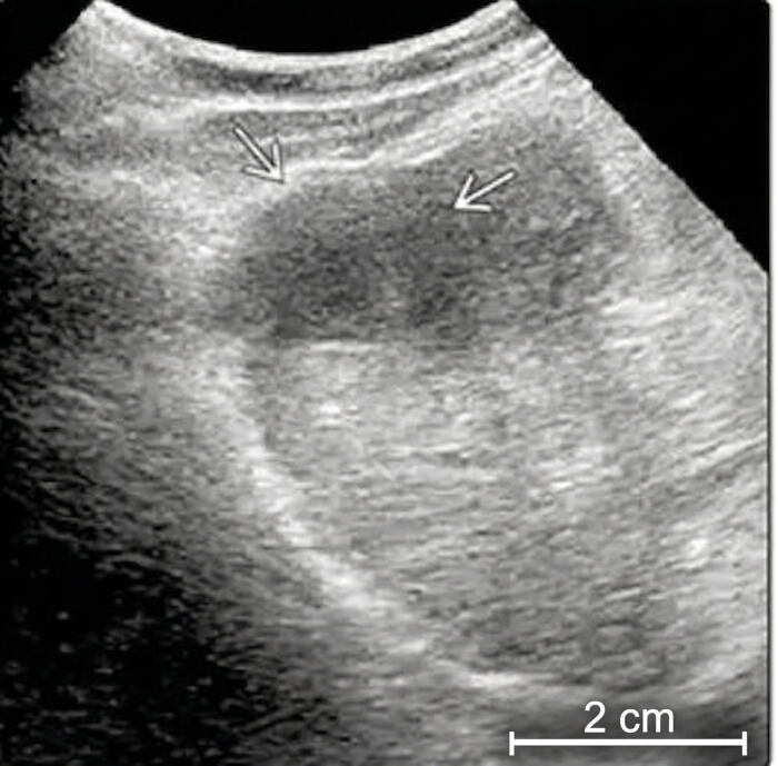
Figure 1: Contrast-enhanced ultrasound imaging of hepatocellular carcinoma. The image demonstrates the typical arterial phase enhancement of a hepatic malignancy. CEUS allows for real-time visualization of tumor microvascularization, which is critical for accurate boundary delineation and real-time monitoring during interventional ablation procedures. (Note: A scale bar is provided in the lower right corner). Abbreviation: CEUS = Contrast-enhanced ultrasound. Please click here to view a larger version of this figure.
The development of ultrasound elastography technology has further enriched diagnostic information. Studies have demonstrated that combining ultrasound elastography techniques to construct prediction models for liver fibrosis yields an area under the receiver operating characteristic curve (AUC) greater than 0.800, providing a validated, non-invasive diagnostic tool13.
The introduction of artificial intelligence-assisted diagnostic systems marks a new stage of diagnostic precision. Recent studies have developed deep learning models for the differential diagnosis of focal liver lesions based on ultrasound images, demonstrating diagnostic performance comparable to that of senior sonographers14. Yao et al.15 established an AI model for the detection, segmentation, and classification of thyroid nodules using ultrasound image data from 10,023 nodules across 208 institutions, improving diagnostic accuracy and consistency.
Expansion of therapeutic areas: From minimally invasive drainage to tumor ablation
The role of ultrasound intervention in therapy has expanded, forming a diversified therapeutic spectrum (Table 1). From traditional sclerotherapy for cysts to complex tumor ablation, ultrasound intervention has developed into a minimally invasive treatment approach comparable to traditional surgical methods.
| The evolving therapeutic role | Evidence Level and Guideline Endorsement | ||
| Thermal ablation for benign nodules and strictly selected microcarcinomas | Level I / Strong Recommendation (e.g., ATA, ETA Guidelines) | ||
| Tumor thermal ablation, portal vein manometry | Level I / Strong Recommendation (e.g., EASL, AASLD Guidelines) | ||
| Tumor thermal ablation, percutaneous nephrostomy | Level II / Recommended Alternative Option | ||
| Abscess drainage,celiac plexus block | Level II / Standard Minimally Invasive Option | ||
| Venous embolization and aneurysm treatment | Level IIb-III / Emerging Clinical Application | ||
Table 1: Evolution of the therapeutic application of ultrasound intervention in non-breast superficial and abdominal diseases. This table outlines the expansion from diagnostic biopsy to radical therapeutic modalities. Evidence levels are categorized according to standardized frameworks (e.g., Level I: Strong recommendation based on international guidelines such as EASL or ATA; Level II: Emerging clinical evidence).
Ultrasound interventions in superficial organs (e.g., thyroid, parotid)
In superficial organs, thermal ablation for thyroid nodules serves as a key clinical model for this transition. Large systematic reviews and meta-analyses, such as Zhao et al.16, have confirmed the safety, volume reduction, and clinical efficacy of thermal ablation for benign thyroid nodules. The safety and efficacy of minimally invasive ablation have also been explored for other head and neck masses17. For symptomatic benign nodules and selected low-risk papillary thyroid microcarcinoma, thermal ablation can achieve outcomes comparable to surgery while preserving thyroid function and avoiding surgical scars (Figure 2 and Figure 3). It is increasingly considered an alternative for patients who are not candidates for or decline surgery.
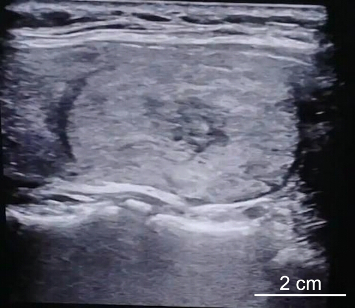
Figure 2: High-resolution B-mode ultrasound of a solid thyroid nodule. The image displays morphological features, such as hypoechogenicity and irregular margins, which are essential for preoperative risk stratification and for determining the need for ultrasound-guided fine-needle aspiration or thermal ablation. (Note: A scale bar is provided in the lower right corner). Please click here to view a larger version of this figure.
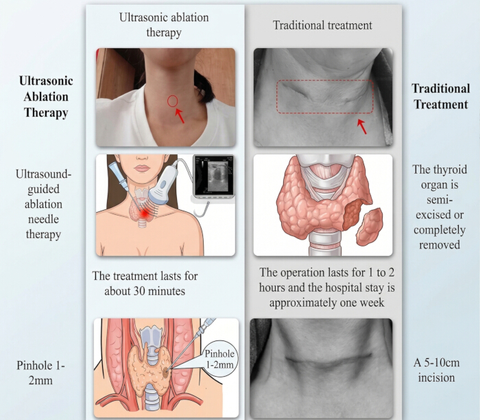
Figure 3: Comparative diagram of ultrasound-guided thermal ablation versus traditional surgical resection for thyroid nodules. This composite figure illustrates the minimally invasive nature of thermal ablation, characterized by localized treatment and the absence of significant surgical scarring, as compared to the anatomical disruption of open surgery. (Note: Written informed consent was obtained from the patient for the publication of the anonymized clinical photographs. This is an original figure prepared for this paper). Please click here to view a larger version of this figure.
Ultrasound interventions in non-superficial abdominal organs (e.g., liver, kidney, pancreas)
In abdominal solid organs, ultrasound intervention has expanded from cystic lesions to solid tumors. Studies have shown that microwave ablation of intrahepatic cholangiocarcinoma can achieve overall survival rates similar to hepatectomy under appropriate conditions18. For early-stage hepatocellular carcinoma, thermal ablation is recommended by EASL and AASLD guidelines as a potentially curative option, particularly for patients with small tumors or those who are not surgical candidates.
The application scope has also expanded to other complex scenarios. In portal hypertension, ultrasound-guided puncture of portal vein branches for pressure measurement provides a basis for surgical decision-making. Ultrasound-guided drainage of complex intra-abdominal fluid collections has demonstrated high technical success and safety in experienced centers19.
In complex abdominal diseases, endoscopic ultrasound (EUS) has expanded therapeutic options. EUS-guided embolization of abdominal aneurysms avoids radiation exposure associated with conventional vascular intervention. For pancreatic pseudocysts and abscesses, EUS-guided drainage has become a first-line minimally invasive option. EUS-guided gastrojejunostomy20 has also been applied to restore gastrointestinal continuity in advanced malignancies (Figure 4).
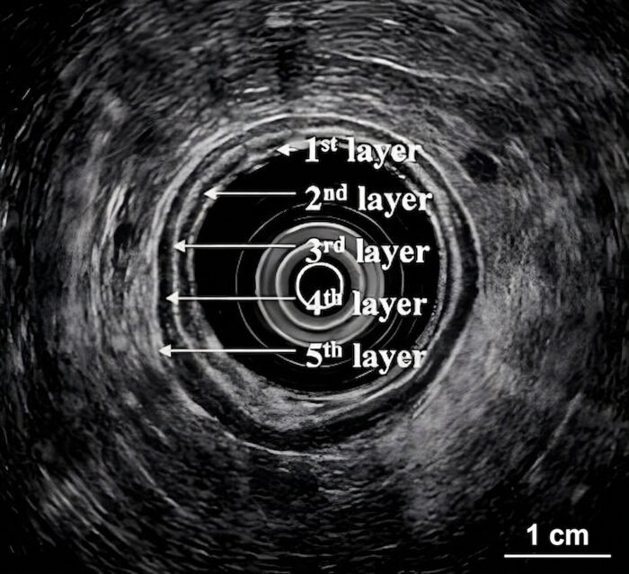
Figure 4: Endoscopic Ultrasound demonstrating the gastrointestinal wall architecture. The image identifies the characteristic five-layer structure: (1st layer) superficial mucosa, (2nd layer) deep mucosa, (3rd layer) submucosa, (4th layer) muscularis propria, and (5th layer) serosa. Precise identification of these layers is vital for safe EUS-guided drainage and stent placement in complex abdominal conditions. (Note: A scale bar is provided in the lower right corner). Abbreviation: EUS = Endoscopic Ultrasound. Please click here to view a larger version of this figure.
The emergence of combined surgical approaches represents a further advancement. Studies have reported the use of laparoscopic resection combined with ultrasound-guided microwave ablation for complex liver tumors21, enabling treatment of lesions in both lobes within a single procedure.
Technological empowerment: Multimodal image fusion and artificial intelligence-driven precise intervention
Technological integration is a key driver of this paradigm shift (Figure 5). The integration of contrast-enhanced ultrasound, elastography, fusion navigation, and artificial intelligence has expanded the capabilities of traditional ultrasound. CEUS enables real-time visualization of blood perfusion and active regions of lesions, allowing biopsy to move from "blind puncture" to "targeted" and ablation from "experience-based" to "precision-guided". Fusion imaging combines the real-time nature of ultrasound with the anatomical detail of CT/MRI, overcoming limitations such as gastrointestinal gas and bone interference, and extending intervention to previously inaccessible areas10. The application of artificial intelligence has shown potential in standardized operation, decision support, and prognosis prediction, suggesting further development toward more automated and intelligent systems.
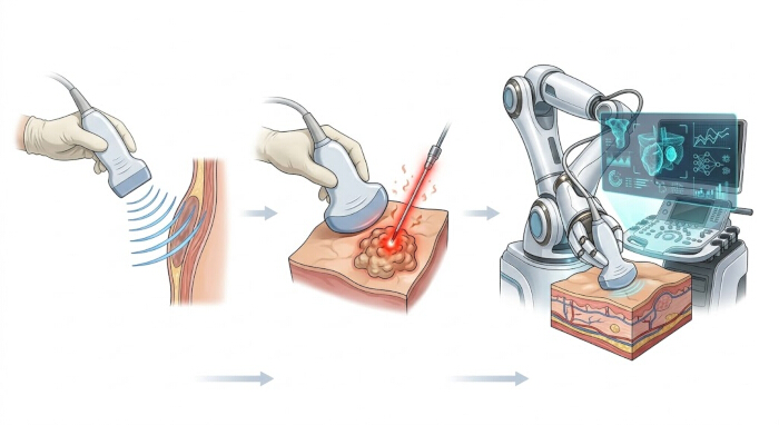
Figure 5: Conceptual evolution of interventional ultrasound from diagnostic to therapeutic and advanced guidance applications. This schematic illustrates three stages of progression: (Left) diagnostic ultrasound imaging; (Middle) image-guided therapeutic intervention using thermal ablation; and (Right) integration of advanced technologies such as robotic assistance and artificial intelligence-supported guidance. This transition reflects ongoing development toward more precise and minimally invasive interventions. Please click here to view a larger version of this figure.
Contrast-enhanced ultrasound technology: Precise navigation from morphology to functionality
The development of contrast-enhanced ultrasound technology marks a transition from morphological assessment to functional navigation. Its core value lies in the real-time display of microvascular perfusion using blood pool tracers. During interventions, CEUS can delineate tumor-feeding vessels and active regions, enabling targeted procedures. Urhuț et al.22 reported that CEUS-guided biopsy can increase diagnostic yield by approximately 15% by avoiding necrotic tissue. In tumor ablation, CEUS can monitor energy deposition in real time and assess treatment completeness. You et al.23 demonstrated that CEUS has accuracy comparable to enhanced MRI in evaluating radiofrequency ablation outcomes, allowing detection of residual disease and guiding additional treatment.
Fusion imaging technology: Breaking through the barriers of image modalities
Fusion imaging combines real-time ultrasound with preacquired CT, MRI, or PET-CT images through spatial registration and synchronized display24. This approach addresses limitations of ultrasound in regions obscured by gas or bone.
In clinical practice, fusion imaging is particularly useful for deep or poorly visualized lesions. For example, liver tumors located near the diaphragm or gastrointestinal tract may not be clearly visualized with ultrasound alone25. Fusion with CT or MRI enables real-time visualization of lesion contours, facilitating precise needle placement and reducing the risk of injury to adjacent structures. Nayak et al.10 reported improved puncture success and ablation completeness in such cases.
Four-dimensional ultrasound and hemodynamic imaging: Spatiotemporal expansion of the interventional field of view
Recent advancements in four-dimensional ultrasound and novel microvascular imaging techniques have provided enhanced spatiotemporal information for interventional procedures. For instance, the team led by Song et al.26,27 developed a super-resolution ultrasound imaging technology that enables dynamic visualization of microcirculation in large organs at millisecond and submillimeter resolutions. This allows visualization of both vascular morphology and hemodynamic changes in real time. As these technologies move toward clinical application, they may expand current capabilities. In liver intervention, they may help identify tumor-feeding vessels and support embolization planning28; in renal ablation, they may support visualization of the tumor and adjacent vascular structures, thereby enhancing treatment precision while preserving function29. This development reflects a transition from "static anatomical navigation" toward "dynamic functional guidance".
Artificial intelligence technology: The core engine of standardization and intelligence
Within interventional ultrasound, artificial intelligence (AI) aims to reduce operator dependence and improve procedural standardization. AI technologies, particularly deep learning, are being explored to address variability and reproducibility across centers. The application of AI spans preoperative, intraoperative, and postoperative stages. During preoperative planning, AI algorithms can assist in lesion identification, segmentation, and characterization, supporting intervention planning. The 2D and 3D deep learning models developed by Baydoun et al.30 demonstrated accurate prediction of clinically significant prostate cancer using standardized ultrasound videos, with performance approaching that of experienced experts. During intraoperative navigation, AI-based tracking and augmented visualization can assist in needle localization and path planning, potentially reducing procedural difficulty31. AI may also support real-time identification of anatomical structures and the generation of risk alerts. In postoperative assessment, Fu et al.32 demonstrated the potential of AI models combining ultrasound and pathological data for breast cancer characterization. Emerging research has also explored integration with 3D US-CT/MRI fusion, electromagnetic (EM)-tracked navigation, and robotic-assisted guidance. If validated in larger studies, these approaches may reduce operator dependence and improve precision in complex anatomical settings33. Concurrently, AI-based decision support and automated lesion detection are being explored for planning and risk assessment34,35. Together, these developments indicate a trend toward more integrated interventional systems.
Clinical integration: From specialized operations to multidisciplinary diagnosis and treatment platforms
Clinical evidence supports the expanding role of ultrasound intervention. Previous studies, including randomized controlled trials and large-scale cohort studies, have demonstrated its efficacy and safety in selected indications. For example, ablation therapy for thyroid nodules, early-stage liver cancer, and kidney cancer has been evaluated in multiple studies11,36 and incorporated into international guidelines, progressing from alternative options to recommended treatments in selected scenarios. This reflects the transition of ultrasound intervention from an emerging technique to an established component of multidisciplinary tumor management. Its application has expanded beyond dedicated interventional suites to include intraoperative and critical care settings37,38.
The scope of intraoperative ultrasound continues to expand. Combined approaches using laparoscopic resection for accessible lesions and ultrasound-guided microwave ablation for deep tumors allow treatment within a single procedure39. In emergency settings, point-of-care ultrasound supports rapid diagnosis and minimally invasive management of acute conditions such as fluid collections or bleeding40. In intensive care, ultrasound-guided catheterization and drainage procedures provide bedside management options while reducing risks associated with patient transport41. Multidisciplinary teams increasingly integrate interventional ultrasound into combined treatment planning for complex malignancies42.
However, several challenges and limitations remain. First, technical standardization and training systems require further development. Operator dependence remains a key limitation, and standardized training, credentialing, and quality control are needed to ensure consistency across centers43. Second, long-term efficacy data remain limited. Although short- and mid-term outcomes are encouraging, longer follow-up is required for definitive evaluation44,45. Third, indications require careful definition. For example, in papillary thyroid microcarcinoma, while short-term outcomes are promising, further data are needed on high-risk variants and long-term outcomes46,47.
Fourth, comprehensive reporting of complication and failure rates remains important for clinical decision-making. A comparative analysis of outcomes and complications across major indications (liver, thyroid, kidney) is summarized in Table 2. Finally, integration of AI and related technologies introduces ethical considerations, including data privacy, algorithmic bias, and clinical accountability, which require appropriate regulatory oversight.
| Clinical Indication | Interventional Modality | Efficacy / Clinical Outcome | Major Complication Rate | References |
| Hepatocellular carcinoma (Early-stage, tumor size ≤3 cm) | Thermal ablation (RFA, MWA) | Complete ablation rate > 90%; 5-year overall survival comparable to surgical resection | < 3% (e.g., hemorrhage, liver abscess, bile duct injury, non-target thermal injury) | [1, 6] |
| Symptomatic benign thyroid nodules | Thermal ablation (RFA, MWA) | Volume reduction rate (VRR) > 60% at 6 months; significant cosmetic and symptom improvement | < 2% (e.g., transient hoarseness, hematoma, nodule rupture) | [16] |
| Papillary thyroid microcarcinoma (PTMC, strictly selected T1aN0M0) | Thermal ablation (RFA, MWA) | Complete tumor disappearance rate > 95%; very low local recurrence and lymph node metastasis rates | 1-3% (e.g., recurrent laryngeal nerve thermal injury) | [44, 45] |
| Small renal cell carcinoma (Stage T1a, selected patients) | Percutaneous thermal ablation (RFA, Cryoablation) | 5-year cancer-specific survival rate > 90%; excellent preservation of renal function | < 5% (e.g., perinephric hematoma, urine leak, stricture) | [40, 46] |
Table 2: Comparative analysis of clinical outcomes and complication rates across major indications. This table summarizes comparative efficacy (e.g., complete ablation rates) and safety profiles (e.g., rates of bleeding or non-target thermal injury) for hepatic, renal, and thyroid interventions.
Access restricted. Please log in or start a trial to view this content.
Conclusions
Ultrasound intervention has established its position as a diagnosis and treatment platform in the management of non-breast superficial and abdominal diseases. Through continuous technological innovation and clinical research, it continues to expand its capabilities, offering patients treatment approaches with less trauma and faster recovery.
In summary, ultrasound intervention has transitioned from a passive observational modality to a minimally invasive therapeutic platform, demonstrating eff...
Access restricted. Please log in or start a trial to view this content.
Disclosures
The authors have no conflicts of interest to disclose.
Acknowledgements
The authors thank Qinghai Red Cross Hospital and the Department of Ultrasound for their institutional support during the preparation of this manuscript. We also express our sincere gratitude to the multidisciplinary clinical teams in hepatology, thyroid surgery, and interventional oncology for their valuable professional insights and academic discussions that informed the framework and scope of this review.
Access restricted. Please log in or start a trial to view this content.
References
- Dietrich, C. F., Nolsøe, C. P., Barr, R. G. WFUMB guidelines and recommendations on interventional ultrasound: part iv - ultrasound-guided treatment of focal liver lesions (update 2024). Ultrasound Med Biol. , (2024).
- Sidhu, P. S., Cantisani, V., Dietrich, C. F. EFSUMB clinical practice guidelines for interventional ultrasound: focused update on safety and quality standards. Ultraschall Med. , (2025).
- Liang, P. State-of-the-art and development trend of interventional ultrasound in china. Adv Ultrasound Diagn Ther. , (2023).
- Popa, A. Indications for abdominal imaging: when and what to choose? J Med Life. , (2020).
- Jennings, J. W. Musculoskeletal interventional oncology: a contemporary review. AJR Am J Roentgenol. , (2023).
- Puijk, R. S., Geboers, B., Nieuwenhuizen, S. Cirse standards of practice on thermal ablation of liver tumours. Cardiovasc Intervent Radiol. , (2024).
- Goldberg, S. N., Ahmed, M. Global trends and future directions in interventional oncology: a ten-year perspective (2016–2026). J Vasc Interv Radiol. , (2025).
- Iglesias-Garcia, J. Advances in endoscopic ultrasound-guided shear wave elastography: a comprehensive review of its clinical applications. World J Gastroenterol. , (2025).
- Zampieri, F. Pocus for acute abdominal pain: practical scan protocols on gastrointestinal diseases and an evidence review. Intern Emerg Med. , (2025).
- Nayak, G., Bolla, V., Balivada, S., P, P. Technological evolution of ultrasound devices: a review. Int J Health Technol Innov. , (2022).
- Dietrich, C. F., et al. History of ultrasound in medicine from its birth to date (2022), on occasion of the 50 years anniversary of EFSUMB. a publication of the European Federation of Societies for Ultrasound in Medicine and Biology (EFSUMB), designed to record the historical development of medical ultrasound. Med Ultrason. , (2022).
- Chinese Medical Association Ultrasound Medicine Branch. Six major advances in the field of ultrasound medicine in 2023. Chin Med Inf Bull. , (2024).
- Wang, K., Lu, X., Zhou, H. Deep learning radiomics of shear wave elastography significantly improved diagnostic performance for assessing liver fibrosis in chronic hepatitis b: a prospective multicentre study. Gut. , (2019).
- Wu, P., Peng, Q., Zhang, Z. Deep learning radiomics for focal liver lesions diagnosis on long-range contrast-enhanced ultrasound and clinical factors. Quant Imaging Med Surg. , (2022).
- Yao, Q. L., Ye, J. J., Gao, C. X. Research progress of deep learning in ultrasound diagnosis of thyroid nodules. J Mol Imaging. , (2024).
- Zhao, X., et al. The long-term effects of thermal ablation for benign thyroid nodules: a systematic review and meta-analysis. Eur Radiol. , (2025).
- Chen, Y., Li, X., Zheng, J. Ultrasound-guided percutaneous thermal ablation of parotid tumors: experience from two-centers. Int J Hyperthermia. , (2023).
- Song, M., et al. Efficacy of microwave ablation for intrahepatic cholangiocarcinoma: a systematic review and meta-analysis. Quant Imaging Med Surg. , (2025).
- Gomaa, S., Farouk, H., Ali, A. Ultrasound guided drainage and aspiration of intra-abdominal fluid collections. Benha J Appl Sci. , (2023).
- Miller, C., et al. Eus-guided gastroenterostomy vs. surgical gastrojejunostomy and enteral stenting for malignant gastric outlet obstruction: a meta-analysis. Endosc Int Open. , (2023).
- Muglia, R., et al. Technical and clinical outcomes of laparoscopic–laparotomic hepatocellular carcinoma thermal ablation with microwave technology: case series and review of literature. Cancers (Basel). , (2024).
- Urhuț, M. C., et al. Diagnostic performance of an artificial intelligence model based on contrast-enhanced ultrasound in patients with liver lesions: a comparative study with clinicians. Diagnostics (Basel). , (2023).
- You, Y., et al. Feasibility of 3D US/CEUS-US/CEUS fusion imaging-based ablation planning in liver tumors: a retrospective study. Abdom Radiol (NY). , (2021).
- Teng, F., Liu, C., Feng, Z., et al. Application of CT/MRI fusion imaging in the diagnosis and staging of liver cancer: a comparative study. Int J Radiat Res. , (2025).
- Rai, P., et al. Efficacy of fusion imaging for immediate post-ablation assessment of malignant liver neoplasms: a systematic review. Cancer Med. , (2023).
- Song, P., Rubin, J. M., Lowerison, M. R., Yin, T., Chen, S. Super-resolution ultrasound microvascular imaging: is it ready for clinical use? Z Med Phys. , (2023).
- Kaiser, U., et al. Super-resolution contrast-enhanced ultrasound examination down to the microvasculature enables quantitative analysis of liver lesions: first results. Life. , (2025).
- Paeng, D. G., Lee, C. A., Imtiaz, C. Principles of Doppler ultrasound and emerging blood flow imaging. Ultrasonography. , (2025).
- Golemati, S., Cokkinos, D. D. Recent advances in vascular ultrasound imaging technology and their clinical implications. Ultrasonics. , (2022).
- Baydoun, A., et al. Artificial intelligence applications in prostate cancer. Prostate Cancer Prostatic Dis. , (2024).
- D'Amore, B., Smolinski-Zhao, S., Daye, D., Uppot, R. N. Role of machine learning and artificial intelligence in interventional oncology. Curr Oncol Rep. , (2021).
- Fu, Y., et al. Integrating multimodal ultrasound imaging and machine learning for predicting luminal and non-luminal breast cancer subtypes. Front Oncol. , (2025).
- Laakso, I., Paulides, M. M., Kodera, S. Roadmap towards personalized approaches and safety considerations in non-ionizing radiation: from dosimetry to therapeutic and diagnostic applications. Phys Med Biol. , (2026).
- Lachenmayer, A., Tinguely, P., Maurer, M. H. Robotic assistance in percutaneous liver ablation therapies: a systematic review and meta-analysis. Ann Surg. , (2024).
- Wang, Y., Li, X., Zheng, J. Deep learning-based real-time ultrasound navigation for percutaneous needle placement. Med Image Anal. , (2024).
- Iacob, R., et al. Evaluating the role of breast ultrasound in early detection of breast cancer in low- and middle-income countries: a comprehensive narrative review. Bioengineering (Basel). , (2024).
- Diaz-Gomez, J. L., Sharif, S., Ablordeppey, E. Guidelines on adult critical care ultrasonography: focused update 2024. Crit Care Med. , (2025).
- DeWitt, J. M., Arain, M., Chang, K. J. Interventional endoscopic ultrasound: current status and future directions. Clin Gastroenterol Hepatol. , (2021).
- Wu, H. R., Bu, H., Liu, Y. Y. Intraoperative laparoscopic ultrasound-guided resection and microwave ablation for colorectal liver metastases. World J Gastrointest Oncol. , (2025).
- Barbic, D. Point-of-care ultrasound for diagnosis of abscess in skin and soft tissue infections. Acad Emerg Med. , (2017).
- Milojevic, I., Lemma, K., Khosla, R. Ultrasound use in the ICU for interventional pulmonology procedures. J Thorac Dis. , (2021).
- Knavel Koepsel, E. Interventional oncology: a primer for clinicians on the role of ablation and embolization for solid tumors. Cancer Med. , (2024).
- Laakso, I., Paulides, M. M., Kodera, S., et al. Roadmap towards personalized approaches and safety considerations in non-ionizing radiation: from dosimetry to therapeutic and diagnostic applications. arXiv. , (2025).
- Battais, A., et al. Fast and selective ablation of liver tumors by high-intensity focused ultrasound using a toroidal transducer guided by ultrasound imaging: the results of animal experiments. Ultrasound Med Biol. , (2020).
- Ma, L., et al. Augmented reality navigation with ultrasound-assisted point cloud registration for percutaneous ablation of liver tumors. Int J Comput Assist Radiol Surg. , (2022).
- Yu, J., Wang, S., Li, X. Thermal ablation for papillary thyroid carcinoma in the isthmus: a systematic review and meta-analysis. Gland Surg. , (2024).
- Teng, D., Mauri, G., Baek, J. H. 2024 international expert consensus on US-guided thermal ablation for T1N0M0 papillary thyroid cancer. Radiology. , (2025).
Access restricted. Please log in or start a trial to view this content.