This review synthesizes current evidence on 3D-printed porous titanium cages in lumbar interbody fusion, integrating material design principles, biomechanical performance, and clinical outcomes. Given increasing clinical adoption and heterogeneous reporting, a critical evaluation of existing data is timely and clinically relevant.
Method Article
3D-Printed Porous Titanium Cages in Lumbar Interbody Fusion
Opens in a new tab
In This Article
Summary
Loading...
$$\rightleftharpoonup{xx}$$
$$\longleftharp{xx}$$,
$$\longrightharp{xx}$$,
Abstract
Loading...
$$\rightleftharpoonup{xx}$$
$$\longleftharp{xx}$$,
$$\longrightharp{xx}$$,
Lumbar degenerative disease is one of the most common indications for spinal surgery, and lumbar interbody fusion (LIF) remains a central technique in its management. The performance of an interbody fusion cage largely depends on its material composition and structural design, which influence segmental stability and the quality of bony fusion. This review summarizes the material characteristics, biomechanical rationale, biological behavior, and clinical applications of three-dimensional–printed porous titanium (3D-pTi) interbody cages. Advances in additive manufacturing enable precise control of pore size, porosity, and lattice architecture, allowing the elastic modulus of 3D-pTi cages to be tuned to more closely approximate that of cancellous bone. These optimizations may improve load transfer, reduce stress shielding, and promote a favorable environment for osseointegration. The interconnected porous structure and roughened surface support osteoblast adhesion, bone ingrowth, and early vascularization. Preclinical studies consistently demonstrate that 3D-pTi cages provide higher bone–implant contact and bone volume fraction compared with solid titanium and polyetheretherketone (PEEK) implants. Early clinical findings across transforaminal lumbar interbody fusion (TLIF), lateral lumbar interbody fusion (LLIF), and anterior lumbar interbody fusion (ALIF) suggest that 3D-pTi cages can achieve high radiographic fusion rates, maintain intervertebral height, and exhibit an acceptable short-term safety profile. However, existing studies are limited by small sample sizes, methodological heterogeneity, and relatively short follow-up durations. Overall, 3D-pTi cages represent a promising biomechanically compatible and osteoconductive option for LIF in degenerative conditions. Nevertheless, their long-term clinical benefits, cost-effectiveness, and optimal indications require further investigation in rigorously designed studies.
Introduction
Loading...
$$\rightleftharpoonup{xx}$$
$$\longleftharp{xx}$$,
$$\longrightharp{xx}$$,
With population aging and lifestyle changes, lumbar degenerative diseases such as degenerative disc disease, lumbar spinal stenosis, and degenerative spondylolisthesis have become among the most common conditions encountered in spinal surgery practice1,2,3. For patients with radiographically evident segmental instability, significant neural compression, or failure of conservative management, lumbar interbody fusion (LIF) serves as an important surgical option to restore spinal stability, correct deformity, and relieve neural compression4. Commonly used LIF techniques include transforaminal lumbar interbody fusion (TLIF), posterior lumbar interbody fusion (PLIF), anterior lumbar interbody fusion (ALIF), and lateral lumbar interbody fusion (LLIF)5,6,7,8. Regardless of the approach, achieving reliable and durable interbody fusion remains a key determinant of clinical outcomes.
In lumbar fusion procedures, the material, size, structural design, and placement angle of the interbody cage influence segmental stability, endplate stress distribution, and ultimately the quality of fusion9,10,11,12,13. Early clinical practice relied heavily on autologous iliac crest bone graft or allograft for interbody support; however, these approaches are associated with donor-site morbidity, higher complication rates, limited structural strength, and a substantial risk of graft collapse. Subsequent adoption of solid titanium cages provided high mechanical strength and structural stability, helping maintain disc height during fusion procedures14. However, the elastic modulus of solid titanium greatly exceeds that of cancellous bone, which has been associated with stress shielding and may contribute to endplate subsidence under certain conditions15. Polyetheretherketone (PEEK) cages, with an elastic modulus closer to that of cortical bone and radiolucency on imaging, have therefore gained widespread use. Yet, PEEK is generally considered biologically inert and exhibits limited intrinsic capacity to promote osteogenic differentiation of human mesenchymal stem cells. In contrast, titanium alloys have been shown to enhance cell adhesion, alkaline phosphatase activity, and calcium deposition in vitro16. To address these limitations, newer implants such as titanium-coated PEEK cages, expandable cages, and additively manufactured or porous PEEK implants have been introduced, aiming to improve bone–implant interface bonding while preserving favorable mechanical characteristics. Comparative evidence among conventional PEEK, additively manufactured PEEK, and 3D-printed titanium cages remains evolving, and no universally accepted “ideal” interbody cage material has been established.
In recent years, three-dimensional–printed porous titanium (3D-printed porous titanium, 3D-pTi) cages have emerged as a major focus of lumbar fusion research. Preliminary clinical studies have reported encouraging radiographic fusion patterns and acceptable clinical outcomes in both anterior and lateral lumbar interbody fusion procedures17,18,19,20. Nevertheless, their comparative performance relative to conventional and additively manufactured PEEK cages, particularly with regard to subsidence risk, reoperation rates, and outcomes across different surgical techniques and patient subgroups, remains under active investigation. Existing studies are frequently limited by small sample sizes, methodological heterogeneity, and relatively short follow-up durations. Although several recent systematic reviews and meta-analyses have summarized clinical outcomes associated with 3D-printed titanium cages, most have focused primarily on comparative radiographic or clinical endpoints, treating these implants mainly as alternative cage materials rather than products of design-driven technological evolution. Increasing evidence suggests that the clinical performance of 3D-pTi cages may be influenced by factors such as lattice architecture, elastic modulus matching, biological osseointegration mechanisms, and patient-specific structural optimization—elements that remain insufficiently integrated in previous evidence syntheses.
Therefore, a more comprehensive understanding of the mechanical and biological characteristics of 3D-pTi cages, as well as their interaction with surgical techniques and biological augmentation strategies, is needed to better inform clinical decision-making in lumbar degenerative disease. On this basis, the present narrative review provides an integrated synthesis of current knowledge, encompassing material development, biomechanical and biological mechanisms, clinical evidence, and emerging translational perspectives, including patient-specific implant design and data-driven optimization strategies. By bridging engineering advances with clinical considerations, this review aims to provide a balanced and clinically relevant framework that extends beyond previously published outcome-focused reviews.
Protocol
Loading...
$$\rightleftharpoonup{xx}$$
$$\longleftharp{xx}$$,
$$\longrightharp{xx}$$,
Literature search strategy
This narrative review was conducted using a structured literature search to ensure comprehensive coverage of relevant evidence. Electronic databases, including PubMed, Web of Science, Embase, and Scopus, were searched for studies published between January 2010 and December 2025. The search strategy included combinations of the following keywords: “3D-printed titanium cage,” “porous titanium,” “lumbar interbody fusion,” “spinal fusion,” “additive manufacturing,” and “osseointegration.” Original clinical studies, biomechanical investigations, animal experiments, and relevant review articles focusing on lumbar interbody fusion using 3D-printed porous titanium implants were considered. Studies unrelated to spinal applications, non-English publications, conference abstracts without full data, and duplicate reports were excluded. Articles were selected based on their relevance to the biomechanical characteristics, biological performance, clinical outcomes, and translational perspectives of 3D-printed porous titanium cages.
Design and characteristics of 3D-pTi cages
Design concept and manufacturing processes
The structural flexibility of 3D-pTi cages is enabled by additive manufacturing (AM) technologies rather than being inherent to titanium itself. AM has also been applied to other biomaterials, including PEEK and cobalt–chromium alloys, allowing geometric customization at the design stage. Using techniques such as selective laser melting (SLM) or electron beam melting (EBM), both macroscopic cage geometry and microscopic lattice architecture can be predefined during computer-aided design21,22. By adjusting printing parameters and applying topology design algorithms, structural strength and elastic modulus can be modulated while preserving channels for bone ingrowth23,24. Although SLM and EBM differ in processing characteristics, current evidence does not clearly favor one technique over the other for spinal interbody applications. The fundamental design principles underlying 3D-printed porous titanium interbody cages are illustrated in Figure 1.
Biomechanical characteristics
From a biomechanical perspective, one of the main design goals of 3D-pTi cages is to approximate the overall elastic modulus to that of human cancellous bone, with the aim of mitigating stress concentration at the bone–implant interface23,25. By increasing porosity and optimizing the orientation and cross-sectional morphology of supporting struts, the effective elastic modulus can be substantially lowered while maintaining adequate compressive strength and fatigue performance, resulting in a more homogeneous load distribution across the vertebral endplate26. Finite element analyses and in vitro experimental studies have shown that well-designed porous titanium structures are more effective than solid titanium or other high-stiffness metallic cages in attenuating local stress concentrations and reducing the risk of endplate collapse27. In addition, the internal porous network provides space for bone ingrowth and the formation of a “bone–metal composite” biological internal fixation construct. As newly formed bone gradually fills the pores, load sharing within the cage–endplate system is expected to shift from being predominantly implant-dependent to a combined “implant + new bone” construct, which may enhance long-term mechanical stability28,29.
Biological characteristics and osseointegration
Biologically, the surface roughness and microarchitecture of porous titanium may promote adhesion, migration, and osteogenic differentiation of osteoblasts and bone marrow–derived mesenchymal stem cells30,31,32. The porous network provides a three-dimensional scaffold that can guide cells to migrate and proliferate along the strut surfaces, facilitating extracellular matrix deposition and mineralization and potentially leading to continuous trabecular bone traversing the interior and exterior of the cage33. In vitro cell studies and selected animal models have reported higher bone–implant contact and bone volume fraction at the interface for porous titanium implants compared with smooth-surface controls, suggesting enhanced osseointegration under experimental conditions34,35. At the same time, interconnected pores allow endothelial cells to infiltrate the interior of the implant, promoting microvascular network formation and thereby supporting the nutrition and metabolism of newly formed bone36,37. Some studies have also reported upregulation of osteogenesis-favoring cytokines on porous titanium surfaces, further supporting its capacity to enhance bone integration38,39.
Current applications in lumbar interbody fusion
With advances in manufacturing techniques and design concepts, 3D-pTi interbody cages have been increasingly adopted in TLIF, LLIF, ALIF, and other lumbar fusion procedures19,40,41. However, the currently available clinical evidence is largely derived from single-center studies with relatively small sample sizes and short follow-up durations. In addition, there is considerable heterogeneity in comparator materials and surgical techniques, and findings regarding subsidence rates, reoperation rates, and improvements in segmental sagittal alignment remain inconsistent across studies.
Biomechanical and biological evidence for 3D-pTi
Biomechanical simulations and in vitro mechanical testing
Finite element analyses and in vitro mechanical tests suggest that 3D-pTi cages, through tailoring porosity and lattice architecture, can reduce the effective elastic modulus to a range closer to that of cortical or cancellous bone while maintaining sufficient mechanical strength, thereby potentially alleviating stress shielding42,43. Zhang et al. analyzed additively manufactured porous lumbar cages and reported that, compared with conventional solid designs, porous configurations reduced both endplate stress and cage stress while improving load distribution44. Zou et al. further demonstrated that lowering cage stiffness significantly decreased peak endplate stress under various loading conditions, which may translate into a reduced risk of subsidence45.
A recent finite element analysis (FEA) comparing porous titanium cages incorporating a biomimetic Voronoi lattice with traditional cage structures found that the Voronoi design could provide an additional reduction of approximately 50% in segmental range of motion and 40%–60% in cage/endplate stresses, suggesting that an appropriate three-dimensional lattice architecture may achieve a more favorable balance between “stability” and “stress reduction”46. From a material standpoint, the effective elastic modulus of 3D-printed porous Ti-6Al-4V alloy (Ti6Al4V) approaches that of cortical bone and better matches the physiological loading characteristics of the anterior spinal column than bulk titanium47. Given the substantial variability in lattice topology and porosity design among 3D-printed cages, quantitative biomechanical findings should be interpreted within specific structural contexts. Accordingly, representative quantitative biomechanical parameters reported in the literature are summarized in Table 1 to facilitate comparison of structural design characteristics and mechanical performance across different 3D-printed porous titanium cages26,48,49,50,51,52,53,54.
Large-animal lumbar fusion models
In a mature ovine lumbar fusion model, McGilvray et al. compared three types of cages—PEEK, plasma-sprayed titanium, and 3D-pTi—implanted at L2–3 and L4–5 via an anterior approach, with follow-up at 8–16 weeks. Segments in the 3D-pTi group exhibited significantly reduced range of motion, and both bone volume fraction and bone–implant contact were markedly higher than in the PEEK group. Extensive new bone and vascular ingrowth were observed within the porous structure, whereas PEEK cages were predominantly surrounded by fibrous tissue49. However, differences in implant surface characteristics and the absence of identical graft conditions across groups should be considered when interpreting these findings. In a similar ovine model, Laratta et al. directly compared “3D-pTi without bone graft” with “PEEK combined with iliac crest autograft,” and found that 3D-pTi achieved superior bone volume fraction and bone bonding at 4 and 8 weeks55. Given the asymmetry in graft supplementation and early evaluation time points, direct attribution of these outcomes solely to cage structure warrants caution.
Using a lateral lumbar fusion model, Walsh et al. further reported that 3D-printed titanium cages provided stable segmental stiffness and reliable radiographic fusion without compromising safety, supporting their application in procedures such as LLIF56. Notably, differences in surgical approach, loading conditions, and endpoint definitions across studies limit strict cross-model comparison. Earlier canine and ovine studies of porous bioactive titanium also demonstrated that pore sizes of approximately 300–600 µm with good interconnectivity can induce robust bone ingrowth57. These findings suggest that pore architecture and surface properties, rather than additive manufacturing itself, may be primary determinants of biological integration. Collectively, although large-animal models support the potential of porous titanium constructs to enhance bone–implant interaction under experimental conditions, variability in animal species, surgical technique, graft use, and outcome assessment methods limits direct extrapolation to clinical fusion performance.
Cellular and molecular evidence
In vitro studies at the cellular and molecular levels have helped elucidate the biological basis underlying the favorable osseointegration of 3D-pTi. 3D-printed porous Ti6Al4V exhibits a rough surface and an interconnected three-dimensional pore network58. Compared with smooth titanium or PEEK, this material has been shown to support adhesion, spreading, and proliferation of osteoblasts and mesenchymal stem cells more effectively, and to upregulate osteogenic markers such as alkaline phosphatase and osteopontin, thereby promoting calcium deposition59. In vitro experiments further demonstrate that, on 3D-pTi scaffolds with trabecular bone–mimicking lattice designs, cells tend to align along the lattice struts, with prominent stress fibers and mineralization bands, and the level of osteogenic differentiation is comparable to or even better than that observed on PEEK controls60,61. Animal studies likewise show that porous titanium can achieve high bone volume fraction and extensive direct bone–cage contact37. Taken together, these findings suggest that porous titanium may enhance osseous integration through a combination of improved mechanical compatibility and a favorable microenvironment for bone formation, potentially reducing fibrous encapsulation and promoting stable bone integration.
Design heterogeneity of 3D-printed porous titanium cages
Despite often being discussed as a single implant category, 3D-printed porous titanium cages demonstrate substantial heterogeneity in structural design parameters. Variations exist in porosity distribution, lattice topology, pore size, surface modification strategies, and additive manufacturing techniques. These design differences directly influence elastic modulus, load transfer behavior, bone ingrowth potential, and implant–endplate interactions. For example, uniform lattice structures primarily aim to achieve global modulus reduction, whereas gradient or biomimetic architectures may further optimize stress distribution and biological integration. Similarly, surface micro- or nano-texturing has been shown to enhance cellular attachment and osseointegration independently of bulk porosity characteristics. Therefore, biomechanical and clinical outcomes reported across studies should be interpreted in the context of specific cage design parameters rather than attributing observed benefits uniformly to all 3D-printed porous titanium implants.
Clinical performance and application of 3D-pTi cages
Representative clinical studies assessing the safety and effectiveness of 3D-printed porous titanium cages across different lumbar fusion techniques are summarized in Table 2. To facilitate contextual interpretation of radiographic fusion outcomes, the graft materials used in each study are also reported, given their potential influence on fusion biology62,63,64,65,66.
Indications and surgical scenarios
3D-pTi cages have been applied across multiple lumbar interbody fusion techniques, although their clinical application has been more frequently reported in several specific scenarios. First, they are commonly used in LLIF and ALIF procedures that require robust anterior column support and a large endplate contact area. Second, they are often selected for revision surgery in patients with prior fusion failure or poor bone quality, with the aim of potentially enhancing bone ingrowth and improving the likelihood of successful fusion. Third, they have been applied in multilevel degenerative disease, where surgeons seek to reduce the risk of intervertebral height loss while maintaining a balance between deformity correction and fusion quality.
Fusion rates and radiographic outcomes
Multiple comparative studies and systematic reviews suggest that 3D-pTi cages may provide advantages over PEEK or conventional solid titanium cages with respect to lumbar fusion rates and radiographic fusion characteristics. Patel et al. conducted a systematic review of seven comparative studies involving in vitro, animal, and human data, including 299 human subjects. Six of these studies favored 3D-pTi over PEEK in terms of fusion or bone–implant bonding quality and subsidence-related outcomes, without a clear increase in revision rates67. This trend has also been observed in several prospective clinical investigations.
In a single-blind randomized controlled trial, investigators compared 3D-printed micro–nano-textured porous titanium cages (3DPPT) with PEEK cages in one- to two-level TLIF. The 3DPPT group demonstrated a higher early fusion success rate at six months, suggesting a potential advantage in accelerating both the rate and quality of fusion18,68.
For anterior and lateral approaches, a single-center retrospective cohort study reported that the use of 3D-pTi cages in ALIF/LLIF procedures achieved nearly 100% segmental fusion at 1-year CT follow-up without adjunctive recombinant human bone morphogenetic protein–2 (rhBMP-2)17. Another LLIF study found that porous 3D-printed titanium cages were associated with a higher proportion of anterior interbody bone bridging and greater restriction of facet motion compared with traditional threaded titanium cages69. In a prospective comparative study, Deng et al. reported that 3D-printed titanium cages demonstrated greater bone–implant interface contact, higher bone density within the fusion area, and more favorable subsidence grading than PEEK, although differences in pain scores and functional outcomes were not statistically significant68. Although several studies report favorable radiographic fusion outcomes, these findings should be interpreted cautiously in light of study design limitations, variability in graft use, and differences in imaging assessment criteria. Overall, the current evidence base remains heterogeneous and is predominantly derived from relatively small, non-randomized cohorts.
Methodological considerations in fusion assessment
Direct comparison of fusion outcomes across preclinical and clinical studies should be interpreted cautiously due to substantial methodological heterogeneity. Variations in graft materials, cage dimensions, surgical techniques, and study endpoints may significantly influence reported outcomes and complicate attribution of fusion results solely to cage design10,70. In particular, biological augmentation strategies, including the use of autograft, allograft, demineralized bone matrix (DBM), or bone morphogenetic proteins (BMPs), may independently enhance fusion rates and therefore represent an important confounding factor when evaluating implant performance71.
In addition, radiographic assessment of fusion remains technically challenging. Metallic artifacts associated with titanium implants may obscure visualization of bone formation within the cage, potentially leading to underestimation of fusion. Conversely, mineralized graft materials or residual bone substitutes may be difficult to distinguish from newly formed bone on imaging, which may result in overestimation of fusion rates. Future studies may benefit from standardized imaging protocols and complementary assessment methods, such as histologic or biomechanical evaluation in preclinical models, to improve the reliability and interpretability of fusion assessment.
Subsidence and safety outcomes
Vertebral endplate collapse is one of the key complications affecting the long-term success of lumbar fusion72. For lateral approaches, a 2024 systematic review and meta-analysis of LLIF, including three studies and 265 patients (441 segments in total), reported that overall subsidence rates were significantly lower in the 3D-pTi group than in the PEEK group (odds ratio [OR] ≈ 0.25), with a marked reduction in severe subsidence (OR ≈ 0.17)69. In early single-arm LLIF series using 3D-pTi cages, segmental subsidence rates were approximately 8%, and only about 1.8% of segments required reoperation for severe collapse, which appears more favorable than previously reported rates for solid titanium or PEEK cages73.
In the TLIF setting, several cohort and database studies suggest that 3D-pTi cages may reduce the risk of reoperation related to pseudarthrosis74. Levy et al. reported a trend toward lower pseudarthrosis-related reoperation rates in TLIF performed with 3D-pTi cages compared with nonporous PEEK cages, although the absolute difference was small and requires confirmation with longer-term follow-up41. Real-world data from a large United States administrative database comparing PEEK and 3D-printed titanium cages in lumbar and lumbosacral fusion showed that both groups had low cumulative rates of reoperation and revision within 3 and 12 months (approximately 1%–2%), with no substantial between-group differences41.
To date, there is no convincing evidence that 3D-pTi cages are associated with novel material-specific severe complications. Although radiographic artifacts remain a consideration, the porous structure may partially mitigate imaging obscuration compared with traditional solid metal cages. However, high-quality long-term data regarding wear particles, metal ion release, and adjacent segment degeneration remain limited and should be a focus of future research.
Patient-specific implants and limitations of current evidence
In addition to “off-the-shelf” standard-sized cages, additive manufacturing has enabled the exploration of patient-specific implants (PSIs) in lumbar interbody fusion. In multicenter prospective trials using patient-specific 3D-printed titanium cages for LIF, follow-up demonstrated significant improvements in pain and Oswestry Disability Index (ODI) scores, along with satisfactory fusion rates18,75. Systematic reviews comparing these approaches with conventional fusion techniques have suggested that patient-specific 3D-printed titanium cages are at least non-inferior to standard implants in terms of symptom relief and health-related quality of life76,77.
However, most available studies have employed single-arm prospective designs and lack direct randomized comparisons with standard 3D-pTi or PEEK cages, making it difficult to isolate the incremental benefit attributable specifically to individualized design. In patients with predominantly degenerative lumbar pathology, 3D-pTi cages may potentially provide improved radiographic fusion characteristics and reduce the risk of intervertebral height loss or pseudarthrosis-related revision, without clear evidence of increased complications or costs. Nevertheless, any potential advantages in long-term clinical outcomes and cost-effectiveness remain to be confirmed in larger randomized controlled trials with extended follow-up. To clarify the conceptual positioning and novelty of the present review relative to recently published evidence syntheses78,79,80,81, a comparison of representative reviews is summarized in Table 3.
Limitations and future research directions
Limitations in evidence quality and generalizability
Despite encouraging findings reported in preclinical and early clinical studies, the overall level of evidence supporting 3D-pTi interbody cages remains limited. Most clinical studies are single-center, retrospective, or small-sample prospective cohorts, with follow-up periods typically ranging from 6–24 months; only a minority consist of prospective trials or systematic reviews65. In addition, substantial heterogeneity exists across studies with respect to surgical approach (TLIF/LLIF/ALIF), number of fused levels, presence of spondylolisthesis or deformity, use of autograft or bone morphogenetic proteins, and the specific structural parameters of the cages.
Endpoints are likewise inconsistent: some studies use CT-based Brantigan–Steffee fusion grading or interbody bone bridging as primary outcomes, whereas others emphasize subsidence grading or reoperation rates. Even the definition and grading criteria for “subsidence” are not uniform. Consequently, the currently available data are more suitable for identifying signal-level trends rather than establishing robust causal inferences. From an imaging perspective, metallic artifacts associated with titanium implants are well recognized in spinal radiology and may interfere with precise evaluation of bone formation on CT imaging56. Beam-hardening and scatter effects can obscure the bone–implant interface, particularly within porous structures. In large-animal models, porous titanium constructs may appear radiodense on imaging, making it difficult to distinguish newly formed bone from the implant lattice. Conversely, residual graft material or mineralized bone substitutes may also be difficult to differentiate from true osseous bridging.
These imaging-related factors introduce uncertainty in radiographic fusion assessment and may lead to either underestimation or overestimation of actual fusion status. Collectively, these considerations suggest that reported radiographic advantages of 3D-pTi cages should be interpreted cautiously in light of imaging limitations and variability in study design.
Lack of a unified “optimal” material and structural design
From a materials and structural engineering standpoint, the key advantage of 3D-pTi lies in the ability to achieve a “customizable” elastic modulus and bone ingrowth channels by adjusting porosity, pore size, and lattice topology. However, there is currently no consensus on which porosity range (e.g., 50%–80%), pore size (e.g., 300–600 µm), or lattice type (e.g., Voronoi, octet truss) is most suitable for the loading environment and bone quality of lumbar endplates. Importantly, it remains uncertain whether a single “optimal” scaffold design exists, or whether multiple structural configurations may achieve adequate mechanical stability and biological integration under different clinical conditions. Recent mechanical testing suggests that an optimized 3D-Ti cage with approximately 70% porosity and an anatomically conforming outer shape can substantially enhance resistance to subsidence26. However, such findings should be interpreted as model-specific rather than universally generalizable.
At the same time, while a more porous structure enhances bone ingrowth potential, it also raises concerns about fatigue strength and long-term durability. Excessively high porosity may compromise fatigue resistance and increase the risk of microcrack formation and structural failure, whereas porosity that is too low may be unfavorable for bone ingrowth and load sharing. Most existing studies have been conducted in healthy animal models or in patients with relatively preserved bone quality. There is a paucity of systematic data to determine whether 3D-Ti designs should be adjusted in a patient-specific manner for older individuals with osteoporosis, metabolic bone disease, or long-term glucocorticoid use. Future research may benefit from focusing on defining “appropriate design ranges” for specific biomechanical and biological contexts rather than identifying a universally optimal scaffold architecture.
Economic, regulatory, and implementation challenges
Additive manufacturing of spinal implants is generally associated with higher production costs and greater technical complexity compared with conventional standardized devices. Patient-specific 3D-printed implants also introduce additional considerations related to regulatory approval pathways, quality control requirements, and post-market surveillance responsibilities. At present, formal health economic evaluations specifically assessing the cost–effectiveness or cost–utility of 3D-pTi cages in lumbar fusion remain limited. Available real-world data suggest that short-term hospitalization costs and perioperative resource utilization are broadly comparable to those of conventional implants; however, robust long-term economic analyses are still lacking. Furthermore, reimbursement policies and guideline recommendations for 3D-printed spinal implants have not yet been standardized across healthcare systems. Taken together, these factors highlight the need for clearer economic evaluation and regulatory frameworks before broader clinical adoption can be fully assessed.
Priority areas for future research and clinical translation
Although preclinical and early clinical studies suggest potential advantages of 3D-printed porous titanium cages, the current evidence base remains heterogeneous and limited in terms of long-term follow-up. Future investigations would benefit from more standardized reporting of structural design parameters and outcome definitions to improve comparability across studies. In addition, longer-term clinical data and appropriately designed comparative studies will be necessary to clarify durability, subsidence behavior, and functional outcomes. Emerging technologies, including patient-specific implant design strategies and data-driven modeling approaches, remain exploratory and require further validation before routine clinical integration can be considered.
Results
Loading...
$$\rightleftharpoonup{xx}$$
$$\longleftharp{xx}$$,
$$\longrightharp{xx}$$,
3D-printed porous titanium interbody cages represent a recent technological development in lumbar fusion surgery and have been investigated in preclinical and early clinical studies. Available evidence suggests that their lattice architecture may facilitate mechanical compatibility and bone–implant interaction under certain conditions. However, current comparative data versus conventional titanium, conventional PEEK, and additively manufactured PEEK cages remain limited and heterogeneous, preventing definitive conclusions regarding relative clinical performance. Long-term durability, cost–effectiveness, and standardized outcome assessment require further investigation. At present, 3D-pTi cages should be viewed as one option among several available interbody implant technologies, with their precise clinical role to be clarified by future high-quality comparative research.
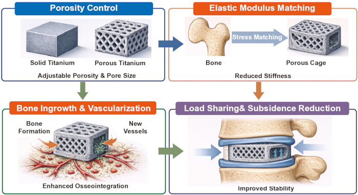
Figure 1: Design principles of 3D-printed porous titanium interbody cages. Controlled porosity and lattice architecture enable adjustment of the elastic modulus toward that of cancellous bone, facilitating more physiological load transfer and potentially reducing stress shielding. The interconnected porous network may support bone ingrowth and vascularization, contributing to improved osseointegration and long-term implant stability while potentially lowering the risk of cage subsidence. Please click here to view a larger version of this figure.
| Title | Study | Year | Study type | Model | Key findings | |||
| Guyer et al. (48) | 2016 | Biomechanical animal study | Porcine model | Porous titanium scaffold vs PEEK vs allograft | Porous titanium scaffold showed significantly stronger bone–implant interface and better osseointegration. | |||
| McGilvray et al. (49) | 2018 | Comparative animal study | Ovine lumbar fusion model (sheep) | PEEK cage; Plasma-sprayed porous titanium coated PEEK (PSP); 3D-printed porous titanium alloy cage (PTA) | 3D-printed porous titanium alloy cages showed significantly greater bone ingrowth, reduced range of motion, and increased construct stiffness compared with PEEK and PSP cages, indicating superior osseointegration and fusion potential. | |||
| Carpenter et al. (50) | 2018 | Finite element analysis | Computational FE model based on micro-CT implant geometry | Porous PEEK vs. porous 3D-printed titanium (Ti) implant structures | Porous PEEK significantly increased load sharing with ingrown bone compared with porous titanium (e.g., ~66% vs ~13% load transfer at 4 weeks under compression) and generated higher physiological strains in adjacent bone, suggesting a more favorable mechanical environment for bone maintenance and osseointegration. | |||
| Zhang et al. (51) | 2020 | Controlled animal experiment | Rhesus macaque lumbar interbody fusion model (OLIF) | 3D-printed porous Ti6Al4V cage; cages filled with simvastatin/poloxamer 407 hydrogel vs hydrogel only vs blank cage | Simvastatin-loaded hydrogel in porous titanium cages significantly increased bone ingrowth, osteoblastic activity (PET/CT SUV), bone volume fraction, and reduced ROM, indicating improved spinal fusion and mechanical stability. | |||
| Yee-Yanagishita et al. (52) | 2022 | In vitro biomechanical mechanical testing | Polyurethane foam block model simulating vertebral endplates | Porous titanium cage (body lattice + microporous endplates); porous PEEK cage; truss titanium cage | Porous titanium cage showed the best subsidence resistance with highest block stiffness and lowest dynamic subsidence displacement compared with porous PEEK and truss titanium cages, indicating that cage design plays a greater role than material alone in preventing subsidence. | |||
| Fogel et al. (26) | 2022 | Comparative preclinical study (in vitro mechanical testing + in vivo animal study) | Polyurethane foam block subsidence model (ASTM F2267) and ovine lumbar interbody fusion model | 3D-printed porous Ti-6Al-4V cage with stress-optimized body lattice and microporous endplates (Modulus XLIF) | Porous titanium cage design reduced implant stiffness, demonstrated high subsidence resistance (block stiffness ≈1976.8 N/mm), and showed progressive bone ingrowth and fusion in an ovine model with fusion rates of 87.5% at 12 weeks and 100% at 26 weeks. | |||
| Xu et al. (53) | 2022 | In vitro + in vivo animal experiment | MC3T3-E1 osteoblast culture and rabbit femoral defect model | 3D-printed porous Ti6Al4V scaffolds with hollow hexagonal prism vs hollow triangular prism pore structures | Hexagonal prism porous titanium scaffold showed greater cell adhesion, higher ALP activity, and better bone integration and new bone formation compared with triangular prism structure. | |||
| Johnson et al. (54) | 2023 | Preclinical animal study with biomechanical, μCT, and histomorphometry analyses | Ovine lumbar interbody fusion model (6 sheep; L2–L3 and L4–L5) | Additively manufactured porous Ti6Al4V lattice cages: body lattice vs body + surface lattice design | Both 3D-printed porous titanium cage designs achieved positive fusion outcomes; body lattice cages showed significantly higher bone-in-contact than surface lattice cages, and overall fusion performance was superior to previously reported PEEK cages. | |||
Table 1: Summary of representative biomechanical and biological studies on 3D-printed porous titanium cages used in lumbar interbody fusion. This table summarizes representative experimental studies investigating the biomechanical and biological performance of porous or 3D-printed titanium interbody cages used in lumbar fusion research. The included studies encompass finite element analyses, in vitro mechanical testing, cellular experiments, and preclinical animal fusion models. Reported outcomes include implant stiffness, subsidence resistance, bone–implant contact, bone ingrowth, and fusion-related biomechanical stability. Collectively, these studies provide mechanistic evidence supporting the potential advantages of porous titanium structures in optimizing load distribution, reducing stress shielding, and promoting osseointegration. Please click here to download this Table.
| Study | Year | Approach | Study design | Sample size | Follow-up | Radiographic outcome | Comparator |
| Adl Amini D. et al (62) | 2021 | LLIF | Retrospective comparative | 113 patients (186 levels) | 6–12 mo | Severe subsidence: 3.0% (Ti) vs 18.5% (PEEK) | 3D-printed Ti vs PEEK |
| Corso KA. et al (63) | 2022 | ALIF / PLIF | Retrospective descriptive cohort | 93 patients per group | 6 mo | Revision: 0% (Ti) vs 0.5% (PEEK) | 3D-printed Ti vs PEEK |
| Alan N. et al (64) | 2023 | LLIF | Retrospective cohort with propensity matching | 97 vs 97 levels | 12 mo | Subsidence: 27% vs 8% | pTi vs PEEK |
| Segi N. et al (65) | 2023 | XLIF | Retrospective comparative study | 32 patients | 3 mo | VEC: 8% vs 45% | 3D-printed Ti vs PEEK |
| Donaldson C. et al (66) | 2024 | ALIF / LLIF | Retrospective cohort | 50 patients (66 levels) | 12 mo | Subsidence 3.03% | None (single-arm) |
Table 2: Key clinical studies evaluating the safety and effectiveness of 3D-printed porous titanium cages in lumbar interbody fusion. This table summarizes representative clinical studies investigating the radiographic and clinical outcomes associated with porous or 3D-printed titanium interbody cages in lumbar interbody fusion procedures, including lateral lumbar interbody fusion (LLIF), anterior lumbar interbody fusion (ALIF), posterior lumbar interbody fusion (PLIF), and extreme lateral interbody fusion (XLIF). The table reports study design, sample size, follow-up duration, and major radiographic outcomes such as cage subsidence, vertebral endplate cyst formation, and revision rates. Both comparative studies (porous titanium vs PEEK cages) and single-arm clinical series are included to provide an overview of current clinical evidence regarding implant stability and short-term safety. Please click here to download this Table.
| No | Author | Year | Review Type | Focus of Review | Biomechanics | Clinical outcomes | Design / manufacturing | Conceptual framework | ||
| 1 | Duan Y (78) | 2024 | Systematic review & meta-analysis | Clinical comparison of 3D-printed Ti vs PEEK cages | No | Yes | Limited | No | ||
| 2 | Pairojboriboon S (79) | 2024 | Network meta-analysis | Fusion outcomes across different cage materials | No | Yes | Limited | No | ||
| 3 | Lee JJ (80) | 2025 | Narrative review | Historical evolution of titanium cages and 3D-printing technology | Limited | Limited | Yes | No | ||
| 4 | This review | / | Narrative integrative review | Mechanistic synthesis of design principles, biomechanical performance, osseointegration, and clinical outcomes of 3D-printed porous titanium cages | Yes | Yes | Yes | Yes | ||
Table 3: Comparison of recent reviews on 3D-printed titanium interbody cages and the scope of the present review. This table compares representative recent reviews on 3D-printed titanium interbody cages with the present review in terms of review type, primary research focus, and thematic coverage. Key aspects evaluated include whether the reviews addressed biomechanical mechanisms, clinical outcomes, implant design, and additive manufacturing principles, and broader conceptual frameworks integrating engineering and clinical perspectives. This comparison highlights the expanded scope of the present review, which integrates biomechanical, biological, design, and clinical evidence to provide a more comprehensive conceptual synthesis. Please click here to download this Table.
Disclosures
Loading...
$$\rightleftharpoonup{xx}$$
$$\longleftharp{xx}$$,
$$\longrightharp{xx}$$,
All authors declare that they have no conflicts of interest related to the design, analysis, or publication of this work. No financial relationships, advisory roles, patent applications, or other competing interests are associated with this manuscript. Conceptualization: Peng Sun and Kai Su; Literature review: Kai Su; Writing—original draft: Kai Su; Writing—review and editing: Peng Sun; Supervision: Peng Sun.
Acknowledgements
Loading...
$$\rightleftharpoonup{xx}$$
$$\longleftharp{xx}$$,
$$\longrightharp{xx}$$,
The authors extend their sincere appreciation to the colleagues who provided support in literature retrieval, data organization, and academic discussions. We also thank our institution for offering a supportive research environment and administrative assistance, which enabled the smooth completion of this work. Generative artificial intelligence was used for language editing and polishing of the manuscript. The authors take full responsibility for the content and interpretation of the work.
References
Loading...
$$\rightleftharpoonup{xx}$$
$$\longleftharp{xx}$$,
$$\longrightharp{xx}$$,
- Kim, H. S., Wu, P. H., Jang, I. T. Lumbar degenerative disease part 1: anatomy and pathophysiology of intervertebral discogenic pain and radiofrequency ablation of basivertebral and sinuvertebral nerve treatment for chronic discogenic back pain. Int J Mol Sci. 21 (4), (2020).
- Fakhoury, J., Dowling, T. J. Lumbar spinal stenosis. , StatPearls Publishing. Treasure Island, FL. (2025).
- Young, J. J., et al. Prevalence of multimorbid degenerative lumbar spinal stenosis with knee and/or hip osteoarthritis: Protocol for a systematic review and meta-analysis. Syst Rev. 9 (1), 232(2020).
- Fortunati, M., Rossi-Mossuti, F., Muroi, C. Degenerative lumbar spinal disorders and their treatment options. Praxis Bern. 109 (2), 87-95 (2020).
- Mehren, C., Ostendorff, N., Schmeiser, G., Papavero, L., Kothe, R. Do TLIF and PLIF techniques differ in perioperative complications? Comparison of complication rates of two high-volume centers. Global Spine J. 15 (1), 84-93 (2025).
- Yunga Tigre, J., et al. Risk factors for vascular injuries in anterior lumbar interbody fusion: a single-institution retrospective study. J Neurosurg Spine. 41 (1), 17-23 (2024).
- Shi, J., et al. Meta-analysis of the efficacy and safety of OLIF and TLIF in the treatment of degenerative lumbar spondylolisthesis. J Orthop Surg Res. 19 (1), 242(2024).
- Ogata, Y., et al. The optimal patient position on the surgical table for lateral lumbar interbody fusion in adult spinal deformity using three-dimensional computed tomography. Spine Surg Relat Res. 8 (2), 180-187 (2024).
- Segi, N., et al. Trabecular bone remodeling after posterior lumbar interbody fusion: comparison of osseointegration in porous titanium cages and polyether-ether-ketone cages. Global Spine J. 15 (1), 66-75 (2025).
- You, K. H., et al. Effect of cage material and size on fusion rate and subsidence following biportal endoscopic transforaminal lumbar interbody fusion. Neurospine. 21 (3), 973-983 (2024).
- Talukdar, R. G., Dhara, S., Gupta, S. Bone ingrowth in randomly distributed porous interbody cage during lumbar spinal fusion. Med Eng Phys. 133, 104248(2024).
- Liu, J., Xue, K., Li, S., Zhang, Y., Cheng, J. Whole-lesion histogram analysis of T1- and T2-weighted imaging for differentiating adrenal adenoma and pheochromocytoma using support vector machine. Can Assoc Radiol J. 72 (3), 452-459 (2021).
- Liang, Y., et al. Effects of different orientations of cage implantation on lumbar interbody fusion. World Neurosurg. 140, e97-e104 (2020).
- Mokawem, M., Katzouraki, G., Harman, C. L., Lee, R. Lumbar interbody fusion rates with 3D-printed lamellar titanium cages using calcium phosphate bone graft. J Clin Neurosci. 68, 134-139 (2019).
- Tan, J. H., Cheong, C. K., Hey, H. W. D. Titanium cages may be superior to PEEK cages in lumbar interbody fusion: systematic review and meta-analysis. Eur Spine J. 30 (5), 1285-1295 (2021).
- Ragni, E., et al. Superior osteo-inductive and osteo-conductive properties of trabecular titanium versus PEEK scaffolds on human mesenchymal stem cells. Int J Mol Sci. 22 (5), 2379(2021).
- Calek, A. K., Hochreiter, B., Buckland, A. J. 3D-printed titanium cages for anterior and lateral lumbar interbody fusion result in excellent fusion rates one year after surgery. Global Spine J. 16 (1), 341-348 (2025).
- Weinberg, J. H., et al. Lumbar fusion with micro- and nano-textured porous titanium versus PEEK interbody cages in TLIF: Randomized controlled trial. Global Spine J. 16 (1), 434-445 (2025).
- Liu, S. X., et al. 3D-printed porous titanium versus PEEK cages in lateral lumbar interbody fusion: systematic review and meta-analysis. Front Med Lausanne. 11, 1389533(2024).
- Wang, H., Zhang, H., Xiao, C., Zhang, K., Qi, L. Clinical and radiological outcomes of titanium cage versus PEEK cage in lumbar interbody fusion: Systematic review and meta-analysis. Neurosurg Rev. 48 (1), 295(2025).
- Smit, T., Aage, N., Haschtmann, D., Ferguson, S. J., Helgason, B. Patient-specific spinal fusion cages designed by full-scale topology optimization. J Mech Behav Biomed Mater. 159, 106695(2024).
- Pan, C. T., et al. Design of customized interbody fusion cages of Ti64ELI with gradient porosity by selective laser melting process. Micromachines Basel. 12 (3), 307(2021).
- Huang, S. F., et al. Biomechanical evaluation of an osteoporotic anatomical 3D printed posterior lumbar interbody fusion cage with lattice design. Int J Bioprint. 9 (3), 697(2023).
- Ho, N. C., et al. Evaluation of topology optimization using 3D printing for bioresorbable fusion cages: a biomechanical study in a porcine model. Spine Phila Pa 1976. 48 (4), E46-E53 (2023).
- Mushtaq, R. T., et al. Optimization of 3D-printed bio-based super tough PLA scaffolds for cancellous bone regeneration. Int J Biol Macromol. 316 (1), 144466(2025).
- Fogel, G., et al. Subsidence and fusion performance of a 3D-printed porous interbody cage with stress-optimized lattice. Spine J. 22 (6), 1028-1037 (2022).
- Liu, B., et al. Structural design and finite element simulation analysis of graded porous titanium implant. Int J Mol Sci. 23 (17), 10090(2022).
- Lei, H., et al. Structural optimization of 3D-printed porous titanium implants promotes bone regeneration for enhanced biological fixation. ACS Appl Mater Interfaces. 17 (12), 18059-18073 (2025).
- Sadighi, A., et al. Prediction of bone ingrowth into porous swelling bone anchors using an osteoconnectivity-based adaptive finite element algorithm. Med Biol Eng Comput. 63, 2923-2942 (2025).
- Idaszek, J., et al. Mimicking nature: Effect of architectural design inspired by cancellous bone on the biological response of hMSC cultured on titanium scaffolds fabricated by laser beam powder bed fusion. J Biomed Mater Res A. 113 (1), e37860(2025).
- Guo, W., et al. Multiscale hierarchical micro- and nano-surface induced by high-repetition-rate femtosecond laser promote peri-implant osseointegration. ACS Appl Bio Mater. 8 (2), 1621-1634 (2025).
- Tambun, D. V., et al. Titanium surface roughness mediated macrophage polarization influences osteogenic differentiation of periodontal ligament-derived mesenchymal stromal cells. Eur J Dent. 20 (1), 105-113 (2025).
- Chu, S., et al. Hierarchical interconnected porous scaffolds with regulated interfacial nanotopography exhibit antimicrobial, alleviate inflammation, promote neovascularization, and tissue integration for bone regeneration. Biomaterials. 318, 123186(2025).
- Zhang, T. M., et al. Effects of 3D-printed porous Ti-6Al-4V scaffold pore structure and micro-nano surface topography on the repair of maxillofacial bone defects. Materials & Design. 257, 114376(2025).
- Shu, T., et al. Microscale bone interlocking enhances osseointegration strength on the rough surface of 3D-printed titanium implants: Experimental and finite element analysis. BMC Oral Health. 25 (1), 208(2025).
- Deering, J., et al. Osseointegration of functionally graded Ti6Al4V porous implants: Histology of the pore network. Biomater Adv. 155, 213697(2023).
- Wu, M. H., et al. In vitro and in vivo comparison of bone growth characteristics in additive-manufactured porous titanium, nonporous titanium, and porous tantalum interbody cages. Materials Basel. 15 (10), 3670(2022).
- Wang, X., Nie, Z., Chang, J., Lu, M. L., Kang, Y. Multiple channels with interconnected pores in a bioceramic scaffold promote bone tissue formation. Sci Rep. 11 (1), 20447(2021).
- Greer, A. I. M., et al. Nanopatterned titanium implants accelerate bone formation in vivo. ACS Appl Mater Interfaces. 12 (30), 33541-33549 (2020).
- Farber, S. H., et al. Computed tomography assessment of long-term fusion and subsidence for anterior lumbar interbody fusion performed at the lumbosacral junction. Clin Spine Surg. 39 (1), E38-E44 (2025).
- Levy, H. A., et al. Do three-dimensional printed porous titanium relative to polyetheretherketone interbody cages reduce complications and revisions after transforaminal lumbar interbody fusion. Global Spine J. 16 (1), 185-195 (2025).
- Zhang, C., Wang, Y. Biomechanical analysis of axial gradient porous dental implants: a finite element analysis. J Funct Biomater. 14 (12), 557(2023).
- Chowdhury, S., Anand, A., Singh, A., Pal, B. Evaluation of mechanical properties of Ti-25Nb BCC porous cell structures and their association with porosity: A finite element and analytical study. Proc Inst Mech Eng H. 235 (7), 827-837 (2021).
- Zhang, Z., et al. Biomechanical analysis of porous additive manufactured cages for lateral lumbar interbody fusion: a finite element analysis. World Neurosurg. 111, e581-e591 (2018).
- Zou, D., et al. Biomechanical analysis of lumbar interbody fusion cages with various elastic moduli in osteoporotic and non-osteoporotic lumbar spine. Global Spine J. 14 (7), 2053-2061 (2024).
- Zhu, C., Deng, K., Shao, Z., Wang, Y. Biomechanical optimization of lumbar fusion cages with a porous design: a finite element analysis. Appl Sci. 15 (10), 5384(2025).
- Zhang, H., et al. Biomaterials for interbody fusion in bone tissue engineering. Front Bioeng Biotechnol. 10, 900992(2022).
- Guyer, R. D., Abitbol, J. J., Ohnmeiss, D. D., Yao, C. Evaluating osseointegration into a deeply porous titanium scaffold: a biomechanical comparison with PEEK and allograft. Spine Phila Pa 1976. 41 (19), E1146-E1150 (2016).
- Mcgilvray, K. C., et al. Bony ingrowth potential of 3D-printed porous titanium alloy: A direct comparison of interbody cage materials in an ovine lumbar fusion model. Spine J. 18 (7), 1250-1260 (2018).
- Carpenter, R. D., et al. Effect of porous orthopaedic implant material and structure on load sharing with simulated bone ingrowth: A finite element analysis comparing titanium and PEEK. J Mech Behav Biomed Mater. 80, 68-76 (2018).
- Zhang, W., et al. 3D-printed porous titanium cages filled with simvastatin hydrogel promote bone ingrowth and spinal fusion in rhesus macaques. Biomater Sci. 8 (15), 4147-4156 (2020).
- Yee-Yanagishita, C., et al. Biomechanical comparison of subsidence performance among three modern porous lateral cage designs. Clin Biomech Bristol. 99, 105764(2022).
- Xu, Z., et al. In vitro and in vivo analysis of the effects of 3D-printed porous titanium alloy scaffold structure on osteogenic activity. Biomed Res Int. 2022, 8494431(2022).
- Johnson, J. W., et al. Novel 3D printed lattice structure titanium cages evaluated in an ovine model of interbody fusion. JOR Spine. 6 (3), e1268(2023).
- Laratta, J. L., et al. 3D-printed titanium cages without bone graft outperform PEEK cages with autograft in an animal model. Spine J. 22 (6), 1016-1027 (2022).
- Walsh, W. R., et al. Preclinical evaluation of lateral interbody fusions using 3D printed PEEK or 3D printed titanium cages. N Am Spine Soc J. 23, 100756(2025).
- Loenen, A. C. Y., et al. Early bone ingrowth and segmental stability of a trussed titanium cage versus a polyether ether ketone cage in an ovine lumbar interbody fusion model. Spine J. 22 (1), 174-182 (2022).
- Wang, C., et al. Improving osteoinduction and osteogenesis of Ti6Al4V alloy porous scaffold by regulating the pore structure. Front Chem. 11, 1190630(2023).
- Yu, M., et al. 3D printed Ti-6Al-4V implant with a micro/nanostructured surface and its cellular responses. ACS Omega. 5 (49), 31738-31743 (2020).
- Papaefstathiou, S., et al. Three-dimensional printing of biomimetic titanium mimicking trabecular bone induces human mesenchymal stem cell proliferation: An in vitro analysis. Spine Phila Pa 1976. 47 (14), 1027-1035 (2022).
- Taylor, B. L., et al. Three-dimensional porous trabecular scaffold exhibits osteoconductive behaviors in vitro. Regen Eng Transl Med. 6 (3), 241-250 (2020).
- Adl Amini, D., et al. Evaluation of cage subsidence in standalone lateral lumbar interbody fusion: novel 3D-printed titanium versus polyetheretherketone cage. Eur Spine J. 30 (8), 2377-2384 (2021).
- Corso, K. A., et al. Early revision events among patients with a three-dimensional printed cellular titanium or PEEK spinal cage for single-level lumbar spinal fusion. Expert Rev Med Devices. 19 (2), 195-201 (2022).
- Alan, N., et al. Graft subsidence and reoperation after lateral lumbar interbody fusion: A propensity score–matched cost analysis of PEEK versus 3D-printed porous titanium interbodies. J Neurosurg Spine. 39 (2), 187-195 (2023).
- Segi, N., et al. Vertebral endplate concavity in lateral lumbar interbody fusion: Tapered 3D-printed porous titanium cage versus squared PEEK cage. Medicina Kaunas. 59 (2), 372(2023).
- Donaldson, C., Santro, T., Awad, M., Morokoff, A. 3D-printed titanium alloy cage in anterior and lateral lumbar interbody fusion for degenerative lumbar spine disease. J Spine Surg. 10 (1), 22-29 (2024).
- Patel, N. A., et al. Three-dimensional printed titanium versus polyetheretherketone cages for lumbar interbody fusion: Systematic review of comparative in vitro, animal, and human studies. Neurospine. 20 (2), 451-463 (2023).
- Deng, Z., et al. Comparison between three-dimensional printed titanium and PEEK cages for cervical and lumbar interbody fusion: A prospective controlled trial. Orthop Surg. 15 (11), 2889-2900 (2023).
- Velluto, C., et al. Radiological evaluation of fusion patterns after lateral lumbar interbody fusion with 3D-printed porous titanium cages versus conventional titanium cages. Front Surg. 11, 1446792(2024).
- Parajón, A., et al. Minimally invasive transforaminal lumbar interbody fusion: Meta-analysis of fusion rates and optimal graft material. Neurosurgery. 81 (6), 958-971 (2017).
- Chen, C., et al. Self-assembled nanocomposite hydrogels as carriers for demineralized bone matrix particles and enhanced bone repair. Adv Healthc Mater. 13 (10), e2303592(2024).
- Chang, C. C., et al. Long-term radiographic outcomes of expandable versus static cages in transforaminal lumbar interbody fusion. J Neurosurg Spine. 34 (3), 471-480 (2021).
- Alan, N., et al. Subsidence after lateral lumbar interbody fusion using a 3D-printed porous titanium interbody cage: Single-institution case series. J Neurosurg Spine. 37 (5), 663-669 (2022).
- Tao, X., et al. TLIF is associated with lower rates of adjacent segment disease and complications compared to ALIF: A matched cohort analysis. Spine Phila Pa 1976. 48 (19), 1335-1341 (2023).
- Corso, K. A., et al. Evaluation of healthcare outcomes of patients treated with 3D-printed titanium and PEEK cages during lumbar fusion procedures. Med Devices Auckl. 18, 37-51 (2025).
- Seex, K. A., et al. Clinical outcomes of 3D-printed titanium patient-specific implants in lumbar interbody fusion: A prospective clinical trial with systematic review. J Pers Med. 15 (7), 320(2025).
- Turlip, R. W., et al. Evaluating the efficacy of a novel titanium cage system in ALIF and LLIF: a retrospective clinical and radiographic analysis. J Clin Med. 14 (16), (2025).
- Duan, Y., et al. Comparison of lumbar interbody fusion with 3D-printed porous titanium cage versus PEEK cage in lumbar degenerative disease: systematic review and meta-analysis. World Neurosurg. 183, 144-156 (2024).
- Pairojboriboon, S., et al. Comparison of TLIF cage material on fusion rates: Systematic review and network meta-analysis. World Neurosurg X. 23, 100392(2024).
- Lee, J. J., Jacome, F. P., Hiltzik, D. M., Pagadala, M. S., Hsu, W. K. Evolution of titanium interbody cages and current uses of 3D-printed titanium in spine fusion surgery. Curr Rev Musculoskelet Med. 18 (12), 635-644 (2025).
- Liu, D., et al. Radiographic and clinical comparison of polyetheretherketone versus 3D-printed titanium cages in lumbar interbody fusion. J Clin Med. 14 (6), 1813(2025).
Reprints and Permissions
Request permission to reuse the text or figures of this JoVE article
Request Permission