Laparoscopic retromuscular mesh repair (LRMR) technique offers a clear view of the abdominal wall anatomy and can be effectively combined with transversus abdominis release (TAR), allowing for tension-free repair and placement of large meshes extending beyond the semilunar lines, particularly in patients with multiple abdominal wall hernias.
Method Article
Laparoscopic Retromuscular Mesh Repair for the Management of Multiple Abdominal Wall Hernias
Opens in a new tab
In This Article
Summary
Loading...
$$\rightleftharpoonup{xx}$$
$$\longleftharp{xx}$$,
$$\longrightharp{xx}$$,
Abstract
Loading...
$$\rightleftharpoonup{xx}$$
$$\longleftharp{xx}$$,
$$\longrightharp{xx}$$,
Laparoscopic retromuscular mesh repair (LRMR) is a minimally invasive and effective approach for treating multiple abdominal wall hernias. The goal of this protocol is to provide a reproducible surgical technique for the management of multiple abdominal wall hernias in the era of minimally invasive surgery. Here, we present a protocol to describe laparoscopic retromuscular mesh repair for the management of multiple abdominal wall hernias, enabling tension-free reconstruction and adequate mesh placement through a minimally invasive approach. The key procedural steps include: (1) creation of the retro-rectus space; (2) expansion of the retro-rectus space with exposure of the hernia defects; (3) crossing the midline to develop the contralateral retro-rectus space; (4) transversus abdominis release to further expand the retromuscular plane when necessary; (5) closure of the hernia defects with reconstruction of the posterior rectus sheath; and (6) retromuscular mesh placement with adequate overlap to ensure durable repair. This technique offers a practical and efficient alternative for the repair of multiple incisional hernias and facilitates tension-free anatomical reconstruction. In a representative case involving multiple abdominal wall hernias, the procedure was completed successfully without complications and was associated with favorable postoperative recovery, demonstrating the versatility and feasibility of the approach and highlighting its potential applicability in complex clinical scenarios.
Introduction
Loading...
$$\rightleftharpoonup{xx}$$
$$\longleftharp{xx}$$,
$$\longrightharp{xx}$$,
Large population-based studies have reported that primary ventral hernias occur in approximately 20% of adults, whereas incisional hernias—a postoperative subtype of ventral hernia—develop in up to 30% of midline abdominal incisions1. LRMR has been applied in clinical practice for the treatment of ventral hernias2,3. The key concept underlying this technique is to disrupt anatomical boundaries, connect the retromuscular planes, and create adequate space for mesh placement4. This approach preserves the principles of retromuscular reconstruction while avoiding the limitations associated with intraperitoneal mesh placement and extensive open dissection.
Current surgical approaches for multiple abdominal wall hernias include open preperitoneal repair (Stoppa), intraperitoneal onlay mesh (IPOM) technique, and anterior subcutaneous rectus sheath approach, each with specific limitations5,6,7. Open preperitoneal repair provides clear anatomical layers but is associated with higher rates of surgical site infection and longer recovery compared with minimally invasive approach8,9,10. Although minimally invasive, the IPOM technique requires intraperitoneal mesh placement, which is associated with concerns regarding postoperative adhesions, visceral complications, and the need for expensive coated meshes11,12,13. The anterior subcutaneous approach requires extensive dissection between the subcutaneous tissue and rectus sheath, increasing the risk of seroma formation and wound complications14. In contrast, LRMR allows retromuscular mesh placement, facilitates tension-free anatomical reconstruction, and avoids direct contact between the mesh and abdominal viscera.
In recent years, minimally invasive retromuscular approaches, including enhanced-view totally extraperitoneal (eTEP) repair, transabdominal retromuscular (TARM) repair, and TAR-based reconstruction techniques, have gained increasing attention in abdominal wall surgery4,15,16. LRMR shares the same anatomical principles of extraperitoneal dissection and retromuscular mesh placement while providing a laparoscopic alternative for complex abdominal wall reconstruction. This technique may be particularly suitable for patients with multiple abdominal wall hernias requiring wide retromuscular dissection, adequate mesh overlap, and tension-free anatomical reconstruction through a minimally invasive approach.
Currently, reports on the surgical management of patients with multiple abdominal wall hernias are limited; only one study has described the use of the IPOM technique for the treatment of a right inguinal hernia, a left Spigelian hernia, an umbilical hernia, and an obturator hernia17. In this article, we present a case of laparoscopic repair of multiple incisional hernias combined with a femoral hernia using a retromuscular mesh repair, with a focus on the technical workflow, anatomical considerations, and clinical applicability of the procedure. Based on this case and a review of the relevant literature, this article presents a reproducible LRMR technique for the management of multiple abdominal wall hernias.
Protocol
Loading...
$$\rightleftharpoonup{xx}$$
$$\longleftharp{xx}$$,
$$\longrightharp{xx}$$,
The protocol follows the guidelines of the Human Research Ethics Committee of Nanchong Central Hospital of North Sichuan Medical College (University). Written informed consent was obtained from the patient for the procedure and the use of data.
1. Preoperative preparation (Figure 1, Figure 2)
- Measure the size of the hernia defects by preoperative computed tomography (CT) in centimeters (cm). Also, record the quantity of hernia.
- Hold a detailed discussion with the patient and their family to obtain informed consent.
- Perform routine skin preparation, urinary catheterization, and establishment of intravenous access as required.
- Select an appropriately sized mesh to ensure adequate coverage of the hernia defects. In case the retromuscular space was not wide enough to accommodate a mesh, extend the dissection to the preperitoneal space with TAR, a posterior component separation technique (CST).
2. Creation of the retro-rectus space (Figure 3, Figure 4)
- Under general anesthesia, place the patient in the supine position.
- Make a 10-mm incision in the left subcostal (left upper quadrant) region. Carry out the dissection through the subcutaneous tissue to the anterior rectus sheath. Incise the anterior rectus sheath, and retract the rectus muscle laterally to expose the posterior rectus sheath.
- Enter the retro-rectus space bluntly, and insert a 10-mm trocar to establish the space at a pressure of 12 mmHg.
- Introduce a laparoscope, and expand the retro-rectus space using a pushing technique. Place two 5-mm trocars in the left abdominal wall to provide additional access to the retro-rectus space.
3. Dissection and expansion of the retro-rectus space and exposure of the hernia sac (Figure 5)
- Under direct vision, dissect the loose connective tissue in the retro-rectus space using blunt and sharp dissection, with electrocautery for hemostasis.
- Proceed with the dissection into the Retzius space, exposing the pubic symphysis and Cooper's ligament.
- Continue the dissection laterally along the inferior epigastric vessels to the Bogros space, reaching the anterior superior iliac spine and exposing the iliopubic tract.
- Identify the internal ring of the femoral hernia, and dissect the hernia sac carefully.
- Ligate the right round ligament of the uterus and divide it with clips, and fully dissect the lateral space.
4. Crossing the midline and expanding the contralateral space (Figure 4)
- Incise the posterior rectus sheath approximately 5 mm lateral to the linea alba. Incise the contralateral posterior rectal sheath to access the contralateral retro-rectus space.
- Place a 10-mm trocar and two 5-mm trocars on the right side, and repeat the dissection as described previously.
- Incise the posterior sheath around the internal ring, and extend the dissection cranially to the xiphoid process and caudally to the pubic symphysis, creating a wide bilateral retro-rectus space.
5. Transversus abdominis release (TAR) (Figure 5)
NOTE: TAR was necessary to achieve sufficient lateral dissection, ensure proper mesh placement with adequate coverage, and restore abdominal wall integrity without excessive tension.
- Using sharp and blunt dissection with electrocautery, identify the transversus abdominis muscle and incise it longitudinally approximately 0.5–1 cm medial to the linea semilunaris, allowing entry into the lateral retromuscular plane.
- Carefully identify and preserve the segmental neurovascular bundles. Gently sweep these neurovascular bundles posteriorly along with the muscle.
- Continue dissection laterally in the plane between the transversus abdominis muscle and the underlying transversalis fascia/peritoneum, creating sufficient space for mesh placement with a minimum overlap of 5 cm beyond the defect edges bilaterally.
- Repeat the procedure on the contralateral side to create a symmetrical, extensive retromuscular space incorporating both the retro-rectus and transversus abdominis release planes for mesh placement.
6. Closure of the hernia defect and reconstruction of the posterior rectus sheath (Figure 6, Figure 7)
- Reconstruct the linea alba, and close the hernia defects using interrupted 0 nonabsorbable sutures.
NOTE: Continuous suturing can also be performed. - Reconstruct the posterior rectus sheath with a continuous 2-0 nonabsorbable suture to restore anatomical integrity.
7. Mesh placement (Figure 8)
- Place two 20 cm × 15 cm titanized meshes (weight: light 35 g/m2) to cover the incisional hernia defects and one 10.8 cm × 16 cm knitted polypropylene pre-formed mesh (a standard-weight polypropylene 3D anatomical mesh was used) for the right femoral hernia through the 10-mm port.
- Ensure that each mesh extends at least 5 cm beyond the edges of the respective hernia defects to achieve adequate overlap.
NOTE: One big mesh can also be chosen for incisional hernias. - Position the meshes flat within the retromuscular space, with edges extending at least 5 cm beyond the hernia defects.
8. Mesh fixation and wound closure (Figure 9)
- Secure the meshes using interrupted nonabsorbable sutures, with approximately 4–6 fixation points per mesh, depending on the size and location of the defect.
- For incisional hernia repair, anchor the meshes to the posterior rectus sheath and adjacent fascial structures within the retromuscular space to ensure stable positioning.
- For the femoral hernia repair, secure the mesh medially to Cooper's ligament, taking care to avoid injury to neurovascular structures.
- Achieve additional stabilization through intra-abdominal pressure and tissue apposition within the retromuscular plane.
- Place a negative-pressure drainage tube.
- Release the extraperitoneal insufflation, and close all trocar sites in layers.
9. Postoperative management and follow-up (Table 1)
- Closely monitor the drainage volume, color, and quality. Remove the negative-pressure drainage tube on postoperative day 5.
- Monitor patients for postoperative complications, including bleeding, infection, and chronic pain.
- Conduct postoperative follow-up to assess recovery and long-term outcomes.
Results
Loading...
$$\rightleftharpoonup{xx}$$
$$\longleftharp{xx}$$,
$$\longrightharp{xx}$$,
In the representative case, preoperative CT and intraoperative assessment identified the following hernia defects: the midline epigastric incisional hernia (3.5 cm × 4.0 cm), midline hypogastric incisional hernia (2.5 cm × 3.0 cm), drain-site incisional hernia (1.5 cm × 1.5 cm), and right femoral hernia (internal ring diameter 2.0 cm) (Figure 1). The operative time was 320 min, with an estimated blood loss of 40 mL. No postoperative complications occurred. The patient resumed a fluid diet 6 h after surgery and was discharged on postoperative day 5, following an uneventful hospital stay. At 3 months postoperatively, the pain score was 0. During a 2-year follow-up, the patient remained free of hernia recurrence and chronic pain.
Preoperative planning was guided by precise abdominal skin markings, delineating the locations of the midline epigastric and hypogastric incisional hernias, the drain site hernia, and the right femoral hernia (Figure 2). Strategic port placement enabled optimal access to the retro-rectus space bilaterally, with a 10-mm port in the left subcostal region and two 5-mm ports on each side of the abdomen, facilitating instrument triangulation and effective dissection (Figure 3). Establishment of the retro-rectus space was achieved systematically, providing clear visualization of the posterior rectus sheath and surrounding planes (Figure 4). Lateral dissection extended fully to the transversus abdominis muscle, which was incised to allow adequate medial mobilization of the rectus complex (Figure 5). The linea alba was reconstructed using transfacial sutures placed via percutaneous needle passage with extracorporeal knot tying using 0 nonabsorbable sutures, and the hernia ring was closed (Figure 6). Subsequent reconstruction of the posterior rectus sheath using continuous 2-0 nonabsorbable sutures restored the integrity of the abdominal wall and prepared the retromuscular plane for mesh placement (Figure 7). Three meshes were implanted to cover the defects, ensuring sufficient overlap and minimizing potential tension at the repair sites (Figure 8). Postoperative abdominal image indicating a negative-pressure drainage tube (red arrow) is shown in Figure 9. Table 1 summarizes the intra- and postoperative outcomes.
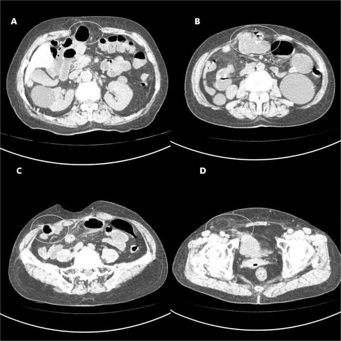
Figure 1: Preoperative CT. (A–D) The white circles in panels A–D indicate, respectively, a midline epigastric incisional hernia (3.5 cm × 4.0 cm), a midline hypogastric incisional hernia (2.5 cm × 3.0 cm), a drain-site incisional hernia (1.5 cm × 1.5 cm), and a right femoral hernia (internal ring diameter, 2.0 cm). Please click here to view a larger version of this figure.
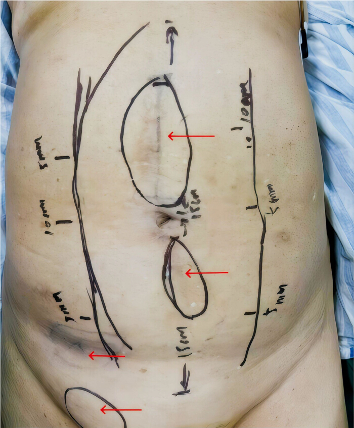
Figure 2: Schematic representation of preoperative abdominal skin markings. The red arrows indicate, from superior to inferior, a midline epigastric incisional hernia, a midline hypogastric incisional hernia, a drain site incisional hernia, and a right femoral hernia. Please click here to view a larger version of this figure.
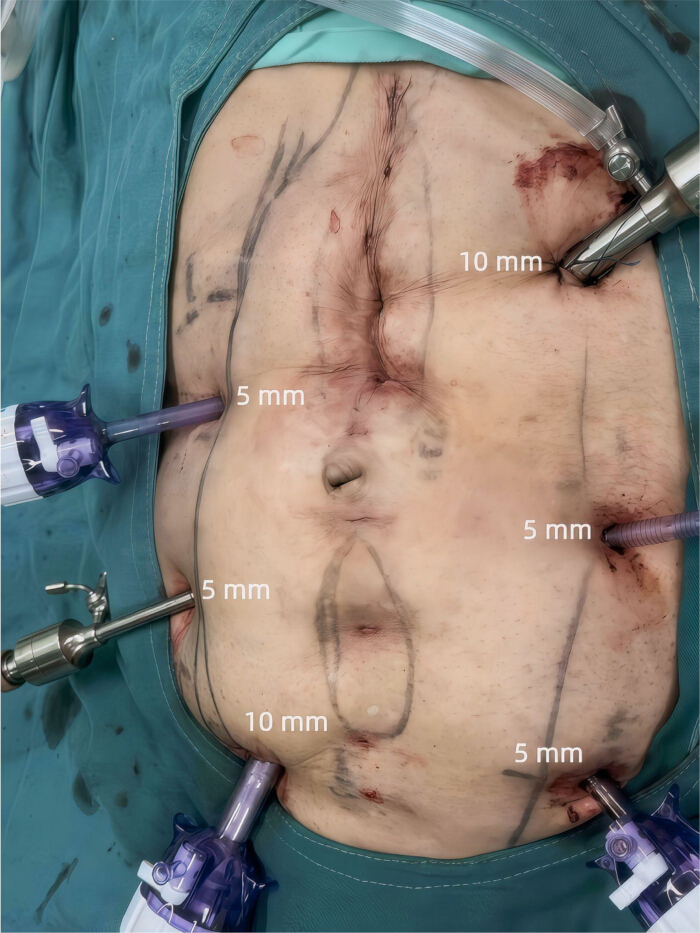
Figure 3: Port placement. A 10-mm port was placed in the left subcostal (left upper quadrant) region, and two 5-mm ports were placed in the left abdominal wall to provide additional access to the retro-rectus space. A 10-mm port and two 5-mm ports were placed on the right side. Please click here to view a larger version of this figure.
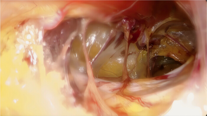
Figure 4: Establishment of the retro-rectus space. Please click here to view a larger version of this figure.
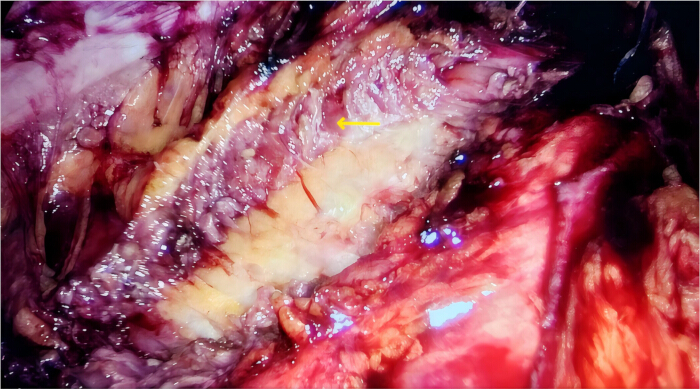
Figure 5: Dissection of the lateral space and incision of the transversus abdominis muscle. The yellow arrow indicates the transversus abdominis muscle. Please click here to view a larger version of this figure.
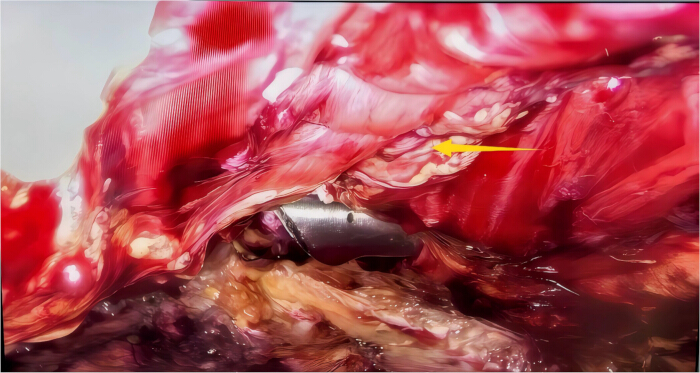
Figure 6: Reconstruction of the linea alba. The linea alba was reconstructed using transfacial sutures placed via percutaneous needle passage with extracorporeal knot tying using 0 nonabsorbable sutures, and the hernia ring was closed. The yellow arrow indicates the linea alba. Please click here to view a larger version of this figure.
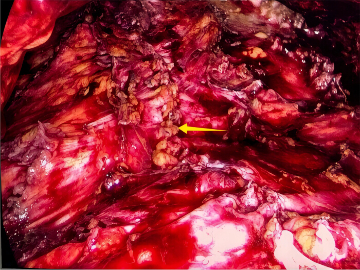
Figure 7: Reconstruction of the posterior rectus sheath. The posterior rectus sheath was reconstructed by continuous suture with 2-0 nonabsorbable suture. The yellow arrow indicates the reconstructed posterior rectus sheath. Please click here to view a larger version of this figure.
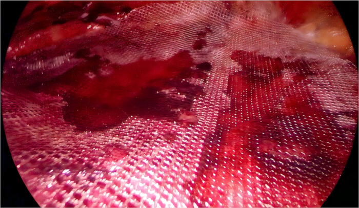
Figure 8: Implantation of the three meshes. Please click here to view a larger version of this figure.
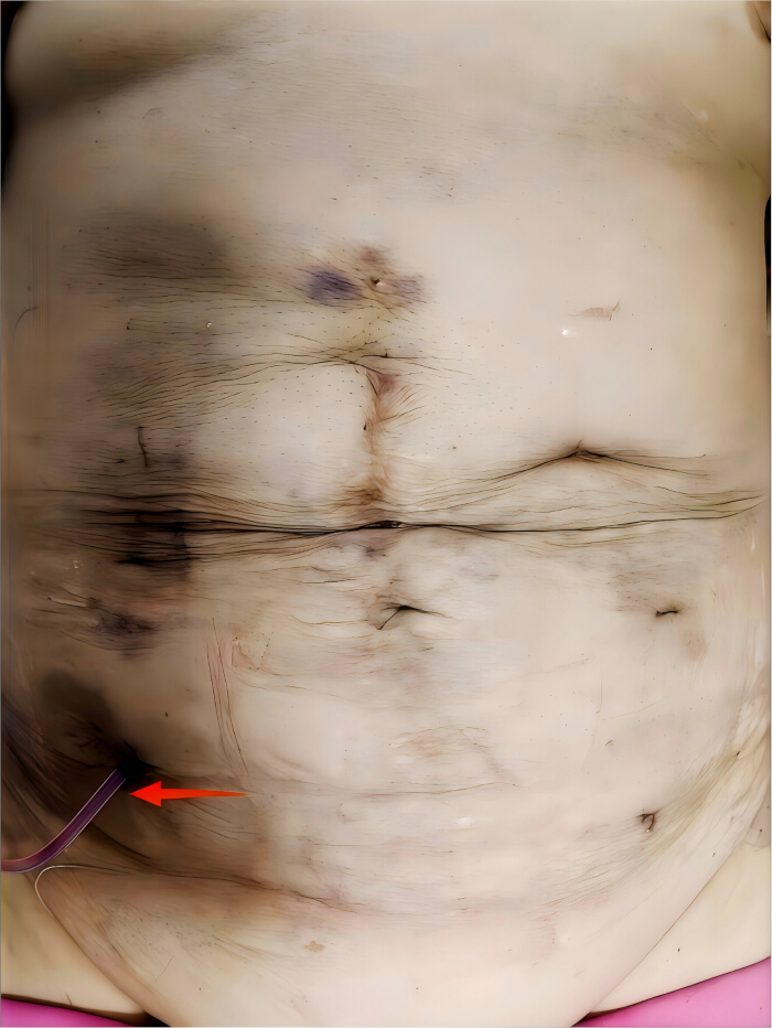
Figure 9: Postoperative abdominal image. The red arrow indicates a negative-pressure drainage tube. Please click here to view a larger version of this figure.
| Variables | Values |
| Operating time (min) | 320 |
| Estimated blood loss (mL) | 40 |
| Postoperative hospital stay (days) | 5 |
| Time to first passage of flatus(days) | 1 |
| Postoperative incision healing | Uninfected |
| VAS pain score at 3 months postoperatively | 0 |
| Telephonic followup | Uneventful |
Table 1: Summary of the intra- and postoperative outcomes.
Discussion
Loading...
$$\rightleftharpoonup{xx}$$
$$\longleftharp{xx}$$,
$$\longrightharp{xx}$$,
This study demonstrates the technical aspects of LRMR for the management of multiple incisional hernias combined with femoral hernia. The primary contribution of this protocol lies in illustrating a standardized, reproducible approach to creating and extending the retromuscular space, enabling adequate mesh coverage under direct endoscopic visualization.
Several technical considerations proved essential for the successful performance of the procedure. First, TAR was employed to expand the retromuscular space and enable tension-free midline approximation18. Second, pneumoperitoneum pressure was lowered to approximately 6–8 mmHg during closure of the linea alba and ventral hernia ring to facilitate tissue approximation and suturing. Third, restoration of the linea alba and closure of the hernia rings could be achieved using percutaneous 0 nonabsorbable sutures. Fourth, one big mesh or multiple meshes can be chosen for multiple ventral hernias, depending on the anatomical characteristics and intraoperative findings. When a single mesh could not provide adequate coverage, multiple meshes were used to ensure sufficient overlap, with mesh borders extending at least 5 cm beyond all defect margins19.
In the present study, fascial closure was technically challenging due to the presence of multiple incisional hernias of varying sizes and locations, compounded by restricted working angles. Under these circumstances, an interrupted suturing technique was preferentially employed, as it allows individualized tension adjustment at each suture point20,21. Continuous suturing, however, offers the advantage of more evenly distributing tension along the suture line and may reduce operative time22. Consequently, the choice between interrupted and continuous suturing should be tailored to the characteristics of the hernia defects, tissue quality, and intraoperative conditions. In the current case, the patient presented with two midline hernias and one lateral hypogastric hernia, resulting in a wide anatomical distribution of defects. Two meshes were utilized to ensure adequate and secure coverage of all defects; alternatively, the use of a single large mesh may be appropriate in selected cases, depending on the anatomical configuration and intraoperative findings.
Minimally invasive treatment of multiple abdominal wall hernias remains technically challenging, particularly when the defects are located at different anatomical levels23. Yang et al. previously described the laparoscopic treatment of four small midline incisional hernias using the intraperitoneal onlay mesh (IPOM) technique, demonstrating that multiple defects can be addressed during a single surgical procedure. The IPOM technique requires intraperitoneal mesh placement with direct contact between the mesh and abdominal viscera24. By establishing a continuous retromuscular space, LRMR provides convenient access to each defect without entering the peritoneal cavity, simplifies the management of multiple incisional hernias, and avoids visceral adhesions associated with intraperitoneal mesh placement25.
In recent years, robotic-assisted approaches have further expanded the application of retromuscular hernia repair. Meta-analyses comparing preperitoneal and retromuscular robotic techniques suggest that retromuscular repair may provide superior mesh positioning and anatomical reconstruction, while maintaining acceptable perioperative outcomes26. Emerging evidence also demonstrates that robotic-assisted transabdominal retromuscular (r-TARM) repair is associated with a definite learning curve, with progressive improvements in operative efficiency and safety27. Furthermore, new generation robotic platforms have been successfully applied in robotic-assisted retromuscular umbilical prosthetic hernia repair (r-TARUP)28. Comparative analyses between robot-assisted eTEP and other retromuscular approaches (e.g., TARM/TARUP) indicate that these techniques share common principles of extraperitoneal dissection and mesh placement, while differing in access strategy and technical complexity29. In addition, systematic reviews of robotic parastomal hernia repair highlight the importance of retromuscular plane development and TAR in achieving durable outcomes30. These findings support the growing role of minimally invasive retromuscular approaches and underscore the relevance of LRMR as a laparoscopic alternative that adheres to the same anatomical and reconstructive principles.
LRMR offers a minimally invasive approach that reproduces the fundamental principles of the Rives-Stoppa technique25,31. Dissection is performed entirely within the retromuscular plane, permitting mesh placement within the retromuscular space while avoiding entry into the peritoneal cavity32. In LRMR, coated mesh is not required, which reduces mesh-related costs, particularly in low-income populations13,25. This technique prevents direct contact between the mesh and the abdominal viscera, thereby reducing the risk of postoperative adhesions, bowel injury, and fistula formation13,25. Another major technical advantage of LRMR is its compatibility with TAR25,33. When midline closure of the posterior rectus sheath is limited by excessive tension or a restricted working space, TAR can be easily performed. This maneuver permits lateral extension, facilitating tension-free reconstruction of the abdominal wall18.
However, LRMR also has its limitations. Firstly, the success of this technique depends heavily on the surgeon's experience with laparoscopic retromuscular dissection and TAR. Inadequate technical proficiency may result in peritoneal breach, neurovascular injury, or insufficient lateral space for mesh placement34. Secondly, wide extraperitoneal dissection may increase the risk of postoperative seroma, necessitating careful postoperative monitoring34.
Several technical challenges may arise during LRMR. Limited retromuscular space can restrict adequate mesh placement and overlap, which may be addressed by extending the dissection or performing a TAR when necessary35. Fascial closure can also be challenging in cases with multiple or widely separated defects; gradual approximation using interrupted sutures, combined with reduced pneumoperitoneum pressure, can facilitate secure closure. Postoperative seroma formation or drainage-related complications may occur and can be effectively managed with negative-pressure drainage and careful postoperative monitoring36.
Future research should aim to validate the applicability and clinical outcomes of LRMR in larger and more diverse patient populations. Comparative studies with other minimally invasive techniques, including eTEP repair and robotic-assisted retromuscular approaches, may help delineate its advantages in terms of operative efficiency, postoperative recovery, and long-term recurrence rates32. Further investigation is also warranted to assess the utility of LRMR in more complex hernia presentations. Standardization of key technical parameters, such as indications for TAR, mesh configuration, and fixation strategies, may enhance procedural reproducibility and facilitate wider clinical adoption. Additionally, long-term studies evaluating functional outcomes, quality of life, and cost-effectiveness are essential to more comprehensively define the role of LRMR in abdominal wall reconstruction.
In conclusion, LRMR is a safe, minimally invasive, and anatomically rational approach for the treatment of multiple abdominal wall hernias. Further refinement and standardization of LRMR protocols may expand their applicability and improve long-term clinical outcomes.
Disclosures
Loading...
$$\rightleftharpoonup{xx}$$
$$\longleftharp{xx}$$,
$$\longrightharp{xx}$$,
The authors declare no competing interests.
Acknowledgements
Loading...
$$\rightleftharpoonup{xx}$$
$$\longleftharp{xx}$$,
$$\longrightharp{xx}$$,
This work was supported by the funding of Key Clinical Specialty in Sichuan Province [ZX-2428-1] and Wu Jieping Medical Foundation [320.6750.2024-07-3].
Materials
| Name | Company | Catalog Number | Comments |
|---|---|---|---|
| Disposable surgical drainage tube | Shandong Branden Medical Devices Co., Ltd. | Round Tube Type, Fr14 | |
| High-definition laparoscopic system | Karl Storz | TC201 | |
| Knitted polypropylene pre-formed mesh | C.R. Bard | 115311 | 10.8 cm × 16 cm |
| Laparoscopic forceps | Tonglu Youshi Medical Instrument Co., Ltd. | 101.052 | 5 mm × 330 mm |
| Laparoscopic high-flow insufflator | Karl Storz | UI400 | |
| Laparoscopic needle holding pliers | Tonglu Youshi Medical Instrument Co., Ltd. | 101.032A | 5 mm × 330 mm |
| Laparoscopic scissors | Tonglu Youshi Medical Instrument Co., Ltd. | 101.022 | 5 mm × 330 mm |
| Single-use trocar for laparoscopy | Surgaid | NGVM-100-1-5; NGVM-100-1-10 | |
| Surgical suture needles with thread | Ningbo Chenghe Microapparatus Factory | 1-0, 80 cm | |
| Surgical suture needles with thread | Shanghai Pudong Jinhuan Medical Products Co., Ltd. | 2-0, 75 cm | |
| Titanized meshes | Pfm Medical Gmbh | 6000609, 6000674 | 20 cm × 15 cm |
Reprints and Permissions
Request permission to reuse the text or figures of this JoVE article
Request Permission