This protocol describes a reproducible rat model for inducing graded segmental ureteral strictures using wire‑calibrated partial ligation via midline laparotomy. A 6‑0 ligature around the ureter and a metal calibration wire generate a defined residual lumen, enabling immediate functional confirmation of obstruction severity.
A subscription to JoVE is required to view this content. Sign in or start your free trial.
Method Article
Wire-Calibrated Induction of Segmental Ureteral Strictures in Rats via Midline Laparotomy
In This Article
Summary
Abstract
Ureteral strictures remain a major challenge in reconstructive urology and can arise from iatrogenic injury, chronic inflammation, trauma, congenital anomalies, or radiation. These lesions frequently lead to progressive obstructive uropathy and loss of renal function. Despite advances in endoscopic and surgical techniques, ureteral strictures remain challenging to address, and their remodeling effects on the renal parenchyma remain the subject of ongoing research, underscoring the need for robust preclinical models. This study establishes a reproducible rat model for controlled induction of various degrees of segmental ureteral strictures using wire‑calibrated partial ligation via midline laparotomy. The protocol enables segmental, non‑transecting constriction of the ureter, intended to approximate fibrotic narrowing at a functional and anatomical level while minimizing evident ischemic damage and maintaining macroscopic tissue viability with ureteral continuity. By providing a reliable experimental platform with low inter‑animal and inter-surgeon variability, this approach supports translational research on biomolecular mechanisms, biomaterials, imaging modalities, and reconstructive techniques for ureteral stricture disease.
Introduction
Ureteral strictures are relatively common and pose a significant challenge in urology. They are reported in up to 4.9% of patients following endoscopic procedures for stone disease1,2,3, with even higher incidences among high-risk populations, such as kidney transplant recipients, where up to 7% may be affected4. Although many strictures are caused by surgical injury or medical interventions, non-iatrogenic causes also comprise an important proportion. These include chronic inflammation, urinary tract infections, physical trauma, congenital abnormalities, and radiation therapy5,6. Each of these etiologies can ultimately result in urinary obstruction, recurrent infections, pain, stone formation, and - if untreated - irreversible loss of renal function. Affected patients often experience morbidity and reduced quality of life, as shown in both institutional cohorts and systematic reviews5,7,8. The long‑term burden is substantial; in a multi‑institutional cohort of patients with ureteral strictures after ureteroscopic stone surgery, nephrectomy of the affected side was required in 25% of cases, underscoring the considerable morbidity of this condition9.
This clinical burden underscores the need for robust preclinical models that can enable research on associated biomolecular processes and inform treatment optimization. The most common treatment modalities are endoscopic methods, such as dilation or laser incision, as well as stenting, with surgical reconstruction reserved for more complex cases10. Despite improved early outcomes, recurrence and restenosis remain problematic. Reported recurrence rates after intervention range from 11% to 40%, and many patients require multiple operations or long‑term stenting for durable relief and organ protection11,12. Treatment success is influenced by stricture site, length, etiology, and details of previous treatments; several recent reviews and large cohort studies underline the need for improved techniques and a deeper mechanistic understanding4,5,13.
Effective translational research requires experimental models that closely replicate the fibrotic mechanisms, histopathologic features, anatomical context, and upstream renal processes of human ureteral stricture disease. Among these, rodent models have become reliable platforms to study ureteral fibrosis and renal functional loss and to evaluate anti‑fibrotic interventions and reconstructive strategies6,14,15,16,17. The pathophysiologic foundation for these models was laid by Chevalier et al., who reviewed different strategies of ureteral obstruction, focusing on unilateral ureteral obstruction (UUO) as a reproducible model of renal interstitial fibrosis and obstructive nephropathy in rats6.
Building on this foundation, contemporary models have refined the level and pattern of obstruction. Vroomen et al. introduced a technically safe approach using irreversible electroporation to induce partial unilateral urinary obstruction with renal scarring14. Similarly, Chen et al. advanced a reversible UUO model enabling dynamic study of fibrosis regression under variable hemodynamic conditions15. Nan et al. and Khater et al. further elucidated the molecular cascades driving ureteral fibrosis progression and therapeutic modulation in partial‑obstruction16,17.
Earlier models that relied on complete ureteral ligation, segmental resection, burying in the psoas muscle, or thermal injury often resulted in uncontrolled, unstandardized degrees of obstruction, extensive ischemia, and necrosis, leading to severe renal parenchymal atrophy rather than graded obstruction. These approaches, as described by Chevalier et al. and Thornhill et al., provided valuable mechanistic insights into obstructive nephropathy, but did not recapitulate the localized fibrotic narrowing characteristic of clinical benign ureteral stricture disease6,18. There remains a lack of simple, standardized rodent models that generate calibrated, non‑transecting segmental strictures with preserved tissue viability and defined degrees of renal urinary obstruction.
To address this gap, this study establishes a standardized surgical protocol for the controlled induction of partial ureteral strictures in rats. The model is designed to be reproducible, anatomically precise, and versatile for use in preclinical research encompassing reconstructive urology, intraoperative imaging evaluation, and tissue‑engineering applications. By minimizing inter‑operator variability and replicating clinically relevant fibrotic narrowing, this model offers a translational platform to advance mechanistic understanding and accelerate therapeutic development for ureteral stricture disease.
In practical use, the degree of obstruction can be tailored by selecting the appropriate calibration wire diameter to match the research question. Larger diameters (e.g., 0.65 mm) approximate mild partial obstruction with near‑physiological urine flow, whereas smaller diameters (0.30–0.20 mm) model high‑grade obstruction with marked flow reduction. Furthermore, this surgical approach can be applied either as an acute terminal model to study immediate hemodynamic and functional changes or adapted for survival experiments to investigate chronic tissue remodeling, fibrosis progression, and long‑term renal adaptation.
In this model, partial obstruction is achieved by tying a 6‑0 ligature around the ureter together with a metal calibration wire of defined diameter and subsequently removing the wire, leaving a residual lumen that corresponds to the wire size. This design enables graded, geometrically controlled narrowing that can be directly correlated with renal functional urinary outflow.
Access restricted. Please log in or start a trial to view this content.
Protocol
All animal activities described here were conducted in accredited facilities and approved by the institutional animal care and use committee (IACUC) of the Baden-Württemberg Regional Council in Karlsruhe, Germany. Experimental animals were handled in accordance with institutional protocols and in compliance with German legislation governing animal welfare, as well as the guidelines set forth by the European Community Council (2010/63/EU) and the Animal Research: Reporting of In Vivo Experiments (ARRIVE) guidelines. Male Sprague Dawley rats (average weight: 400 g) were used following a 7-day acclimatization period in the animal facility under standardized conditions. The reagents and the equipment used are listed in the Table of Materials.
1. Anesthesia and analgesia
- Induce sedation using 4 vol% isoflurane delivered in 100% oxygen via an induction chamber at a flow rate of 5 L/min.
- Upon loss of righting reflex, administer a subcutaneous injection of 100 mg/kg ketamine (10% solution; about 0.35 mL) and 4 mg/kg xylazine (1:10 dilution of 2% solution; about 0.75 mL) for dissociative anesthesia, analgesia, and sedation.
- Confirm anesthetic depth by the absence of response to a firm toe-pinch stimulus using forceps.
- Apply ophthalmic ointment to both eyes to prevent corneal desiccation.
- Achieve additional analgesia through a subcutaneous injection of 5 mg/kg carprofen (1:10 dilution of 5% solution; about 0.35 mL).
- Maintain anesthesia with supplemental 4 vol% isoflurane via a neonatal face mask as needed throughout the procedure.
- Maintain body temperature at 36.5–37.5 °C using a thermostatically controlled warming pad throughout the procedure.
2. Operating site and instrument preparation
- Prepare the scrub table with all required instruments, including atraumatic preparation forceps, blunt overholt clamps, preparation scissors, non-resorbable monofilament 6-0 ligatures, metal calibration wires (alloy with electropolished surface and deburred ends; 0.65, 0.55, 0.30, 0.20 mm in diameter; sterilized (autoclaved) prior to use), humidified cotton wool swabs, gauze swabs, 1 mL syringe with integrated 30 G needle, methylene blue (0.5% solution, undiluted), isotonic sodium chloride solution (0.9% NaCl), and capillary tubes.
- Mount the animal on the rodent surgical exposure apparatus as described in previous reports19,20.
- Shave the abdominal access area and perform alternating triple scrubs with 70% ethanol and povidone-iodine or chlorhexidine-based antiseptic swabs in concentric circles from the incision site outward. Repeat three times.
- Cover the animal with sterile drapes, leaving only the surgical field exposed.
- Handle volatile anesthetics, sharps, and chemical dyes in accordance with institutional biosafety regulations, using appropriate gas scavenging systems, protective equipment, and approved sharps disposal containers.
3. Surgical access via midline laparotomy
- Perform a median abdominal cutaneous incision of approximately 3 cm using fine scissors.
- Incise the linea alba longitudinally in the midline to access the peritoneal cavity.
- Use preparation hooks and moist compresses to retract the abdominal wall and expose the retroperitoneum.
4. Identification and mobilization of the left ureter
- Identify the kidney and retroperitoneal space and gently mobilize the surrounding retroperitoneal fat using blunt dissection with forceps and humidified cotton swabs (Figure 1A).
- Puncture the renal pelvis through the renal parenchyma with a fine 30 G needle (Figure 1B).
- Inject 0.01–0.2 mL of undiluted methylene blue to facilitate visual identification of the ureter (Figure 1B.5).
- Observe progressive dye demarcation to visually track the ureteral course from the renal pelvis towards the bladder (Figure 1B.6–10).
- Gently separate the surrounding connective and adipose tissue from the dye-demarcated ureter using fine forceps and humidified cotton swabs with a plucking motion (Figure 1C). Avoid direct grasping, compression, or excessive traction of the ureteral wall to prevent ruptures or fistulas. Mobilize the ureter until it can be followed continuously from the renal pelvis toward the vesicoureteral junction (Figure 1D).
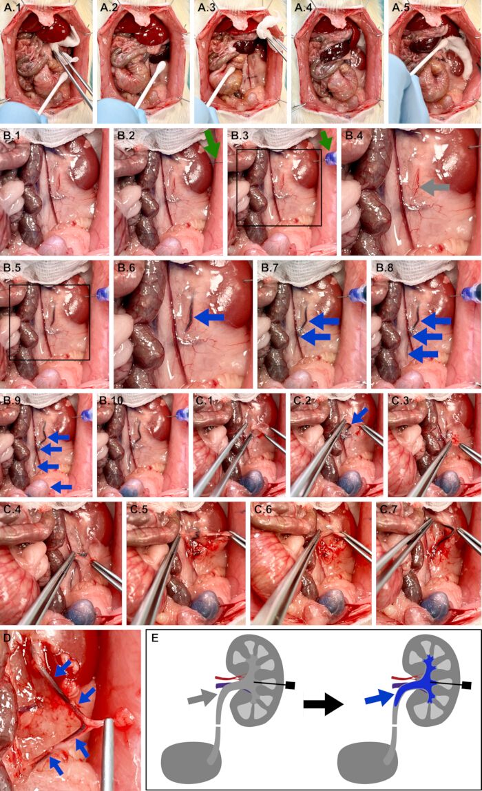
Figure 1: Dye-assisted identification and mobilization of the left ureter. The green arrow depicts the renal injection site. The grey arrow depicts the naïve ureter. The blue arrow depicts a dye‑demarcated ureter. The black box depicts a subsequently magnified area. (A) Left retroperitoneal exposure after midline laparotomy. (B) Dye‑assisted ureter demarcation. (C) Blunt dissection mobilizes the ureter from surrounding tissue. (D) Fully exposed left ureter from renal pelvis to vesicoureteral junction. (E) Schematic illustration of the described steps, including syringe and methylene blue. Please click here to view a larger version of this figure.
5. Distal calibrated partial ligation of the left ureter using a metal wire
- Elevate a mobilized segment of the distal ureter using atraumatic forceps without compressing the ureter (Figure 2A.1–2).
- Pass a 6-0 monofilament suture underneath the ureter using blunt forceps (Figure 2A.3).
- Place a metal calibration wire of the desired diameter (0.65, 0.55, 0.30, or 0.20 mm) parallel to the ureter within the ligature loop (Figure 2B.1–6).
- Tighten the ligature gradually using a sliding knot with several loop throws in the same direction until the suture sling adapts tightly to the ureter‑wire complex without loosening, which requires at least 4 throws with slowly bouncing finger movements to allow for suture settling in between throws.
NOTE: The suturing tension should be just below the tensile breaking strength, which is 5–7 N for a 6-0 non-resorbable suture, so 3 N is recommended. This translates to a pulling weight of about 306 g and can either be measured by using a spring scale or is comparable to the digital strength when holding a regular cell phone with two fingers in a pinch grip. The essential aspect is that the suture is completely closed to the ureter‑wire complex and that there are so many loop throws that they do not become loose before the locking throws follow. Due to the fragile nature of the 6-0 sutures, there is no “too tight” as the suture will break before it would be too tight for the ureter tissue to degenerate. Now secure the knot with 3 counterwise locking loop throws to prevent air knots (Figure 2B.7–10). - Carefully withdraw the calibration wire in a slow, straight motion to avoid shearing injuries to the ureteral wall, leaving a standardized residual ureteral lumen corresponding to the wire diameter (Figure 2C.1–5). The polished wire surface minimizes friction during removal and reduces the risk of tissue damage.
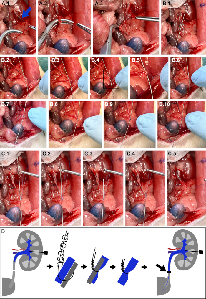
Figure 2: Distal calibrated partial ligation of the left ureter using a metal wire. (A) Distal ureter isolated and elevated with overholt clamps; 6-0 suture placed underneath. (B) Calibration wire positioned parallel to the ureter within the ligature and ligation using a sliding knot. (C) Wire withdrawal, leaving a standardized residual lumen. (D) Schematic illustration of the described steps with the ureter (blue), calibration wire (gray), and ligature (black). Please click here to view a larger version of this figure.
6. Identification and mobilization of the right ureter
- Repeat the steps in analogy to the contralateral side (Figure 3).
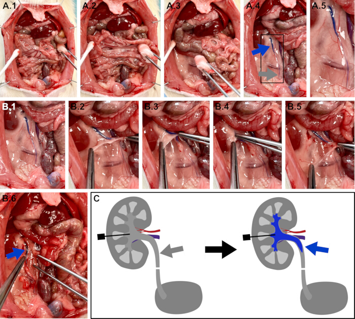
Figure 3: Dye-assisted identification and mobilization of the right ureter. (A) Right retroperitoneal exposure with dye‑assisted ureter demarcation. (B) Blunt dissection mobilizing the ureter from surrounding tissue. (C) Schematic illustration of the described steps. Please click here to view a larger version of this figure.
7. Proximal complete ligation of the right ureter using a sliding knot without wire
- Elevate a mobilized segment of the proximal ureter using atraumatic forceps without compressing the ureter (Figure 4A.1–3).
- Pass a 6-0 non-resorbable monofilament suture underneath the ureter using blunt forceps (Figure 4A.4,5).
- Tighten the ligature gradually using a sliding knot with several loop throws in the same direction until the suture sling tightly ligates the ureter without loosening, as described above. Then, secure with counterwise locking loop throws in order to achieve near-complete luminal closure (Figure 4B.1–4).
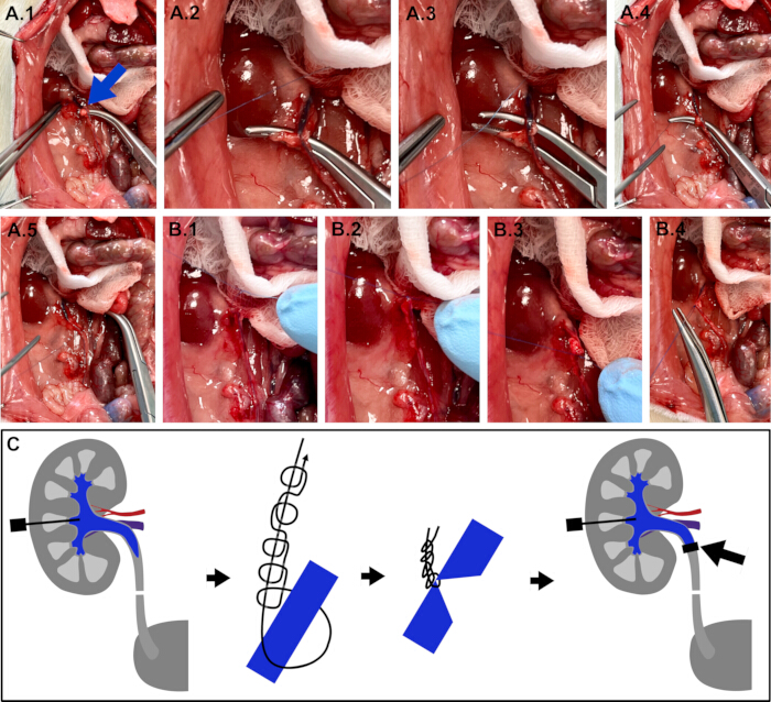
Figure 4: Proximal complete ligation of the right ureter using a sliding knot without wire. (A) Proximal ureter isolated and elevated with Overholt clamps; 6-0 suture placed underneath. (B) Complete ligation without wire using a sliding knot. (C) Schematic illustration of the described steps. Please click here to view a larger version of this figure.
8. Functional validation of partial and complete ureteral obstruction
- For validation of partial obstruction on the left side, inject 0.2 mL undiluted methylene blue transparenchymatously into the renal pelvis using a syringe and observe antegrade filling of the ureter distal to the ligation site (Figure 5A.1–3).
- Inject 1.0 mL saline into the renal pelvis to progressively clear the dye signal by wash-out and confirm continued ureteral patency (Figure 5A.3). To further increase certainty of incomplete ureteral obstruction, continue injection until the bladder volume capacity is surpassed and visible fluid outflow from the urethra can be observed (Figure 5A.4–5).
- For validation of complete obstruction on the right side, inject 0.2 mL undiluted methylene blue transparenchymatously into the renal pelvis and confirm absence of distal dye passage (Figure 5B.1,2).
- Identify proximal ureteral caliber dilation upstream of the ligation site as an indicator of complete outflow block (Figure 5B.3).
- For additional validation beyond the feasible usability of the animal model, transect both ureters distally and position the open ureteral ends directly adjacent to glass capillaries for urinary collection measurements (Figure 5C).
- For validation of this model, quantify the urinary production per kidney over a 30-min period under physiological conditions and after partial ligation using calibration wires of 0.65, 0.55, 0.30, and 0.20 mm diameters (Figure 5C.2).
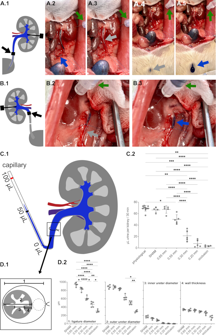
Figure 5: Functional validation of partial and complete ureteral obstruction. (A) Partial obstruction: dye colorization and saline wash-out demonstrate distal ureter filling and urethral outflow, indicating residual patency. (B) Near-complete obstruction: absence of distal dye, with a ballooned proximal ureter upstream of the ligature. (C) Capillary collection of unilateral urinary volume over 30 min per kidney shows a graded reduction of urine output from physiological conditions to complete occlusion. (D) Microscopic measures of (1) ligature diameter, (2) outer ureter diameter, (3) inner ureter diameter, and (4) ureter wall thickness. Asterisks indicate exploratory significance levels. Please click here to view a larger version of this figure.
9. Terminal steps and considerations
- Close the abdominal wall in multiple layers under sterile conditions as described in previous reports19,20,21.
- Following surgery, observe each animal continuously until it regains sternal recumbency and spontaneous mobility. Rehouse animals only after they are fully alert and capable of coordinated movement. Administer postoperative analgesia with subcutaneous carprofen at 5 mg/kg twice daily for 48 h and ensure that veterinary staff conduct daily clinical evaluations to monitor recovery and detect potential complications.
- Document animal welfare using standardized scoring systems such as the Rat Grimace Scale22 and the Body Condition Score23. Humane endpoints include a Rat Grimace Score ≥ 6 or a Body Condition Score of 1 despite adequate analgesia. Terminate animals presenting these findings, as well as those exhibiting postoperative wound infection or abdominal wall dehiscence, immediately according to institutional standards of care.
- For non-survival experiments or for termination after follow-up, euthanize the animal by sharp cardiectomy under deep anesthesia or by cervical dislocation (following institutionally approved protocols). Optionally, resect both kidneys and ureters en bloc for subsequent anatomical inspection, biomolecular analysis, or histopathological imaging.
- Dispose of animal carcasses, resected tissues, sharps, and remaining chemical solutions according to institutional animal facility and biosafety guidelines.
Access restricted. Please log in or start a trial to view this content.
Results
The calibrated ligation technique produced a diameter‑dependent reduction in urine flow across all experimental groups. Urine flow was assessed in a total of 14 rats using both kidneys, resulting in measurements from 5 organs per partial obstruction level (0.65, 0.55, 0.30, and 0.20 mm diameter). Additionally, 4 animals were included that received Sham treatment on one side (the ureter was mobilized, and a loose, non-constricting air-knot ligature was tied without wire) and complete occlusion on the other (a ligatu...
Access restricted. Please log in or start a trial to view this content.
Discussion
This study establishes a reproducible surgical method for inducing unilateral ureteral strictures in rats through midline laparotomy and calibrated partial ligation, resulting in segmental ureteral narrowing with preserved macroscopic tissue viability that functionally and anatomically resembles benign human ureteral strictures.
Clinically, ureteral stricture disease remains a major contributor to upper tract obstruction and renal morbidity with long-term functional loss, frequently arising af...
Access restricted. Please log in or start a trial to view this content.
Disclosures
No conflicts of interest declared. No specific funding was received. Animal use approved by the Baden-Württemberg Regional Council (IACUC 35-9185.81/G-62/23).
Acknowledgements
The authors gratefully acknowledge the data storage service SDS@hd supported by the Ministry of Science, Research, and the Arts Baden-Württemberg (MWK) and the German Research Foundation (DFG) through grant INST 35/1314-1 FUGG and INST 35/1503-1 FUGG. Furthermore, the authors gratefully acknowledge the support from the NCT (National Center for Tumor Diseases in Heidelberg, Germany) through its structured postdoc program and the Surgical Oncology program. We also acknowledge the support through state funds approved by the State Parliament of Baden-Württemberg for the Innovation Campus Health + Life Science Alliance Heidelberg Mannheim from the structured postdoc program for Alexander Studier-Fischer: Artificial Intelligence in Health (AIH) - A collaboration of DKFZ, EMBL, Heidelberg University, Heidelberg University Hospital, University Hospital Mannheim, Central Institute of Mental Health, and the Max Planck Institute for Medical Research. Furthermore, we acknowledge the support through the DKFZ Hector Cancer Institute at the University Medical Center Mannheim.
Access restricted. Please log in or start a trial to view this content.
Materials
| Name | Company | Catalog Number | Comments |
|---|---|---|---|
| atraumatic preparation forceps | Aesculap | FB395R | DE BAKEY ATRAUMATA atraumatic forceps, straight |
| blunt overholt clamps | Aesculap | BJ012R | BABY-MIXTER preparation and ligature clamp, bent, 180 mm |
| cannula | BD (Beckton, Dickinson) | 301300 | BD Microlance 3 cannula 20 G |
| capillary tubes | Sigma Aldrich | P1174 | Microcapillary tube, calibrated 100 µL |
| carprofen | cp pharma | 115 | carprofen 0,5% (5% with 50 mg/mL diluted 1:10) (Carprosol®) |
| cotton whool swaps | Amazon | ASIN: B0FPDB8ZGS | non-sterile cotton whool swaps (autoclave for survival experiments) |
| fixation rods | legefirm | 500343896 | tuning forks used as y-shaped metal fixation rods |
| gauze swaps | Medrull | ASIN: B09164D1JG | sterile gauze swaps |
| heating pad | Royal Gardineer | IP67 | Royal Gardineer Heating Pad Size S, 20 Watt |
| isoflurane | Piramal Critical Care | PZN / EAN 09714675 / 4150097146757 | 100% isoflurane 250 mL |
| isoflurane vaporizer | UNO ROESTVASTSTAAL BV | 180000002 | isoflurane vaporizer |
| isotonic sodiumchloride solution | B. Braun | ASIN: B007PZJOQ4 | 0,9% sodiumchloride solution |
| ketamine | cp pharma | 1202 | ketamine 10% (100 mg/mL) |
| metal calibration wire (0.20 mm diameter) | QUARKZMAN | ASIN: B0DCZBT4LW | Cr20Ni80-Nichrom metal wire (0.20 mm diameter) |
| metal calibration wire (0.30 mm diameter) | Rayher | 4006166052859 | Cr20Ni80-Nichrom metal wire (0.30 mm diameter) |
| metal calibration wire (0.55 mm diameter) | SKYPRO | UPC: 792138793816 | Cr20Ni80-Nichrom metal wire (0.55 mm diameter) |
| metal calibration wire (0.65 mm diameter) | twipp | ASIN: B07WFWNNRM | Cr20Ni80-Nichrom metal wire (0.65 mm diameter) |
| methylene blue 0,5% | Provepharm | PZN / EAN 10179678 / 4150101796787 | METHYLTHIONINIUMCHLORID Proveblue 5 mg/mL in 10 ml vials |
| non-resorbable monofilament 6-0 ligatures | COVIDIEN | VP-889-X | monofilament surgical suture from non-resorbable poly-propylene; needle can be removed |
| ophthalmic ointment | Bayer Vital GmbH | 1578681 | 5% dexpanthenol |
| plastic perfusor tube | M. Schilling GmbH | S702NC150 | connecting tube COEX 150 cm |
| preparation scissors | Aesculap | BC177R | JAMESON preparation scissors, bent, fine model, blunt/blunt, 150 mm (6") |
| steel plate | Maschinenbau Feld GmbH | C010206 | Galvanized sheet plate, 40 x 50 cm, thickness 4.0 mm |
| syringe with integrated needle | BLPRKOT | BL2509L3yici08 | 1 ml syringe with integrated 30 G needle |
| xylazine | cp pharma | 1206 | xylazine 0,2% (2% with 20 mg/mL diluted 1:10) (Xylavet) |
References
- Moretto, S., et al. Ureteral stricture rate after endoscopic treatments for urolithiasis and related risk factors: systematic review and meta-analysis. World J Urol. 42 (1), 234(2024).
- Sahin, C., et al. Ureteral stricture formation after endoscopic removal of obstructing stones: could it be predicted with well-assessed radiological parameters. Urolithiasis. 52 (1), 34(2024).
- Sunaryo, P. L., et al. Ureteral strictures following ureteroscopy for kidney stone disease: A population-based assessment. J Urol. 208 (6), 1268-1275 (2022).
- Minkovich, M., et al. Ureteral strictures post-kidney transplantation: Trends, impact on patient outcomes, and clinical management. Can Urol Assoc J. 15 (10), E524-E530 (2021).
- Rosenfeld, J., et al. A review of complications after ureteral reconstruction. Asian J Urol. 11 (3), 348-356 (2024).
- Chevalier, R. L., Forbes, M. S., Thornhill, B. A. Ureteral obstruction as a model of renal interstitial fibrosis and obstructive nephropathy. Kidney Int. 75 (11), 1145-1152 (2009).
- Reicherz, A., et al. Ureteral obstruction promotes ureteral inflammation and fibrosis. Eur Urol Focus. 9 (2), 371-380 (2023).
- Li, Z., et al. Health-related quality of life (HRQoL), anxiety and depression in patients with ureteral stricture: A multi-institutional study. World J Urol. 41 (1), 275-281 (2023).
- May, P. C., et al. The morbidity of ureteral strictures in patients with prior ureteroscopic stone surgery: multi-institutional outcomes. J Endourol. 32 (4), 309-314 (2018).
- Contreras, P., et al. Ureteral stricture: Endoscopic management. Eur Urol Focus. 11 (5), 730-732 (2025).
- Su, B., et al. Long-term outcomes of Allium ureteral stent as a treatment for ureteral obstruction. Sci Rep. 14 (1), 21958(2024).
- Kowalski, F., et al. Endourological treatment of ureteral strictures with the use of self-expanding stents: Is it possible to completely cure the stricture endoscopically? a retrospective study with two-year follow-up. World J Urol. 43 (1), 201(2025).
- Bourillon, A., et al. Robotic upper urinary tract reconstruction for ureteral stricture: A single-center series. J Robot Surg. 19 (1), 573(2025).
- Vroomen, L., et al. A new intrasurgical technique to safely and reproducibly induce partial unilateral urinary obstruction and renal scarring in a rat model. Int Urol Nephrol. 52 (7), 1209-1218 (2020).
- Chen, T., Zhang, Z., Lai, C. Improvement of the reversible unilateral ureteral obstruction model. Ren Fail. 47 (1), 2454721(2025).
- Nan, Q. Y., et al. Pathogenesis and management of renal fibrosis induced by unilateral ureteral obstruction. Kidney Res Clin Pract. 43 (5), 586-599 (2024).
- Khater, Y., Barakat, N., Shokeir, A., Samy, A., Karrouf, G. Renal fibrosis progression following partial unilateral ureteral obstruction: mechanisms and therapeutic insights. World J Urol. 43 (1), 229(2025).
- Thornhill, B. A., Burt, L. E., Chen, C., Forbes, M. S., Chevalier, R. L. Variable chronic partial ureteral obstruction in the neonatal rat: A new model of ureteropelvic junction obstruction. Kidney Int. 67 (1), 42-52 (2005).
- Studier-Fischer, A., et al. Endotracheal intubation via tracheotomy and subsequent thoracotomy in rats for non-survival applications. J Vis Exp. (205), e66684(2024).
- Studier-Fischer, A., et al. Controlled reversible visceral arterial ischemia, venous congestion and combined malperfusion via midline laparotomy in rats. J Vis Exp. (209), e67171(2024).
- Studier-Fischer, A., et al. 5/6 nephrectomy using sharp bipolectomy via midline laparotomy in rats. J Vis Exp. (218), e67322(2025).
- Sotocinal, S. G., et al. The rat grimace scale: A partially automated method for quantifying pain in the laboratory rat via facial expressions. Mol Pain. 7, 55(2011).
- Hickman, D. L., Swan, M. Use of a body condition score technique to assess health status in a rat model of polycystic kidney disease. J Am Assoc Lab Anim Sci. 49 (2), 155-159 (2010).
- Gild, P., et al. Adult iatrogenic ureteral injury and stricture-incidence and treatment strategies. Asian J Urol. 5 (2), 101-106 (2018).
- Luo, J., et al. marrow mesenchymal stem cells reduce ureteral stricture formation in a rat model via the paracrine effect of extracellular vesicles. J Cell Mol Med. 22 (9), 4449-4459 (2018).
- Maruschke, M., et al. Development of a rat model for investigation of experimental splinted uretero-ureterostomy, ureteral stenting and stenosis. In Vivo. 27 (2), 245-249 (2013).
- Demirhan, K., Saglam, H. S., Cimen, H. I., Elcin, B., Cokluk, E. Effect of the location and severity of partial ureteral obstruction on urinary system stone disease formation. Sci Rep. 15 (1), 11560(2025).
- Wen, J. G., Chen, Y., Frøkiaer, J., Jørgensen, T. M., Djurhuus, J. C. Experimental partial unilateral ureter obstruction I pressure flow relationship in a rat model with mild and severe acute ureter obstruction. J Urol. 160 (4), 1567-1571 (1998).
- Li, P., et al. An immature murine model of reversible unilateral ureteral obstruction. J Vis Exp. (218), e67238(2025).
- Martínez-Klimova, E., Aparicio-Trejo, O. E., Tapia, E., Pedraza-Chaverri, J. Unilateral ureteral obstruction as a model to investigate fibrosis-attenuating treatments. Biomolecules. 9 (4), 141(2019).
- Mohammadi, A., et al. Effects of the surgical ligation of the ureter in different locations on the kidney over time in the rat model. Adv Urol. 2024, 6611081(2024).
- Narváez Barros, A., et al. Reversal unilateral ureteral obstruction: A mice experimental model. Nephron. 142 (2), 125-134 (2019).
- Nakazawa, R., et al. Renal replacement strategies: dual benefits of hydrogen gas inhalation and hydrogen-enriched dialysate. Med Gas Res. 15 (3), 444-445 (2025).
- Cheng, J., Shi, M., Sun, X., Lu, H. Therapeutic effect of hydrogen and its mechanisms in kidney disease treatment. Med Gas Res. 14 (2), 48-53 (2024).
- Liu, B., et al. Hyperbaric oxygen therapy for male infertility: a systematic review and meta-analysis on improving sperm quality and fertility outcomes. Med Gas Res. 15 (4), 529-534 (2025).
Access restricted. Please log in or start a trial to view this content.