This protocol describes reproducible green synthesis methods for fabricating gold and silver nanoparticles using plant extracts, fungal filtrates, and bacterial cultures, followed by nanoparticle confirmation through ultraviolet–visible spectroscopy and transmission electron microscopy.
A subscription to JoVE is required to view this content. Sign in or start your free trial.
Method Article
Green Synthesis of Gold and Silver Nanoparticles Using Plant Extracts, Fungal Filtrates, and Bacterial Cultures
In This Article
Summary
Abstract
Green synthesis of metal nanoparticles (NPs) using biological systems provides a sustainable alternative to conventional chemical fabrication methods. This protocol presents reproducible experimental and analytical procedures for the synthesis of gold (Au) and silver (Ag) NPs using bacterial cultures, fungal filtrates, and plant leaf extracts as reducing and stabilizing agents. AgNPs were synthesized using extracts from Psidium guayaquilensis, Acanthophora spicifera, and Earliella sp., exhibiting characteristic ultraviolet–visible (UV–Vis) absorption bands between 405 and 425 nm that confirmed NP formation. A design-of-experiments approach was implemented to evaluate the influence of environmental factors, including oxygen conditions, pH, Au concentration, cell concentration, electron donor type, and temperature, on the synthesis of AuNPs by Shewanella oneidensis and Cupriavidus metallidurans. Under ideal conditions (0.2 mM Au and pH 5), S. oneidensis produced predominantly spherical AuNPs with an average size of 43.6 ± 11.0 nm and a characteristic absorption peak at 520 nm. NP formation and morphology were confirmed using UV–Vis spectroscopy and transmission electron microscopy. This workflow provides a reproducible platform for biogenic NP synthesis and supports applications in biosensing, bioremediation, and antimicrobial technologies.
Introduction
Metallic nanoparticles (NPs) possess unique physicochemical properties associated with their high surface area, including distinctive electronic, magnetic, and catalytic behaviors, as well as high reactivity. These characteristics have promoted their application across multiple fields, including medicine for disease diagnosis and drug delivery1,2,3,4,5,6, cosmetics for sunscreen formulations7, electronics for transistors and solar cells8, environmental engineering for water treatment2,9,10, and agriculture for fertilizers and pesticides11,12. The continuous expansion of NP applications across disciplines is reflected in the increasing number of related publications reported in recent years13,14.
Synthetic methods for metallic NPs enable precise control over important characteristics such as particle size, morphology, and stability, making the synthesis process a critical determinant of NP performance. Conventional chemical synthesis methods have been extensively optimized; however, they frequently involve toxic reagents, high energy consumption, and elevated production costs14,15. These limitations have motivated the development of greener and more sustainable alternatives. Green synthesis approaches employ biological systems such as plant extracts 16,17, microorganisms including fungi and bacteria18, and algae19 as natural reducing and stabilizing agents. During these processes, metal salts are reduced to zero-valent forms, generating NPs through environmentally friendly and cost-effective pathways. In addition, biologically synthesized NPs are often biocompatible, facilitating their use in medical, agricultural, and environmental applications8,14.
The selection of an appropriate biological route for NP synthesis depends on the intended application, as well as the available infrastructure and operational requirements. Plant-based synthesis is generally preferred for rapid and low-cost NP production because it requires minimal sterile handling; however, variability in extract composition may affect reproducibility20,21. Fungal-mediated synthesis can provide high NP yields and extracellular enzymes that improve NP stabilization and downstream processing, although this approach requires longer cultivation periods and controlled growth conditions22. In contrast, bacterial systems enable tunable synthesis under defined metabolic conditions and rapid biomass production, making them suitable for mechanistic studies and NP-associated applications22. However, bacterial synthesis often requires strict aseptic handling, controlled bioreactor conditions, and additional downstream processing when intracellular NPs are recovered21,22.
Despite the advantages mentioned, green synthesis methods continue to face challenges associated with reproducibility and standardization. Furthermore, detailed visual step-by-step protocols for the green synthesis of metallic NPs remain limited23, restricting the ability of researchers to reproduce and implement existing methodologies successfully. The goal of this work is to present reproducible green synthesis protocols for gold (Au) and silver (Ag) NPs using plant extracts, fungal filtrates, and bacterial cultures. Specifically, aqueous extracts from Psidium guayaquilensis and Acanthophora spicifera, extracellular fungal filtrates from Earliella sp., and bacterial systems involving Shewanella oneidensis and Cupriavidus metallidurans were employed for NP synthesis. The presented protocols emphasize critical steps in biological extract preparation, NP synthesis, and preliminary NP characterization. In addition, the workflow integrates design of experiments, standardized preparation procedures, and in-line characterization to support reproducible and optimizable biogenic NP synthesis.
Access restricted. Please log in or start a trial to view this content.
Protocol
The research was performed in accordance with the institutional guidelines of the Escuela Superior Politécnica del Litoral (ESPOL, Ecuador). The plant, algal, and fungal biomass used in this study were collected from accessible environments and did not involve protected species or restricted areas. According to institutional and national regulations, no specific permits were required for the collection of freely available biomass under these conditions. Refer to Figure 1 for the integrated experimental workflow used for green synthesis of AgNPs and AuNPs using plant, algal, fungal, and bacterial biological systems.
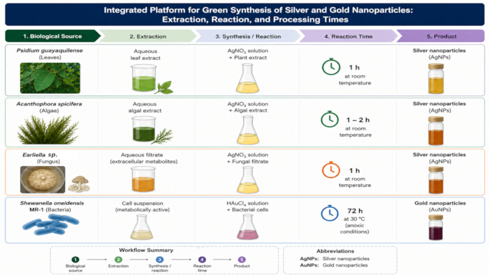
Figure 1: Integrated Workflow for Green Synthesis of Silver (Ag) and Gold (Au) Nanoparticles (NPs) Using Biological Systems. Schematic of the experimental workflow for the green synthesis of AgNPs and AuNPs using plant, algal, fungal, and bacterial systems. The workflow illustrates biological source selection, preparation of extracts or biomass, reaction with metal precursors (AgNO3 or HAuCl4), reaction conditions, and NP formation. The biological systems include Psidium guayaquilensis leaf extract, Acanthophora spicifera algal extract, Earliella sp. extracellular filtrate, and Shewanella oneidensis MR-1 bacterial cultures. Please click here to view a larger version of this figure.
NOTE: Follow all institutional laboratory safety guidelines when handling chemicals and biological materials. Consult safety data sheets for all reagents before use.
1. Synthesis of AgNPs Using P. guayaquilensis Leaves
- Leaf Collection
- Collect fresh leaves of P. guayaquilensis during the dry season in Guayaquil, Guayas Province, Ecuador (2°10′S, 80°0′W).
NOTE: Voucher specimens of this endemic species were deposited at the GUAY Herbarium, Faculty of Natural Sciences, University of Guayaquil, Ecuador, and at the New York Botanical Garden (NYBG) STEERE HERBARIUM, New York, USA. Additional information is available at: http://sweetgum.nybg.org/science/vh/specimen-details/?irn=2602207
- Collect fresh leaves of P. guayaquilensis during the dry season in Guayaquil, Guayas Province, Ecuador (2°10′S, 80°0′W).
- Preparation of Leaf Extract
- Wash the fresh plant material thoroughly with distilled water.
NOTE: Ensure complete removal of surface impurities. - Dry the leaves in a tray dryer with forced airflow at 40°C.
NOTE: Low drying temperatures minimize degradation of thermolabile organic compounds involved in Ag+ reduction. - Grind the dried material using a hand mill to obtain a fine powder.
- Suspend 10 g of powdered material in 200 mL of distilled water.
- Heat the suspension at 70°C under continuous stirring for 1 h.
- Allow the extract to cool to room temperature (RT, ~28°C).
- Filter the extract under vacuum through qualitative filter paper (Grade 1, pore size 11 µm, diameter 110 mm).
NOTE: Remove all solid residues from the plant material. - Store the filtered extract at 4°C in amber bottles until further use.
- Wash the fresh plant material thoroughly with distilled water.
- Fabrication of Phytogenic AgNPs Using Leaf Extracts
NOTE: During optimization experiments, vary one parameter per assay while maintaining the remaining parameters constant. Evaluate silver nitrate (AgNO3) concentration (1–5 mM), extract fraction (10%–50% v/v), reaction time (0.5–1 h), and temperature (25°C–80°C). Optimize synthesis parameters using response surface methodology based on a central composite DoE. Study each factor at five levels (-α, -1, 0, +1, and +α). Use the wavelength of the surface plasmon resonance (SPR) peak (λSPR), which correlates with NP size, as the response variable24. The experimental design consists of 16 factorial points, 8 axial points, and 5 center-point replicates, resulting in 29 total experiments.- Prepare AgNO3 solutions by dissolving the appropriate mass of AgNO3 in distilled water to obtain concentrations between 1 and 5 mM.
CAUTION: AgNO3 is corrosive and a strong oxidizing agent that may cause skin and eye irritation. Wear appropriate personal protective equipment, including gloves and eye protection. Avoid exposure to light and dispose of Ag-containing waste according to institutional safety guidelines. - Mix the AgNO3 solution with the plant extract to obtain extract fractions between 10% and 50% (v/v).
- Heat and stir the reaction mixture at the selected temperature (25°C–80°C) for the desired reaction time (0.5–1 h).
- Allow the reaction mixture to stand at RT for 24 h.
- Centrifuge the brown reaction mixture at 1660 × g for 15 min at RT. Discard the supernatant and collect the solid fraction.
- Dry the recovered NPs in an oven at 60°C.
- Prepare AgNO3 solutions by dissolving the appropriate mass of AgNO3 in distilled water to obtain concentrations between 1 and 5 mM.
2. Synthesis of AgNPs Using A. spicifera Algal Extracts
- Algal Collection
- Collect A. spicifera algae in Valdivia, Santa Elena Province, Ecuador (1°57′23.91′′S, 80°43′51.33′′W).
- Preparation of Algal Extract
- Prepare the algal extract by following the procedure described in Step 1.2. Replace the plant leaves with A. spicifera algal biomass.
- Fabrication of Phycogenic AgNPs Using Algal Extracts
NOTE: Perform a series of 12 experiments by varying one parameter at a time while maintaining the remaining parameters constant. Evaluate AgNO3 concentration (1–5 mM), extract fraction (5%–25% v/v), reaction time (1–4 h), and temperature (25°C–70°C).- Prepare the AgNO3 solutions and algal extract as described in Steps 1.3.1 and 2.2.
- Mix the AgNO3 solution with the algal extract to obtain an extract fraction of 5%–25% (v/v). Heat and stir the reaction mixture at the selected temperature (25°C–70°C) for the desired reaction time (1–4 h).
- Allow the solution to cool to RT for 24 h. Collect the solid fraction after centrifugation and dry it in an oven as described in Steps 1.3.5 and 1.3.6.
3. Synthesis of AgNPs Using Earliella sp.
- Culture Collection and Maintenance
- Transfer actively growing mycelial fragments of Earliella sp. onto Potato Dextrose Agar plates.
NOTE: The fungal strain used in this study was isolated from environmental samples collected on the campus of the ESPOL, Ecuador. The isolate was taxonomically identified based on morphological characteristics and preserved in the culture collection of the Biotechnology Research Center of Ecuador (CIBE, ESPOL) under the internal code E2. As this strain is maintained in an institutional collection and has not been deposited in an international repository, no RRID or external accession number is available. - Incubate the plates at 28°C for 48 h.
- Transfer a mycelial fragment into Potato Dextrose Broth and incubate the culture under constant agitation at 150 rpm for 5 days.
NOTE: This step promotes biomass production and secretion of extracellular metabolites. - Filter the fungal culture through sterile filter paper to separate the mycelial biomass from the extracellular broth. Collect the filtrate for subsequent NP synthesis.
NOTE: The extracellular broth contains biomolecules that function as reducing and stabilizing agents during AgNP synthesis.
- Transfer actively growing mycelial fragments of Earliella sp. onto Potato Dextrose Agar plates.
- Fabrication of Mycogenic AgNPs
NOTE: Generate 14 experimental conditions by varying one parameter per assay while maintaining constant volume ratios between the fungal broth and AgNO3 solution. Evaluate AgNO3 concentration (1–10 mM), pH (4–10 adjusted using HCl or NaOH), reaction time (1–4 h), and temperature (25°C–70°C).
CAUTION: Hydrochloric acid (HCl) and sodium hydroxide (NaOH) are highly corrosive. Wear appropriate personal protective equipment and perform pH adjustments slowly under continuous stirring to avoid splashing and localized overheating. Use 1 M acid or base solutions during pH adjustment.- Mix the fungal extracellular broth with an AgNO3 solution at the desired concentration (1–10 mM) using a volume ratio between 2.5% and 10% (v/v) to obtain a final reaction volume of 50 mL. Adjust the final reaction volume according to the experimental condition being tested.
- Stir the reaction mixture at the selected temperature (25°C–70°C) for the desired reaction time (1–4 h). Allow the solution to cool to RT for 24 h. Collect the solid fraction after centrifugation and dry it in an oven as described in Steps 1.3.5 and 1.3.6.
4. Synthesis of AuNPs Using C. metallidurans and S. oneidensis
- Culture Collection and Maintenance
- Inoculate frozen cell stocks preserved in 20% glycerol at −80°C of either C. metallidurans CH34 or S. oneidensis MR-1 onto tryptic soy agar plates under aerobic conditions. Incubate the plates at 28°C for 24 h.
- Select an individual colony from each plate and inoculate it into 250 mL of tryptic soy broth under aerobic conditions. Incubate the culture at 28°C in a rotary shaker operating at 120 rpm.
- Harvest the cells at the stationary phase after 72 h of incubation, when the optical density at 610 nm (OD610) reaches approximately 1. Centrifuge the cultures at 10000 × g for 10 min at ~20°C.
- Wash the collected pellets twice with sterile 0.9% (w/v) NaCl solution. Concentrate the washed cells to an OD610 value of approximately 7.
- Generate anoxic concentrated cell stocks by degassing the suspensions and filling the headspace with N2 gas.
- Fabrication of Bacteriogenic AuNPs
NOTE: Use a fractional-factorial matrix to evaluate the influence of environmental factors on AuNP synthesis25. Construct the matrix using 7 factors at 2 levels selected according to previous studies26,27. Arrange the experiments in an orthogonal matrix and randomize the experimental order to minimize trial-combination bias. The resulting experimental order is presented in Table 1.- Dissolve gold chloride trihydrate (HAuCl4) in deionized water to prepare a stock solution at 5 g L−1.
CAUTION: HAuCl4 is toxic and a strong oxidizing agent. Handle all solutions inside a certified chemical fume hood while wearing appropriate personal protective equipment, including a lab coat, gloves, and safety goggles. Avoid contact with organic materials and dispose of waste according to institutional hazardous waste protocols. - Prepare the experimental reactors by diluting the stock solution to the target concentration in 0.9% (w/v) NaCl solution and adjusting the pH according to the experimental conditions listed in Table 1.
- Dispense 50 mL of the prepared metal solution into clean 120 mL serum bottles. Immediately seal each bottle using a butyl rubber stopper.
- Secure the stopper using an aluminum crimp seal and a handheld crimper. Sterilize the sealed serum bottles by autoclaving at 121°C for 21 min.
- Allow the bottles to cool to RT and visually inspect each bottle for precipitation.
NOTE: Discard any bottles showing visible precipitation and prepare replacement reactors. - Establishment of Anoxic Conditions
- Connect each sterilized serum bottle to a gas manifold system equipped with a high-purity nitrogen (N2) source and a vacuum pump.
- Generate an anoxic headspace by performing 21 consecutive purge cycles for each reactor requiring anoxic conditions according to Table 1. During each cycle, apply vacuum followed by nitrogen backfilling for approximately 30 s until the internal pressure returns to approximately atmospheric pressure (~1 atm).
NOTE: Direct verification of anoxic conditions is not required; however, headspace gas analysis, such as gas chromatography, may be used to confirm oxygen removal when necessary.
CAUTION: Nitrogen gas may displace oxygen and create an asphyxiation hazard in enclosed spaces. Ensure adequate laboratory ventilation and verify secure gas-line connections before use. - Maintain the reactors under slight positive N2 pressure after the final purge cycle.
- Label the reactors from 1–8 according to the experimental order. For example, label the fifth experimental condition as R5.
- Addition of Electron Donors and Inoculation
- Inject the required volume of sterile anoxic liquid electron donor solution, such as sodium lactate, into the liquid phase of the designated reactors according to Table 1 using a sterile syringe and needle.
- Inject the required volume of gaseous electron donor, such as H2, into the designated reactors headspace using a gas-tight syringe.
- Inoculate all biotic reactors by aseptically injecting the required volume of concentrated anoxic microbial stock culture into the liquid phase.
NOTE: Store concentrated stock cultures at 4°C before inoculation to maintain cell viability.
- Set-Up of Control Experiments
- Prepare an abiotic control by following all steps described in Step 4.2 while replacing the microbial inoculum with an equivalent volume of sterile anoxic culture medium or buffer.
- Prepare a killed-cell control by following all steps described in Step 4.2 using an inoculum sterilized by autoclaving.
NOTE: Use the controls to account for abiotic metal reduction and biosorption. Prepare the killed-cell control by autoclaving viable cell stocks at 121°C for 21 min to evaluate the contribution of active microbial mechanisms.
- Incubation and Reaction Time
- Place all serum bottles in temperature-controlled incubators.
- Adjust the incubation temperature according to the experimental conditions specified in Table 1.
- Place the incubators on rotary shaker platforms and incubate the reactors at 120 rpm.
- Allow the reactions to proceed for 72 h.
NOTE: Pause the experiment after incubation and store the serum bottles at 4°C until sample processing.
NOTE: Optionally quantify residual dissolved metal after NP biosynthesis using inductively coupled plasma optical emission spectroscopy to evaluate metal removal efficiency and distinguish NP formation from residual dissolved species.
- Dissolve gold chloride trihydrate (HAuCl4) in deionized water to prepare a stock solution at 5 g L−1.
- Replication of Optimal Conditions
- Identify the reactor condition associated with the maximum metal removal following primary screening and data analysis.
- Replicate the selected optimal condition in three independent reactor bottles to confirm reproducibility.
- Perform the preparation, reactor set-up, and incubation procedures for the triplicate verification experiments according to Steps 4.1 and 4.2.
NOTE: After completion of the experiments, temporarily store and label all dissolved solutions as aqueous waste until disposal by certified waste-management companies according to the national Organic Code of the Environment and institutional technical guidelines.
| Reactor / Bacteria | Temperature (°C) | Atmosphere | Electron donor | Optical density (OD610) | pH | [Gold] (mM) |
| 1 / CMC | 37 | N2 | H2 | 0.5 | 1 | 2 |
| 2 / CMC | 28 | N2 | C3H5O3− | 1 | 1 | 0.2 |
| 3 / SOM | 37 | O2 | C3H5O3− | 0.5 | 1 | 0.2 |
| 4 / CMC | 28 | O2 | H2 | 1 | 1 | 2 |
| 5 / SOM | 37 | N2 | H2 | 1 | 5 | 0.2 |
| 6 / CMC | 37 | O2 | C3H5O3− | 1 | 5 | 2 |
| 7 / SOM | 28 | N2 | C3H5O3− | 0.5 | 5 | 2 |
| 8 / CMC | 28 | O2 | H2 | 0.5 | 5 | 0.2 |
Table 1: Fractional factorial experimental design used for bacteriogenic AuNP synthesis. This table summarizes the experimental conditions evaluated for AuNP synthesis using S. oneidensis MR-1 (SOM) or C. metallidurans CH34 (CMC). Variables included incubation temperature, headspace atmosphere, electron donor, inoculum optical density (OD610), solution pH, and initial Au3+ concentration.
5. Characterization of AgNPs and AuNPs
NOTE: Use ultraviolet–visible (UV–Vis) spectroscopy as an accessible method to confirm NP formation through surface plasmon resonance. Use transmission electron microscopy (TEM) to directly evaluate NP morphology and size. Additional physicochemical characterization may be performed using complementary techniques such as dynamic light scattering (DLS), X-ray diffraction (XRD), Fourier transform infrared (FTIR) spectroscopy, and energy-dispersive X-ray spectroscopy (EDS) to evaluate size distribution, crystallinity, surface chemistry, and elemental composition when more comprehensive characterization is required28,29.
- UV–Vis Spectroscopy
- Instrument Set-Up
- Turn on the UV–Vis spectrophotometer and allow the instrument to warm up for at least 15 min to ensure signal stability.
- Select spectral scanning mode and configure the wavelength range from 300 to 800 nm using a data interval of 1 nm.
- Instrument Set-Up
- Sample Preparation
- Collect aliquots of freshly synthesized AgNP or AuNP suspensions. Dilute the samples with distilled water when necessary to maintain absorbance values within the linear detection range.
- UV–Vis Measurement of NPs
- Perform baseline correction using distilled water as the blank solution.
- Transfer the prepared sample into a cuvette, place the cuvette in the sample holder, and record the UV–Vis spectrum over the 300–800 nm range.
- Identify the SPR band corresponding to NP formation.
NOTE: AgNPs typically exhibit SPR bands between 405 and 425 nm, whereas AuNPs generally exhibit SPR bands between 520 and 550 nm.
- Transmission Electron Microscopy
NOTE: Analyze NP morphology and size using TEM operated at 80 kV and equipped with a high-resolution digital camera.- Transfer 1.5 mL of NP suspension into a centrifuge tube and centrifuge at 5000 × g for 5 min at RT.
- Discard the supernatant and retain the pellet.
- Add 1 mL of fixation solution containing 4% paraformaldehyde and 5% glutaraldehyde prepared in 0.1 M cacodylate buffer to the pellet. Incubate the sample for 25 h at RT. Then, wash the pellet overnight with 0.1 M sodium cacodylate buffer.
- Centrifuge the sample at 5000 × g for 5 min and discard the supernatant.
- Add 1 mL of 1% (w/v) osmium tetroxide (OsO4) prepared in 0.1 M sodium cacodylate buffer for fixation for 3 h.
CAUTION: OsO4 is highly toxic and volatile. Handle all solutions exclusively inside a certified chemical fume hood while wearing appropriate personal protective equipment, including a lab coat, gloves, and safety goggles. - Centrifuge the sample at 5000 × g for 5 min and discard the supernatant.
- Dehydrate the sample using a graded ethanol series. Add 50% ethanol, incubate for 15 min. Repeat the procedure sequentially using 70% and 90%. Complete dehydration with three consecutive incubations in absolute ethanol for 30 min per incubation.
- Embed the pellet in epoxy resin at RT according to the manufacturer’s instructions.
- Prepare semi-thin sections (~1 µm) and stain the sections with toluidine blue for preliminary observation.
- Cut ultrathin sections (~60 nm) using a diamond knife and stain the sections with uranyl acetate followed by lead citrate.
- Mount the sections on TEM grids and load them into the transmission electron microscope.
- Acquire TEM images to evaluate NP morphology and size.
Access restricted. Please log in or start a trial to view this content.
Results
AgNPs were successfully synthesized through green and biosynthetic routes using different biological reducing agents, and ideal synthesis conditions were identified for each method. In the green synthesis mediated by P. guayaquilensis leaf extract, the optimal conditions consisted of an AgNO3 concentration of 5 mM, an extract-to-metal solution ratio of 25% (v/v), a reaction temperature of 80°C, and an agitation time of 60 min followed by a resting period of 24 h. Under these conditions, UV–Vis spectro...
Access restricted. Please log in or start a trial to view this content.
Discussion
When comparing the evaluated synthesis routes in terms of feasibility, NP size control, operational cost, reproducibility, and overall process efficiency, plant-mediated synthesis using P. guayaquilensis leaf extract demonstrated the most favorable balance among the AgNP synthesis methods investigated. This protocol provided improved control over NP size through systematic optimization and direct microscopic characterization while operating under mild conditions without prolonged biological cultivation. In addit...
Access restricted. Please log in or start a trial to view this content.
Disclosures
The authors have nothing to disclose.
Acknowledgements
The authors gratefully acknowledge the co-authors of the original study and the Center for Microbial and Environmental Technologies (CMET, UGent, Belgium), where the foundational work was conducted. Special thanks are extended to Julio de la Paz, Master’s student in Chemical Engineering from ESPOL, for his valuable contributions to the comparative analysis that supported this research.
Access restricted. Please log in or start a trial to view this content.
Materials
| Name | Company | Catalog Number | Comments |
|---|---|---|---|
| Absolute ethanol | Merck | E7023 | TEM dehydration series |
| Aluminum crimp seals | Dwk Wheaton | 224178-01 | For serum bottles |
| Amber glass bottles | Duran | 2180154 | Light-protected storage |
| ATR-FTIR spectrometer | Thermo Scientific | Nicolet iS10 | Spectral range: 300–3600 cm-1 |
| Autoclave | Yamato | SM300 | Sterilization at 121 °C |
| Butyl rubber stoppers | Dwk Kimble | 73827-21 | Gas-tight sealing |
| Centrifuge | BIOBASE | BKC-TH20RL | Bench-top centrifuge |
| Chloroauric acid trihydrate (HAuCl4·3H2O) | Sigma-Aldrich | 27988-77-8 | Gold precursor |
| Diamond knife | Aurion | Dyatom 45 | Ultrathin TEM sectioning |
| Distilled or deionized water | Methrom | N/A | Solution preparation |
| Epoxy resin (EMBED 812/Epon) | Aurion | EMBED 812 | TEM embedding medium |
| Filter paper, qualitative Grade 1 | Merck Whatman | WHA1001110 | Pore size: 11 µm; diameter: 110 mm |
| Fourier transform infrared spectrometer (FTIR) | Thermo Scientific | Nicolet iS10 | Spectral range: 300–3600 cm-1 |
| Gas manifold system | Custom-built | N/A | For purge cycles |
| Gas-tight syringe | Hamilton | 1001 RN | 1 mL |
| Glutaraldehyde | VWR | 219859510 | 5% Glutaraldehyde, 4% paraformaldehyde in 0.1 M Sodium Cacodylate Buffer |
| Glycerol | Sigma-Aldrich | G5516 | 20% stock preservation |
| Handheld crimper | Biomed | USYGQ-20Z | Crimp sealing of serum bottles |
| Hand mill/grinder | Corona | L10000 | Plant biomass grinding |
| Hydrochloric acid (HCl) | Merck | 320331 | pH adjustment |
| Hydrogen (H2) gas | Linde | Industrial grade | Electron donor gas |
| ICP-OES system | Varian | Vista-MPX | Metal quantification |
| Incubator | Memmert | SM 400 | Temperature-controlled incubation |
| Laboratory oven | Barnstead | M100 | Drying nanoparticles |
| Lead citrate | Merck | 7398 | TEM staining |
| Nitric acid (HNO3) | Chem-Lab | CL00.0125 | Acid reagent |
| Nitrogen (N2) gas | Linde | Industrial grade | Anoxic conditions |
| Osmium tetroxide (OsO4) | Merck | 201030 | TEM fixation |
| Paraformaldehyde | VWR | 101176-014 | 5% Glutaraldehyde, 4% paraformaldehyde in 0.1 M Sodium Cacodylate Buffer |
| Potato Dextrose Agar (PDA) | Merck | 110130 | Fungal cultivation |
| Potato Dextrose Broth (PDB) | Merck | P6685 | Microbiological medium |
| Rotary shaker | JoanLab | OS-20Pro | Incubation at controlled agitation |
| Serum bottles (120 mL) | Wheaton | 223748 | Borosilicate glass |
| Silver nitrate (AgNO3) | Sigma-Aldrich | 209139 | ACS reagent, ≥99.0% |
| Sodium cacodylate buffer | VWR | 11650 | 5% Glutaraldehyde, 4% paraformaldehyde in 0.1 M Sodium Cacodylate Buffer |
| Sodium chloride (NaCl) | Carl Roth | 3957.1 | 0.9% washing solution |
| Sodium hydroxide (NaOH) | Sigma-Aldrich | S5881 | pH adjustment |
| Sodium lactate | Sigma-Aldrich | L7022 | Electron donor/carbon source |
| Sterile syringes and needles | BD | Various | Sterile liquid handling |
| Stirring hot plate | Biologix | 01-310X | Heating and agitation |
| Toluidine blue | EMS | DcU-10 | Semi-thin section staining |
| Transmission electron microscope (TEM) | FEI | Tecnai Spirit Twin G20 | Equipped with high-resolution digital camera |
| Tray dryer with forced airflow | Handmade | N/A | Biomass drying at 40 °C |
| Tryptic Soy Agar (TSA) | Oxoid | CM01301 | Microbiological medium |
| Tryptic Soy Broth (TSB) | Carl Roth | X938.1 | Microbiological medium |
| Uranyl acetate | Sigma-Aldrich | 94260 Fluka | TEM staining |
| UV–Vis spectrophotometer | Thermo Scientific | Genesys 10S | UV–Vis spectral analysis |
| Vacuum pump | KNF or equivalent | N 820 G | For anoxic conditions |
| Biological Materials | |||
| Biological Material | Source | Identifier | Comments/Description |
| Acanthophora spicifera | Local collection | N/A | Marine algal biomass |
| Cupriavidus metallidurans CH34 | SCK-CEN | LMG 1195 | Bacterial strain |
| Earliella scabrosa | CIBE-ESPOL | E2 | Institutional fungal collection |
| Psidium guayaquilensis | Local collection | N/A | Wild plant biomass |
| Shewanella oneidensis MR-1 | BCCM/LMG | LMG 19005 | Culture collection strain |
Reprints and Permissions
This article has been published
Video Coming Soon