Bleomycin electrosclerotherapy (BEST) enhances intralesional bleomycin activity through reversible electroporation using an electrical pulse generator. This protocol describes patient selection, preparation, dosing, electrode placement, and electroporation parameters to standardize BEST for the treatment of slow-flow vascular malformations.
Method Article
Current Operating Procedure (COP) for the Treatment of Slow-Flow Vascular Malformations with Bleomycin Electrosclerotherapy (BEST)
Miha Stabuc1 , Rok Dezman2 , Tobian Muir3 , Walter A Wohlgemuth4 , Tine Arnez5 , Orjana Velikonja6 , Masa Omerzel2 , Barbara Lisec2 , Dimitrij Kuhelj1 , Maja Cemazar2,7 , Gregor Sersa2,8
1Institute of Radiology, University Medical Center Ljubljana, 2Department of Experimental Oncology, Institute of Oncology Ljubljana, 3James Cook University Hospital, 4Martin-Luther University Halle-Wittenberg, 5Division of Surgery, University Medical Center Ljubljana, 6Pediatric Clinic, University Medical Center Ljubljana, 7University of Primorska, 8University of Ljubljana
Opens in a new tab
In This Article
Summary
Loading...
$$\rightleftharpoonup{xx}$$
$$\longleftharp{xx}$$,
$$\longrightharp{xx}$$,
Abstract
Loading...
$$\rightleftharpoonup{xx}$$
$$\longleftharp{xx}$$,
$$\longrightharp{xx}$$,
Bleomycin electrosclerotherapy (BEST) is an emerging, minimally invasive treatment for treating slow-flow vascular malformations. Following intralesional administration of bleomycin, reversible electroporation is applied to enhance drug uptake and prolong local exposure due to a vascular lock effect and endothelial disruption. The procedure is based on electrochemotherapy principles, which have shown selective sensitivity of dysplastic and proliferative endothelium. Clinical experience indicates that BEST achieves effective lesion reduction with lower cumulative bleomycin doses and fewer treatment sessions compared to conventional Bleomycin sclerotherapy.
This article presents a standardized protocol based on the current operating procedure (COP) developed by the International Network for Sharing Practices on Electrochemotherapy (InspECT) and collaborating centers. Sections cover patient selection, pre-treatment imaging, anesthesia considerations, lesion access, drug preparation, optimal dosing, electrode selection, electrical parameters, and post-procedural care. The protocol supports ultrasound-guided interventions and uses Cliniporator-validated European Standard Operating Procedures of Electrochemotherapy (ESOPE) pulse parameters.
BEST is especially suitable for venous, lymphatic, capillary, and mixed slow-flow malformations where intralesional injection and safe electrode placement are possible. The method offers benefits for primary, resistant, or recurrent lesions and may significantly reduce disease burden. This protocol is intended to facilitate reproducible implementation of BEST across clinical centers and serves as a foundation for future refinement toward a fully validated standard operating procedure (SOP).
Introduction
Loading...
$$\rightleftharpoonup{xx}$$
$$\longleftharp{xx}$$,
$$\longrightharp{xx}$$,
Slow-flow vascular malformations are benign vascular anomalies, usually caused by somatic mosaic mutations disrupting endothelial signaling. In venous malformations, these mutations most commonly affect PIK3CA or TEK/TIE2 and lead to dysregulation of the PI3K/AKT/mTOR axis. These mutations impair endothelial morphogenesis, vessel maturation, and vascular stability, resulting in dysplastic, dilated vascular channels. Conventional therapies – including sclerotherapy, laser therapy, and surgery – often require repeated interventions and may carry risks of scarring, incomplete response, or recurrence1,2,3,4,5.
Reversible electroporation has proven utility in oncology (electrochemotherapy), where pulsed electric fields transiently permeabilize cell membranes, increasing intracellular delivery of chemotherapeutics such as bleomycin6,7,8,9,10. Electrochemotherapy induces two key vascular effects: temporary cessation of blood flow (vascular lock), which prolongs intralesional drug contact time, and selective endothelial disruption due to increased intracellular drug uptake. Tumor and malformation vessels have higher proliferative activity than normal vasculature, making them more susceptible to these effects11,12,13.
Bleomycin electrosclerotherapy (BEST) applies this principle to vascular malformations by combining intralesional bleomycin with controlled pulse delivery using the generator of electric pulses and a specific electrical pulse generator. BEST has shown promising efficacy and safety as a minimally invasive treatment, achieving substantial lesion reduction and symptom improvement with lower bleomycin doses and fewer side effects compared to conventional therapies, although further studies are needed to establish its role as a first-line treatment14. Case series demonstrate high clinical effectiveness and reduced treatment frequency, even in therapy-resistant lesions. However, many studies were performed before the development of the COP, with wide variations in clinical practice, limiting the broader adoption and reproducibility of BEST15,16,17.
This article presents a structured, reproducible protocol for BEST, trying to enable a consistent clinical application.
Protocol
Loading...
$$\rightleftharpoonup{xx}$$
$$\longleftharp{xx}$$,
$$\longrightharp{xx}$$,
The protocol has been approved by the National Medical Ethics Committee and Agency for Medicinal Products and Medical Devices of the Republic of Slovenia (CTIS No: 2025-524123-45-00).
1. Patient evaluation and preparation
- Assess eligibility.
- Confirm diagnosis of slow-flow vascular malformation in adults (venous, lymphatic, capillary, or mixed) via clinical history and examination, clinical presentation, and ultrasound and/or magnetic resonance imaging (MRI).
- Evaluate suitability for intralesional injection and electrode placement. Consider the size, depth, and geometry of the lesion with no major vessels or nerves in the immediate trajectory.
- Exclude contraindications.
- Rule out pregnancy, lactation, bleomycin hypersensitivity, and excessive prior exposure (>100,000 IU in adults; >1300 IU/kg).
- Assess pulmonary risk with clinical examination (refer to pulmonology if needed):
- Assess pulmonary risk in adults with previous bleomycin exposure with a cumulative dose greater than 100,000 IU. In case of abnormal respiratory findings or chest pathology (including prior severe or long COVID), consult a pulmonologist; provide special care, and may need to contraindicate bleomycin exposure.
- Assess for acute lung infection or severely reduced lung function.
- Assess for bleomycin-related lung toxicity or reduced lung function, which can indicate bleomycin-related lung toxicity.
- Pre-procedure imaging
- Use ultrasound (lesion-dependent settings, mostly linear-array transducer) to map lesion structure and flow status.
2. Anesthesia and patient positioning
- Choose anesthesia.
- Prefer general anesthesia due to discomfort from electrical pulses.
- Apply electrocardiography (ECG) stickers to the axillae and foot; if possible, with a low amount of surgical tape to avoid unnecessary adhesive to minimize bleomycin-related hyperpigmentation.
- Reduce FiO₂ to <30% after bleomycin administration when possible according to the anesthesiologist's protocol.
- Consider special precautions.
- Use ECG synchronization on the device when treating near the left chest wall.
Avoid rubbing in any portion of the body that enhances hyperpigmentation.
- Use ECG synchronization on the device when treating near the left chest wall.
3. Bleomycin preparation
- Prepare the drug solution.
- Reconstitute bleomycin to 1000 IU/mL in 0.9% NaCl.
- For fluoroscopy-guided delivery, dilute 1 part bleomycin with 3 parts contrast medium (final bleomycin concentration thus 250 IU/mL).
- Determine dose.
- Tailor dose to lesion (longest) diameter:
<1 cm: up to 500 IU
–3 cm: 500–1,000 IU
–5 cm: 1000–2,000 IU
>5 cm: 2,000–10,000 IU
NOTE: Do not exceed 10,000 IU/session (adults) or 200 IU/kg. Lifetime dose: ≤100,000 IU adults; ≤1300 IU/kg.
- Tailor dose to lesion (longest) diameter:
4. Intralesional injection
- Establish access.
- Insert needles into all compartments of the malformation (needle row or hexagonal geometry, 1–3 cm in depth depending on lesion size).
- Confirm intralesional position by opacification with contrast to visualize with ultrasound contrast imaging. Evaluate needle position, outflow, compartments of the lesion, and estimate overall lesion volume.
- Inject drug.
- Slowly inject (manually) diluted bleomycin, ensuring filling of all compartments.
- For macrocystic lymphatic lesions, aspirate cysts with a syringe before injection.
5. Electroporation
- Select electrode.
- Use finger, linear (recommended voltage 400 V), or hexagonal (recommended voltage 730) electrodes for superficial lesions, depending on lesion size and depth (Figure 1).
- Use long needle electrodes in triangular or quadratic geometry for deeper and larger lesions; spacing ≤3 cm, field ≤1000 V/cm, or a single bipolar electrode (≤1000 V).
- Deliver pulses
- Start delivering pulses immediately, within minutes (1–3 min) after intralesional bleomycin injection.
- Avoid overlapping electroporated zones to reduce the inadvertent risk of irreversible electroporation and necrosis or hyperpigmentation.
- In venous malformations, begin puncturing from the drainage zone and progress away from it.
- After pulse delivery, apply pressure to reduce bleeding (if needed). Use sterile gauze or cotton and place it directly over the area.
6. Post-operative care
- Monitor for airway compromise, swelling, and pain (24 h).
NOTE: Consider low molecular- weight heparin prophylaxis per local protocol, especially in large volume venous malformations. Provide analgesic regimen if needed. Postprocedural analgesia consisted of intravenous NSAIDs and oral acetaminophen as needed.
7. Follow- up
- Schedule follow-up MRI at 3–6 months; repeat treatments if needed in ≥12-week intervals.
- In suspected acute bleomycin pneumonitis, which typically presents with progressive dyspnea, dry cough, and low-grade fever, as well as tachypnea, hypoxemia, fatigue, and fine inspiratory crackles on lung auscultation, promptly administer high-dose corticosteroids.
Results
Loading...
$$\rightleftharpoonup{xx}$$
$$\longleftharp{xx}$$,
$$\longrightharp{xx}$$,
Treatment of a vascular malformation in the lower part of the triceps muscle of the left arm of a 22-year-old woman was performed using BEST in accordance with the published COP15. Prior to treatment, the patient underwent a comprehensive clinical evaluation and imaging assessment, including MRI, to determine the extent, anatomical localization, and structural characteristics of the vascular malformation (Figure 2). The malformation was relatively large, measuring 47 mm × 25 mm × 85 mm. The procedure was carried out under aseptic conditions, with anesthesia tailored to the patient's age in line with COP guidelines.
Bleomycin in a dose of 1,000 IU/mL was prepared, and 1 part was diluted in 3 parts of contrast agent. Bleomycin (1 mg) was then administered intralesionally under imaging guidance to ensure accurate needle placement and homogeneous distribution of the drug within the malformation. Following bleomycin injection, needle row (N-30-4B) electrodes were positioned within the treated area. Subsequently, eight applications of electric pulses were delivered.
After treatment, the patient was carefully monitored with special attention to procedure-related adverse events for 24 h and was thereafter released from the hospital. A follow-up MRI was performed 3 months after the treatment and was used to assess the therapeutic response by evaluating changes in lesion volume and internal structure.
BEST demonstrates substantial effectiveness in the management of vascular malformations, as evidenced by marked reductions in lesion volume and associated clinical symptoms. In the illustrated presentation of the protocol in a patient with a venous malformation of the left arm, pre-treatment imaging shows an extensive lesion, whereas follow-up MRI performed three months after BEST reveals a pronounced decrease in lesion size; the calculation showed 70% reduction in lesion volume. This response indicates that BEST is effective at inducing regression of vascular malformations within a relatively short time frame, highlighting its therapeutic potential as a minimally invasive treatment.
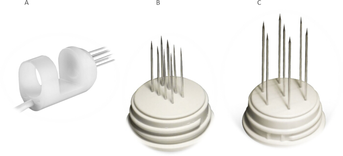
Figure 1: Type of electrodes used for BEST. (A) Finger, (B) linear, or (C) hexagonal electrodes. Please click here to view a larger version of this figure.
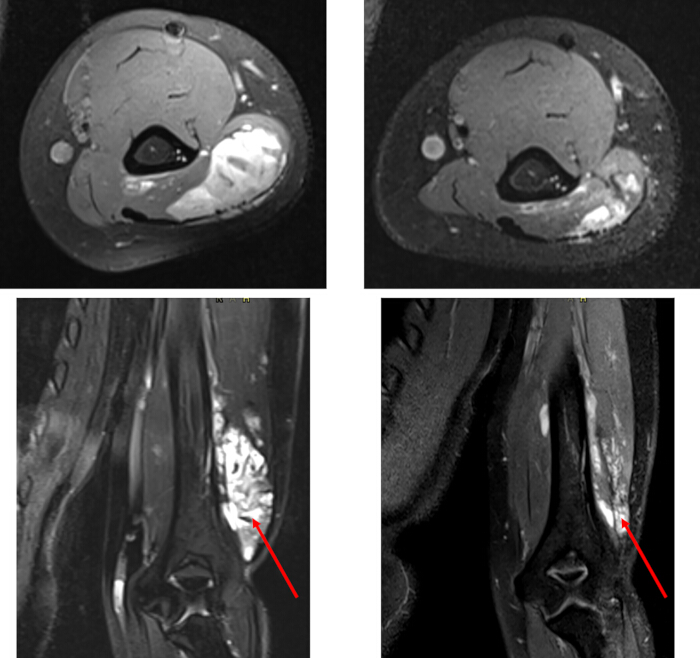
Figure 2: Effectiveness of BEST in the management of vascular malformations. Treatment of a venous malformation in the lower part of the triceps muscle, left arm. The left image shows the vascular malformation before BEST, whilst the right MRI image shows the vascular malformation 3 months after treatment. A significant reduction in lesion volume is observed. Please click here to view a larger version of this figure.
Discussion
Loading...
$$\rightleftharpoonup{xx}$$
$$\longleftharp{xx}$$,
$$\longrightharp{xx}$$,
The protocol standardizes BEST for the treatment of slow-flow vascular malformations by integrating principles of electrochemotherapy with image-guided sclerotherapy into a reproducible clinical workflow.
Key steps within the procedure are critical for achieving consistent therapeutic outcomes. Accurate pre-procedural imaging using ultrasound and MRI is essential to define lesion extent, compartmentalization, venous drainage, and proximity to critical anatomical structures, thereby enabling appropriate treatment planning and safe electrode placement. Equally important is the homogeneous intralesional distribution of bleomycin throughout all lesion compartments prior to electroporation. Incomplete drug distribution may result in heterogeneous electric field coverage and suboptimal endothelial exposure to bleomycin, ultimately reducing treatment efficacy. Immediate or near-immediate pulse delivery after injection is another crucial step, as electroporation transiently increases membrane permeability, enhancing intracellular uptake of bleomycin while simultaneously inducing a vascular lock effect that prolongs local drug retention. Correct electrode geometry, spacing, and avoidance of excessive overlap between electroporated areas are also necessary to minimize the risk of irreversible electroporation, tissue necrosis, bleeding, or post-treatment hyperpigmentation. Previous electrochemotherapy studies have demonstrated that electric field distribution critically influences therapeutic selectivity and tissue response5,6,7,8,9.
Several technical modifications and troubleshooting strategies may be required depending on lesion type, depth, and anatomical location. Superficial and smaller malformations can typically be treated using finger or linear electrodes, whereas deeper or more extensive lesions require long needle electrodes arranged in triangular or hexagonal geometries to ensure adequate electric field coverage. For macrocystic lymphatic malformations, aspiration of cystic fluid before bleomycin administration improves drug distribution and reduces dilution within cystic compartments. Ultrasound- or fluoroscopy-guided visualization of contrast distribution is particularly important in complex or septated lesions, where incomplete filling may necessitate needle repositioning or additional access points. Troubleshooting during the procedure may also include management of intralesional leakage, transient bleeding after electrode removal, inadequate electroporation coverage, or procedure-related edema. Applying gentle manual pressure with sterile gauze after pulse delivery may reduce bleeding from puncture sites, while post-procedural swelling can generally be controlled with analgesics, corticosteroids, or supportive care, depending on lesion location and severity3,4,10,11.
Despite promising clinical outcomes, BEST has several limitations that should be acknowledged. The procedure requires expertise in vascular anomaly imaging, percutaneous access, and electroporation-based therapies. Large, diffuse, or anatomically inaccessible malformations may be difficult to fully cover with electrodes, potentially leading to incomplete treatment responses. Although electroporation enables lower cumulative bleomycin doses compared with conventional sclerotherapy, cumulative lifetime exposure to bleomycin remains an important safety consideration because of the potential risk of pulmonary toxicity. Furthermore, transient edema, pain, skin discoloration, or hyperpigmentation may occur, particularly in superficial lesions or when the electric field overlap is excessive. Another limitation is the current lack of large prospective multicenter randomized studies directly comparing BEST with established therapies. Optimal parameters, including electric pulse coverage thresholds, electrode configurations, treatment intervals, and minimal effective bleomycin doses, also remain incompletely defined and warrant further investigation4,5,6,7,8,9,10,11.
Compared with conventional sclerotherapy, BEST offers several important advantages. Conventional bleomycin sclerotherapy often requires repeated treatment sessions because passive drug diffusion into dysplastic endothelium may be limited. In contrast, BEST substantially increases intracellular bleomycin uptake and prolongs local drug exposure through transient vascular lock effects, thereby improving therapeutic efficiency. In comparison with ethanol sclerotherapy, BEST may reduce the risk of severe tissue necrosis, fibrosis, nerve injury, and scarring. BEST also represents a minimally invasive alternative to surgery, particularly for lesions in anatomically challenging or cosmetically sensitive regions where surgical morbidity may be substantial. Importantly, early clinical studies suggest that BEST may be particularly beneficial in therapy-resistant or recurrent malformations that have responded poorly to conventional approaches3,4,10,11.
Future developments of BEST will likely focus on further optimization and personalization of treatment delivery. In addition, future translational studies investigating endothelial biology, vascular permeability, and molecular responses to electroporation may help define the mechanisms that determine treatment response and support the identification of predictive biomarkers. These studies may also clarify whether BEST can be combined with targeted molecular agents acting on PI3K/AKT/mTOR signaling pathways. Broader multicenter prospective studies, standardized treatment protocols, and long-term follow-up analyses will ultimately be required to validate safety, refine treatment parameters, and establish BEST as a standardized treatment modality for vascular malformations.
Disclosures
Loading...
$$\rightleftharpoonup{xx}$$
$$\longleftharp{xx}$$,
$$\longrightharp{xx}$$,
The authors declare no conflicts of interest.
Acknowledgements
Loading...
$$\rightleftharpoonup{xx}$$
$$\longleftharp{xx}$$,
$$\longrightharp{xx}$$,
The authors gratefully acknowledge financial support from the state budget of the Slovenian Research Agency (ARIS), program P3-0003.
Materials
| Name | Company | Catalog Number | Comments |
|---|---|---|---|
| Bleomycin medac | Medac | Drug | |
| Cliniporator VITAE | IGEA s.r.l. | IG0020B | Electroporation device |
| Electrodes N-30-4B | IGEA s.r.l. | IG0E152 | Needle row electrode |
| Electrodes N-30-HG | IGEA s.r.l. | IG0E104 | Hexagonal electrode |
| Electrodes F-20-NL | IGEA s.r.l. | IG0E380 | Needle row electrode |
| SonoVue | Electrodes F-20-NL | 519526 | Sulfur hexafluoride microbubble ultrasound contrast agent |
Reprints and Permissions
Request permission to reuse the text or figures of this JoVE article
Request Permission