This randomized study evaluates the use of fast-track surgery combined with postoperative targeted training in children with developmental dysplasia of the hip and demonstrates improved balance, motor, and hip function, along with enhanced exercise compliance and rehabilitation satisfaction compared with routine postoperative care.
A subscription to JoVE is required to view this content. Sign in or start your free trial.
Research Article
Fast-Track Surgery Combined with Postoperative Targeted Training in Children With Developmental Dysplasia of the Hip: A Randomized Study
In This Article
Summary
Abstract
Developmental dysplasia of the hip (DDH) is a common congenital musculoskeletal disorder in children, and effective postoperative rehabilitation is important for functional recovery. This randomized study evaluated the application of postoperative targeted training combined with fast-track surgery (FTS) in children with DDH. A total of 80 children with DDH treated between January 2022 and March 2023 were included. All children successfully underwent hip dislocation-related surgery and were randomly divided into two groups. Group A (n = 40) received general nursing combined with routine rehabilitation training, whereas Group B (n = 40) received FTS combined with postoperative targeted training. Balance function was assessed using the Berg Balance Scale (BBS), motor function using the Fugl–Meyer Assessment (FMA) scale, and hip function using the Harris Hip Score (HHS). Compliance with functional exercise and rehabilitation satisfaction were also evaluated. Before intervention, there were no significant differences between the two groups in dynamic or static BBS scores or HHSs (P > 0.05). After intervention, the dynamic and static BBS scores, HHSs, and FMA scores of Group B were significantly higher than those of Group A (P < 0.05). In addition, compliance with functional exercise and rehabilitation satisfaction were significantly better in Group B than in Group A (P < 0.05). These findings suggest that postoperative targeted training combined with FTS may promote the recovery of balance, motor, and hip function in children with DDH while improving exercise compliance and rehabilitation satisfaction.
Introduction
Developmental dysplasia of the hip (DDH) refers to displacement of the femoral head from the acetabulum at or shortly after birth and is accompanied by skeletal and soft tissue dysplasia. It is one of the most common congenital musculoskeletal disorders in children, with posterior dislocation being more common. Surgical intervention is the primary treatment for DDH. However, because children with DDH represent a special patient population, perioperative care and rehabilitation training are particularly important in addition to surgical treatment, as they contribute to surgical success and accelerate postoperative recovery of joint function1,2. With increasing expectations for healthcare quality, the importance of nursing services and rapid rehabilitation strategies has become increasingly recognized. Fast-track surgery (FTS) was introduced into clinical practice in 20023. FTS is a multidisciplinary model that integrates surgery, anesthesia, nursing, and other disciplines. Nursing interventions based on FTS not only facilitate perioperative rehabilitation but may also improve the utilization of medical resources4,5.
For children with DDH, postoperative rehabilitation exercise can promote blood circulation in the affected limb, reduce edema, prevent muscle atrophy and joint stiffness, minimize joint adhesion, and facilitate an earlier return to daily activities6,7. Despite the recognized benefits of both FTS and rehabilitation training8, studies evaluating the combined application of FTS and postoperative targeted training in children with DDH remain limited. Therefore, this study investigated the application of postoperative targeted training combined with FTS in children with DDH to provide a scientific reference for rehabilitation interventions in this population.
Access restricted. Please log in or start a trial to view this content.
Protocol
This study was approved by the Medical Ethics Committee of Shanxi Children’s Hospital (Ethics Approval No. 1027/11/2021). A total of 80 children with DDH were enrolled between January 2022 and March 2023. Written informed consent was obtained from the legal guardians of all participating children before enrollment. All 80 children successfully underwent DDH-related corrective surgery at our hospital. The procedure consisted of open reduction combined with Salter innominate osteotomy in all cases, including 40 children in Group A and 40 children in Group B. Adductor tenotomy and capsulorrhaphy were performed as adjunctive procedures when required according to intraoperative hip stability, soft-tissue tension, and surgeon judgment. All procedures were performed by the same pediatric orthopedic surgical team, and postoperative hip immobilization was applied according to the institutional pediatric orthopedic protocol.
The sample size was estimated based on the primary outcome of hip function, as assessed by the Harris Hip Score after the 8-week intervention. According to preliminary clinical data and previous rehabilitation studies involving children with developmental dysplasia of the hip, a moderate between-group effect size was anticipated. Using a two-sided α level of 0.05, a statistical power of 80%, and a 1:1 allocation ratio, a minimum of 36 participants was required per group. To account for an anticipated dropout rate of approximately 10%, a total of 80 participants were enrolled in this randomized study. The children were randomly divided into two groups, with 40 cases in each group. Randomization was performed using a computer-generated random number sequence at a 1:1 allocation ratio, and the allocation sequence was generated by a researcher who was not involved in participant recruitment, intervention delivery, or outcome assessment. Group assignments were placed in sequentially numbered, opaque, sealed envelopes, which were opened only after eligibility assessment and guardian consent had been completed.
The baseline characteristics of the two groups were comparable (P > 0.05) (Table 1), and the overall study workflow, including participant enrollment, eligibility assessment, randomization, intervention allocation, 8-week follow-up, outcome assessment, and statistical analysis, is shown in Figure 1.
| Characteristic | Category | Group A (n = 40) | Group B (n = 40) | χ²/t | P Value |
| Sex, n (%) | Male | 12 (30.00) | 10 (25.00) | χ² = 0.251 | 0.617 |
| Female | 28 (70.00) | 30 (75.00) | |||
| Age (years) | 5.35 ± 0.48 | 5.43 ± 0.50 | t = 0.730 | 0.468 | |
| Site of involvement, n (%) | Right hip | 10 (25.00) | 11 (27.50) | χ² = 0.065 | 0.799 |
| Left hip | 30 (75.00) | 29 (72.50) | |||
| Tönnis classification, n (%) | Grade I | 8 (20.00) | 7 (17.50) | χ² = 0.114 | 0.99 |
| Grade II | 10 (25.00) | 10 (25.00) | |||
| Grade III | 10 (25.00) | 11 (27.50) | |||
| Grade IV | 12 (30.00) | 12 (30.00) |
Table 1: Baseline Characteristics of the Study Population. Baseline demographic and clinical characteristics of children with developmental dysplasia of the hip (DDH) enrolled in Group A and Group B prior to intervention. Data are presented as mean ± standard deviation (SD) or n (%). No statistically significant differences were observed between groups at baseline (P > 0.05).
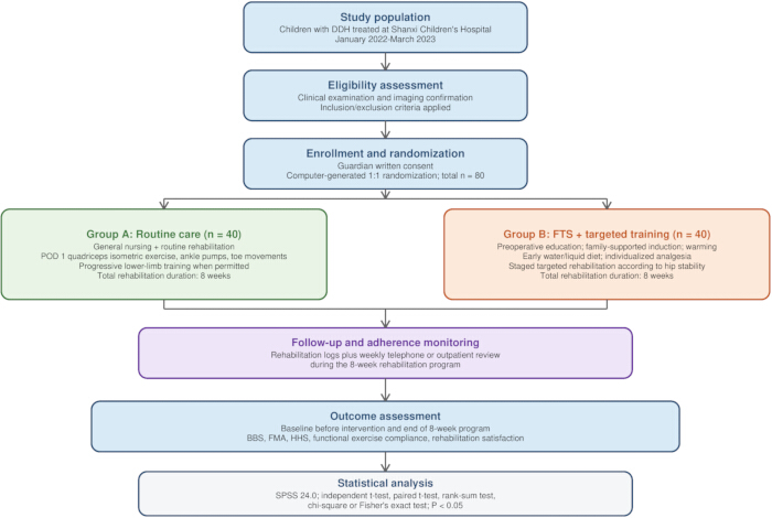
Figure 1. Overall study workflow. Schematic representation of participant enrollment, eligibility assessment, randomization, intervention allocation, follow-up, outcome assessment, and statistical analysis. Children with developmental dysplasia of the hip were screened according to the inclusion and exclusion criteria, enrolled after written guardian consent, and randomly assigned to Group A (general nursing combined with routine rehabilitation training) or Group B (fast-track surgery combined with postoperative targeted training). Both groups completed an 8-week rehabilitation program with adherence monitoring. Outcome measures, including balance function, motor function, hip function, compliance with functional exercise, and rehabilitation satisfaction, were assessed at baseline and at the end of the 8-week intervention period, followed by statistical analysis. DDH, developmental dysplasia of the hip; FTS, fast-track surgery; BBS, Berg Balance Scale; FMA, Fugl–Meyer Assessment; HHS, Harris Hip Score. Please click here to view a larger version of this figure.
Inclusion Criteria
The inclusion criteria were as follows: 1) DDH was confirmed by clinical examination combined with imaging assessment. Clinical evaluation included the Barlow test, Allis sign examination, Ortolani test, limited hip abduction assessment, and the hip and knee flexion abduction test. Imaging confirmation was performed using pelvic anteroposterior radiography for children with ossified femoral heads and hip ultrasonography for younger children, when appropriate. DDH was diagnosed when imaging demonstrated acetabular dysplasia, an abnormal acetabular index, disruption of Shenton’s line, lateral or superior displacement of the femoral head, hip subluxation, or hip dislocation. The final diagnosis was confirmed by an experienced pediatric orthopedic surgeon; 2) The guardian of the child was informed about the study and provided written informed consent; 3) All hip dislocation-related surgical procedures were successfully completed at our hospital.
Exclusion Criteria
The exclusion criteria were as follows: 1) Coagulation dysfunction; 2) Congenital malformations of both lower limbs; 3) Presence of active infectious diseases before surgery, including fever, respiratory tract infection, urinary tract infection, gastrointestinal infection, skin or soft-tissue infection around the surgical area, elevated leukocyte count or C-reactive protein levels suggestive of active infection, or any infection requiring antibiotic treatment before surgery.
Group A
General nursing combined with routine rehabilitation training was used. Family members were informed of the precautions during the perioperative period, including fasting and fluid restriction. After surgery, routine rehabilitation exercises were initiated on postoperative day 1 after stable vital signs were confirmed and the attending surgeon permitted rehabilitation activity. Quadriceps isometric contraction, ankle pump movements, and toe joint movements were performed for 10–15 repetitions per set, 3 sets per day, for approximately 10–15 minutes per session. During hospitalization, the exercises were taught and supervised by rehabilitation nurses or therapists. After discharge, the same exercises were continued at home under guardian supervision.
In the later stage of rehabilitation, generally beginning from postoperative week 6 after wound healing, stable hip reduction, and permission for progressive lower-limb activity had been confirmed by the attending surgeon, children were instructed to perform lower-limb training, including gentle squatting and supine pedaling exercises. These activities were introduced only after the postoperative immobilization status allowed the corresponding range of hip and lower-limb movement. These later-stage exercises were performed for 10–15 minutes per session, 2–3 sessions per day, under therapist instruction during hospitalization and guardian supervision after discharge.
During the training, progression was implemented in a step-by-step manner, and the total exercise duration was 8 weeks. The 8-week rehabilitation program was conducted during hospitalization and continued after discharge. During hospitalization, training was supervised by rehabilitation nurses or therapists. After discharge, guardians recorded daily exercise completion in a rehabilitation log, including exercise type, frequency, duration, and any discomfort. Adherence was checked through weekly telephone follow-up or outpatient review.
Group B
FTS combined with postoperative targeted training was used.
1. Before surgery, children and their families received education regarding the disease using cartoons, videos, and other methods. The treatment plan, surgical procedure, and hospitalization precautions were explained to alleviate anxiety. The educational intervention was delivered by trained nursing staff using standardized cartoon booklets, short videos, face-to-face explanations, and question-and-answer communication. The content included DDH-related knowledge, surgical preparation, anesthesia and perioperative precautions, postoperative pain control, diet, immobilization care, rehabilitation precautions, and home exercise requirements. Education was provided once after admission and once on the day before surgery, with each session lasting approximately 20–30 min. Guardians were encouraged to ask questions, and a teach-back method was used to confirm understanding.
2. Anesthesia induction was performed in the presence of family members to relieve anxiety. During surgery, a thermal blanket was used to maintain body temperature. The infusion temperature and room temperature were maintained at approximately 28°C. These perioperative measures were implemented as part of the FTS bundle and were applied to Group B. Group A received routine perioperative nursing according to the standard institutional protocol. To ensure equitable clinical treatment, both groups were managed by the same surgical and anesthesia teams and received the same basic perioperative safety monitoring, infection-prevention measures, and postoperative clinical observation. The main difference between groups was the structured FTS-based perioperative management and targeted rehabilitation program applied in Group B.
3. Children were allowed to drink water immediately after surgery and received an appropriate liquid diet 2 h later.
4. Postoperative analgesia was selected according to the child’s age, body weight, pain severity, surgical procedure, and anesthesiologist’s assessment. Children with moderate-to-severe postoperative pain or those requiring continuous analgesia received an analgesic pump according to the institutional pediatric postoperative analgesia protocol under anesthesiologist supervision. Children with mild pain or those suitable for oral medication received ibuprofen suspension at 5–10 mg/kg per dose every 6–8 h as needed, without exceeding the maximum daily dose recommended for children. Pain was assessed using the Face, Legs, Activity, Cry, and Consolability scale, and analgesic adjustment was performed when clinically necessary.
For targeted training, family members were informed about the importance of postoperative training, dietary recommendations were provided, exercise precautions were explained, and the limb function of each child was assessed. Limb function was assessed before individualized training according to hip range of motion, lower-limb muscle activation, pain response, wound condition, postoperative immobilization status, ability to cooperate with exercise, and surgeon-confirmed hip stability. The rehabilitation plan was adjusted according to these findings to avoid excessive hip movement, excessive force, or premature weight bearing.
Targeted training was implemented in stages according to postoperative recovery and immobilization status. From postoperative day 1, only distal joint movement, isometric muscle contraction, circulation-promoting exercises, breathing exercises, and guardian-assisted positioning care were performed when vital signs were stable and the attending surgeon permitted activity. Hip-related active exercises, including cross-legged exercise, flexion-extension exercise, and sitting-up exercise, were not performed during the early immobilization period. These exercises were introduced only after wound healing, stable hip reduction, and surgeon permission had been confirmed, generally from postoperative week 6. Weight-bearing walking training was initiated only after the attending surgeon confirmed that partial or full weight bearing was clinically appropriate.
Distal joint movement and isometric muscle contraction were started from postoperative day 1 when permitted, and hip-related active exercises were introduced only after immobilization status, wound healing, pain response, and surgical stability had been reviewed. Targeted training consisted of cross-legged exercises and flexion-extension exercises (30 repetitions/session, 3 sessions/day) and sitting-up exercises (30 repetitions/session, 3 sessions/day). After the child was able to get out of bed, ankle joint, quadriceps femoris, and toe joint exercises were strengthened, and weight-bearing walking training was performed for 30 min/session, 2 sessions/day.
Readiness for out-of-bed activity and weight-bearing walking was determined jointly by the attending surgeon and rehabilitation therapist. Criteria included stable vital signs, controlled pain, absence of wound complications, stable hip reduction on clinical and/or imaging evaluation, adequate lower-limb muscle activation, and permission for weight bearing according to the postoperative immobilization protocol. Weight-bearing training was not initiated until the surgeon confirmed that partial or full weight bearing was clinically appropriate.
Because postoperative immobilization and permitted hip movement varied according to surgical stability and recovery stage, each exercise was performed only when clinically appropriate. During the immobilization period, training focused on distal joint movement, isometric muscle contraction, circulation-promoting exercises, breathing exercises, and guardian-assisted positioning care. Cross-legged exercises, sitting-up exercises, active hip flexion-extension exercises, and weight-bearing walking were introduced only after the attending surgeon confirmed stable hip reduction and permitted the corresponding movement or weight-bearing activity.
Lower extremity joint training was intensified for 2 weeks, and training intensity was gradually increased according to a step-by-step approach. A total of 8 weeks of training was completed. Training progression was based on standardized clinical criteria, including absence of increased pain, no new swelling, no wound abnormality, stable hip position, adequate completion of the previous exercise level, improved cooperation with exercise, and approval from the rehabilitation therapist and attending surgeon. Exercise frequency, duration, range of motion, and weight-bearing level were increased gradually only when these criteria were met.
Both groups were supervised by the same rehabilitation nursing team and pediatric rehabilitation therapists. Before study implementation, all staff received standardized training regarding intervention procedures, exercise instructions, safety precautions, and outcome-recording methods. A standardized rehabilitation checklist was used to ensure consistency of intervention delivery and documentation across participants. During routine care and rehabilitation, clinically evident safety events, including wound abnormalities, suspected hip instability, gastrointestinal intolerance, analgesia-related adverse effects, neurovascular abnormalities, and rehabilitation-related pain, were recorded according to institutional documentation procedures. Rehabilitation progression was permitted only after confirmation of stable vital signs, adequate pain control, absence of wound complications, and surgeon-confirmed hip stability. Safety events were monitored as part of routine clinical care; however, complication rates were not predefined study outcomes and were therefore not included in the formal statistical analyses.
Outcome Measures
All outcome measures were assessed at two time points: before intervention, defined as the baseline assessment before surgery or before initiation of the rehabilitation program, and after intervention, defined as the end of the 8-week rehabilitation program. The prespecified outcomes focused on functional recovery and rehabilitation-related outcomes. FTS-specific perioperative indicators, including length of hospital stay, time to ambulation, perioperative stress-response markers, and postoperative complication rates, were not included as outcome measures.
Balance Function:
Compliance with functional exercise and rehabilitation satisfaction were evaluated at the end of the 8-week rehabilitation period. Balance function was evaluated using the Berg Balance Scale (BBS)9. The scale includes 14 items assessing lower extremity balance function, including independent sitting, standing with eyes closed, sit-to-stand transitions, and stand-to-sit transitions, comprising 6 static and 8 dynamic assessment items. Each item is scored from 0 to 4, with a maximum score of 56. Higher scores indicate better lower extremity balance function.
Motor Function:
Motor function was evaluated using the Fugl–Meyer Assessment (FMA)10. The FMA includes 17 items assessing lower extremity motor function, such as reflex activity and flexor movement, with a maximum score of 34. Higher scores indicate better lower extremity motor function.
Hip Function:
Hip function was evaluated using the Harris Hip Score (HHS)11. The total score is 100 and includes assessments of pain, range of motion, daily activities, and walking distance. Higher scores indicate better hip function.
Compliance with Functional Exercise:
Compliance with functional exercise was assessed according to criteria adapted from the literature12. Complete compliance was defined as completion of the prescribed daily exercise program with adequate quality and quantity. Partial compliance was defined as completion of only part of the exercise program or performance below the required quantity or quality. Noncompliance was defined as refusal to exercise or completion of only a few exercise sessions, resulting in poor exercise outcomes. Exercise compliance was assessed by a rehabilitation nurse who was not directly responsible for daily intervention delivery. Guardians completed a daily rehabilitation log documenting the type, frequency, and duration of exercises performed. During hospitalization, the log was checked by nursing staff. After discharge, compliance was verified through weekly telephone follow-up or outpatient review. The assessor reviewed the rehabilitation logs and follow-up records to classify compliance as complete compliance, partial compliance, or noncompliance.
Rehabilitation Satisfaction:
Rehabilitation satisfaction was assessed using a self-designed rehabilitation satisfaction questionnaire developed by the study team according to the rehabilitation procedures used in this study. The questionnaire contained 10 items covering clarity of rehabilitation education, understanding of exercise methods, perceived exercise safety, comfort during rehabilitation, pain-management satisfaction, communication with medical staff, feasibility of home exercise, guardian participation, perceived recovery support, and overall satisfaction. Each item was scored from 0 to 10, with a total score of 100. Scores ≥90 were classified as very satisfied, scores of 75–89 as satisfied, and scores ≤75 as dissatisfied. Total satisfaction was calculated as (very satisfied + satisfied) / total cases × 100%. The questionnaire was reviewed by five pediatric orthopedic and rehabilitation nursing experts for content relevance before use and was pilot-tested in 20 guardians to confirm clarity and feasibility. The internal consistency of the questionnaire was acceptable, with a Cronbach’s α of 0.86 in the pilot assessment. Formal external validation was not performed, and this was acknowledged as a limitation of the study.
Blinding of Methods
Outcome assessments were performed by trained assessors who were not involved in daily intervention delivery. Group allocation was not actively disclosed to the assessors during functional evaluation. However, because some rehabilitation procedures differed visibly between groups, complete assessor blinding could not be fully guaranteed.
Statistical Analysis
Statistical analyses were performed using SPSS Statistics version 24.0. The Jarque–Bera test was used to assess the normality of measurement data. Normally distributed continuous variables were expressed as mean ± standard deviation (SD). Between-group comparisons were performed using the independent-samples t-test, whereas within-group comparisons between pre-intervention and post-intervention values were performed using the paired-samples t-test. Non-normally distributed continuous variables were analyzed using nonparametric tests. Ordinal data were analyzed using the Mann–Whitney U test or rank-sum test, as appropriate. Categorical data were expressed as n (%) and analyzed using the chi-square (χ2) test or Fisher’s exact test, as appropriate. A two-sided P value of <0.05 was considered statistically significant.
Access restricted. Please log in or start a trial to view this content.
Results
Balance Function
Balance function was assessed before intervention and after completion of the 8-week rehabilitation program. There was no significant difference in dynamic or static BBS scores between the two groups before intervention (P > 0.05). After the 8-week intervention, the dynamic and static BBS scores of Group B were significantly higher than those of Group A (P < 0.05), indicating better balance function in children who received FTS combin...
Access restricted. Please log in or start a trial to view this content.
Discussion
DDH is commonly associated with unequal lower limb length, a waddling gait, limping, and other functional abnormalities. If timely intervention is not provided, the disease may progress, involving the acetabulum, joint capsule, ligaments, and surrounding muscles, thereby aggravating joint instability and dislocation and adversely affecting the quality of life and mental health of affected children13. Therefore, effective perioperative management and rehabilitation strategies are important componen...
Access restricted. Please log in or start a trial to view this content.
Disclosures
Conflict of Interest:
The authors declare that they have no competing financial interests related to this work.
Materials
| Name | Company | Catalog Number | Comments |
|---|---|---|---|
| Analgesic infusion pump | ICU Medical / Smiths Medical | CADD-Solis Ambulatory Infusion Pump, Model 2110 | Multimodal postoperative pain management |
| Anti-slip rehabilitation mat | AIREX AG | AIREX Elite Balance Pad; SKU 2970000879 | Balance and gait training |
| Berg Balance Scale (BBS) assessment form | Rehabilitation Medicine Standard Tool | N/A | Used for evaluation of static and dynamic balance function |
| Educational cartoons/videos | Hospital-prepared materials | N/A | Preoperative education for children and families. |
| Fugl–Meyer Assessment (FMA) scale | Rehabilitation Medicine Standard Tool | N/A | Lower-extremity motor function assessment |
| Harris Hip Score (HHS) form | Orthopedic Clinical Assessment Tool | N/A | Evaluation of postoperative hip function |
| Ibuprofen suspension (oral) | Aurohealth LLC | NDC 58602-002-24 | Postoperative analgesia in children |
| Pediatric thermal blanket | Solventum / 3M Bair Hugger | Global Catalog Number 55000; Product ID 7100212597 | Intraoperative temperature maintenance |
| Printed rehabilitation satisfaction questionnaire | Self-designed | N/A | Assessment of rehabilitation satisfaction. |
| SPSS Statistics software (Version 24.0) | IBM Corp., USA | N/A | Statistical data analysis |
| Standard pediatric hospital bed | Stryker | Cub Pediatric Crib, Model FL19H | Used for postoperative positioning and rehabilitation training |
References
- Myers CA, et al. Effect of intraoperative treatment options on hip joint stability following total hip arthroplasty. J Orthop Res. 2022;40(3):604-613.
- Nerakh G, et al. Microcephalic primordial dwarfism with predominant Meier-Gorlin phenotype, ichthyosis, and multiple joint deformities-Further expansion of DONSON cell cycle-opathy phenotypic spectrum. Am J Med Genet A. 2022;188(7):2139-2146.
- Kehlet H, Wilmore DW. Multimodal strategies to improve surgical outcome. Am J Surg. 2002;183(6):630-641.
- Silva MS, et al. Radiography, CT, and MRI of hip and lower limb disorders in children and adolescents. Radiographics. 2019;39(3):779-794.
- Resnick MJ. Effect of multimodal prehabilitation vs postoperative rehabilitation on 30-day postoperative complications for frail patients undergoing resection of colorectal cancer: A randomized clinical trial. J Urol. 2020;204(4):869-870.
- Rao L, et al. Effect of nursing intervention to guide early postoperative activities on rapid rehabilitation of patients undergoing abdominal surgery: A protocol for systematic review and meta-analysis. Medicine (Baltimore). 2021;100(12):24776.
- Ji X, Ding H. The efficacy of enteral nutrition combined with accelerated rehabilitation in non-small cell lung cancer surgery: A randomized controlled trial protocol. Medicine (Baltimore). 2020;99(48):23382.
- Ding XF, et al. Short-term efficacy evaluation of open reduction and Salter innominate osteotomy for the treatment of developmental dislocation of the hip in children. Orthopedic Journal of China. 2013;21(9):879-882.
- Weng CS, et al. Assessments of internal reliability and concurrent validity of Berg Balance Scale in stroke patients. Chinese Journal of Rehabilitation Medicine. 2007;22(8):688-690.
- Lundquist CB, Maribo T. The Fugl-Meyer assessment of the upper extremity: Reliability, responsiveness and validity of the Danish version. Disabil Rehabil. 2017;39(9):934-939.
- Huang BL, Yu NS. Harris score after total hip arthroplasty. Journal of Modern Clinical Medical Bioengineering. 2004;10(1):44-46.
- Si ZX, et al. Reliability and validity of the modified Morisky Medication Adherence Scale applied to patients on warfarin therapy after mechanical heart-valve replacement. Journal of Nursing Science. 2012;27(22):23-26.
- Sioutis S, et al. Developmental dysplasia of the hip: A review. J Long Term Eff Med Implants. 2022;32(3):39-56.
- Zhang C, Xiao J. Application of fast-track surgery combined with a clinical nursing pathway in the rehabilitation of patients undergoing total hip arthroplasty. Int J Clin Med Res. 2020;48(1):1-13.
- Irie T, et al. Three-dimensional hip joint congruity evaluation of the borderline dysplasia: Zonal-acetabular radius of curvature. J Orthop Res. 2020;38(10):2197-2205.
- Koene S, et al. Hearing loss, cleft palate, and congenital hip dysplasia in female carriers of an intragenic deletion of AMMECR1. Am J Med Genet A. 2022;188(5):1578-1582.
- Li X, et al. Application of situational adaptation training combined with childlike nursing for children undergoing tonsillectomy or adenoidectomy. Int J Pediatr Otorhinolaryngol. 2021;145:110707.
- Stringer MR, et al. Periprosthetic fractures of the femur in primary total hip arthroplasty: A New Zealand Joint Registry analysis. ANZ J Surg. 2021;91(3):404-408.
- Zhang L, et al. Influence of continuing nursing care team mode on the rehabilitation of patients with pancreatic cancer after postoperative daily chemotherapy. Support Care Cancer. 2022;30(11):9003-9009.
- Gaffney et al. Effect of simulated rehabilitation on hip joint loading during single limb squat in patients with hip dysplasia. J Biomech. 2021;22(116):132-138.
- Freke M, et al. Acute and subacute changes in dynamic postural control after hip arthroscopy and postoperative rehabilitation. J Athl Train. 2022;57(5):494-501.
- Gao C, et al. The efficacy of bosentan combined with vardenafil in the treatment of postoperative pulmonary hypertension in children with congenital heart disease: A protocol of randomized controlled trial. Medicine (Baltimore). 2021;100(1).
- Li J, et al. Efficacy of surgery combined with postoperative 125I interstitial brachytherapy for treatment of acinic cell carcinoma of the parotid gland in children and adolescents. Pediatr Blood Cancer. 2020;67(7).
- Eiermann J, et al. Recommendations for rehabilitation after surgical treatment of cranial cruciate ligament disease in dogs: A 2017 survey of veterinary practitioners. Vet Surg. 2020;49(1):80-87.
- Zhou K, et al. Effects of progressive upper limb exercises and muscle relaxation training on upper limb function and health-related quality of life following surgery in women with breast cancer: A clinical randomized controlled trial. Ann Surg Oncol. 2019;26(7):2156-2165.
- Ramirez JL, et al. Depression predicts non-home discharge after abdominal aortic aneurysm repair. Ann Vasc Surg. 2021;74(41):131-140.
- Lähdeoja T, et al. Subacromial decompression surgery for adults with shoulder pain: A systematic review with meta-analysis. Br J Sports Med. 2020;54(11):665-673.
- Zacharias A, et al. Comparison of gluteus medius and minimus activity during gait in people with hip osteoarthritis and matched controls. Scand J Med Sci Sports. 2019;29(5):696-705.
- Yang L, Li J. Neuraxial anesthesia compared to general anesthesia in subjects with hip fracture surgery: A meta-analysis. Int J Clin Med Res. 2023;15(2):66-76.
- Lin Y, et al. A tri-channel oxide transistor concept for the rapid detection of biomolecules including the SARS-CoV-2 spike protein. Adv Mater. 2022;34(3).
- Zhu MZL, et al. Traumatic fracture dislocation of the thoracic spine sparing both the spinal cord and aorta. ANZ J Surg. 2019;89(5):212-213.
- Hao JZ, Wang K, Feng J. Effects of early postoperative rehabilitation training on pain severity, limb function and quality of life of children with developmental dislocation of hip. Modern Journal of Integrated Traditional Chinese and Western Medicine. 2022;31(6):848-851.
- Osama H, Saeed H, Nicola M. Neuraxial anesthesia compared to general anesthesia in subjects with hip fracture surgery: A meta-analysis of randomized clinical trials. Int J Clin Med Res. 2023;1(2):37-48.
- Chen J, Chen TT. Effects of lower extremity rehabilitation training based on rapid rehabilitation surgery on lower extremity motor function of children with developmental dislocation of hip joint. Maternal and Child Health Care of China. 2022;37(11):2005-2008.
Access restricted. Please log in or start a trial to view this content.