This retrospective study evaluates the feasibility and safety of ultrasound-guided vacuum-assisted breast biopsy for resecting breast nodules ≤ 1.5 cm with BI-RADS 3–4a in patients with retroglandular breast implants, demonstrating high resection success and low complication rates.
A subscription to JoVE is required to view this content. Sign in or start your free trial.
Research Article
Feasibility of Ultrasound-Guided Vacuum-Assisted Breast Biopsy for Breast Nodules in Patients with Retroglandular Breast Implants
* These authors contributed equally
In This Article
Summary
Abstract
Ultrasound-guided Vacuum-Assisted Breast Biopsy has been widely utilized as a key modality for breast nodule resection. Nevertheless, its application remains relatively contraindicated in patients who have undergone breast prosthesis placement. This study aimed to evaluate the feasibility and safety of ultrasound‑guided vacuum‑assisted breast biopsy (US‑VABB) for the resection of breast nodules ≤ 1.5 cm with BI-RADS 3–4a in patients with retroglandular breast implants. A single-center retrospective observational study was conducted on patients with breast nodules who underwent US-VABB in the Department of Ultrasound, Beijing Friendship Hospital, Capital Medical University from January 2020 to October 2024. A total of 18 patients (18 nodules) who had previously undergone breast implant placement with implants located behind the glandular tissue were included. The complete nodule resection rate, implant-related complications, postoperative pathological results, and short-term follow-up data were analyzed. Primary outcomes included complete resection rate and implant-related complications, while secondary outcomes included pathological findings and short-term follow-up results. The maximum diameter of the nodules in the 18 patients ranged from 0.5 to 1.5 cm (mean: 1.1 ± 0.4 cm), and all nodules were completely resected in a single operation, with no evidence of residual lesions on imaging. No serious complications such as breast implant rupture or leakage occurred during or after the operation. The implant rupture rate was 0% (0/18), and minor complications occurred in 16.7% of patients. Postoperative pathology revealed fibroadenoma (n=11), breast adenosis (n=3), intraductal papilloma (n=2), atypical ductal hyperplasia (n=1), and ductal carcinoma in situ (DCIS, n=1). The patient with DCIS underwent additional resection, with no residual tumor detected. These findings demonstrate that US-VABB is a safe and feasible minimally invasive approach for patients with retroglandular breast implants presenting with breast nodules ≤1.5 cm and BI-RADS 3–4a, with high resection success and low complication rates.
Introduction
The use of breast implant surgery in aesthetic and reconstructive procedures has increased significantly in recent years1. Accordingly, the detection rate of breast nodules in patients with breast implants has increased due to widespread imaging screening. Achieving accurate diagnosis and minimally invasive treatment of breast nodules while preserving implant integrity remains a significant clinical challenge.
Currently, ultrasound-guided core needle biopsy (CNB) is a commonly used diagnostic method for breast nodules that require further clarification of their pathological characteristics2. However, for small or deep-seated nodules, CNB may result in insufficient sampling, missed targeting, and pathological underestimation. In patients with breast implants, limited operating space and implant position restrict the puncture pathway, increasing procedural difficulty and risk. Patients with benign nodules may still request resection due to psychological concerns, cosmetic reasons, or lesion growth3. However, traditional surgical resection is invasive, and the presence of implants increases procedural complexity and complication risk, limiting its use2.
As an interventional technique combining accurate biopsy and minimally invasive resection, Ultrasound-guided Vacuum-Assisted Breast Biopsy (US-VABB) has the advantages of small incision, sufficient sampling, high complete resection rate, and good postoperative aesthetic effect4,5. In recent years, it has been widely used in the diagnosis and treatment of benign breast lesions and some low-risk lesions6,7,8. However, due to repeated rotational resections under negative pressure, this technique carries potential risks of implant damage and incomplete lesion removal3. Therefore, breast implants are considered a relative contraindication, limiting the application of this technique in such patients.
To date, limited clinical evidence is available regarding the safety and feasibility of ultrasound-guided vacuum-assisted breast biopsy (US-VABB) in this patient population. In this study, we retrospectively analyzed 18 patients to evaluate the feasibility and safety of US‑VABB in patients with retroglandular breast implants presenting with breast nodules ≤ 1.5 cm and BI‑RADS 3–4a and to identify key operative strategies. This study thus seeks to provide clinical evidence for the minimally invasive diagnosis and treatment of such breast nodules in breast implant recipients. We hypothesize that US‑VABB can be safely and effectively performed in these selected patients with breast implants without increasing implant‑related complications.
Access restricted. Please log in or start a trial to view this content.
Protocol
Ethical statement
This study was approved by the Ethics Committee of Beijing Friendship Hospital (approval number: NO.2022-P2-060-01). As a retrospective study, re-obtaining written informed consent was waived. All procedures involving human participants were performed in accordance with institutional and national research ethics guidelines and the Declaration of Helsinki. This was a single-center retrospective descriptive study. The data of patients with breast nodules who underwent US-VABB in the Department of Ultrasound, Beijing Friendship Hospital from January 2020 to October 2024 were retrospectively analyzed.
A total of 20 patients were screened, of whom 18 met the inclusion criteria and were included in the final analysis. Among them, 18 female patients who had previously undergone breast implant placement with implants located behind the glandular tissue were included, with a total of 18 breast nodules. The age of the patients ranged from 33 to 55 years, with an average of (44.5 ± 7.8) years. All patients were confirmed to have breast nodules based on preoperative ultrasound and magnetic resonance imaging (MRI).
Study population and selection criteria
Patients were included if they underwent preoperative evaluation using both ultrasound and magnetic resonance imaging (MRI), had breast nodules classified as BI-RADS category 3 with a maximum diameter of approximately 1.0–1.5 cm and a clear desire for therapeutic resection, or BI-RADS category 4a with a maximum diameter of approximately 0.5–1.0 cm requiring diagnostic excisional biopsy. All included patients had breast implants located behind the glandular tissue. These strict and specific selection criteria were critical to the favorable safety profile and low complication rate observed in this study.
Patients were excluded if imaging suggested a high suspicion of malignancy (BI-RADS category 4b or higher), if nodules were adjacent to the implant or located behind the nipple preventing the establishment of a safe operating space, if the patient was pregnant, or if the patient had severe coagulation disorders or other contraindications to surgery.
Instruments and equipment
A vacuum-assisted breast biopsy system with 7G and 10G rotational resection needles was used. The ultrasound equipment was a Mindray R9 color Doppler ultrasound diagnostic instrument with a linear array probe frequency of 9-12 MHz.
Surgical methods
Patients were placed in the supine position. Prior to surgery, a standard ultrasound examination was conducted for two core purposes: first, to identify the body surface location of breast nodules; second, to evaluate key nodule characteristics including size, shape, depth, and spatial relation to the implants. After routine disinfection and draping, a 23G needle was used under ultrasound guidance to inject a local anesthetic mixture (5 mL of 2% lidocaine + 0.1 mL of 1:1000 epinephrine + normal saline supplemented to 30 mL) at the skin incision and nodule needle tract.
To ensure the completeness of lesion resection, appropriate diameter of rotational resection needle was selected according to the specific characteristics of the nodules in this study:
The selection of 7G or 10G needles was based on nodule diameter and length‑to‑short‑diameter ratio, and these criteria were predefined in clinical practice and uniformly applied to all patients in this study. For lesions with a maximum diameter ≤ 1 cm or a length/short diameter ratio > 2, a 10G rotational resection needle was used. Its needle slot size was better adapted to the volume of such lesions, and resection could be completed by a single or a small number of multiple incisions9. For lesions with a maximum diameter > 1 cm and a length/short diameter ratio ≤ 2, a 7G rotational resection needle was adopted to enhance resection efficiency by virtue of its larger needle slot capacity. Concurrently, this approach reduced tissue irritation surrounding the implant caused by repeated needle insertion and withdrawal, thereby minimizing the risk of indirect implant injury. During the resection operation, incisions were uniformly started from below the nodules, and the natural tension of the tissues was used to facilitate the entry of the lesions into the needle slot. For superficial nodules, an additional skin isolation zone was established, and the angle of the needle slot was adjusted to avoid pulling the implant when the needle penetrates the superficial tissues during resection. Ultrasound imaging was used for real-time multi-directional monitoring to assess lesion reduction and ensure complete resection, and to dynamically evaluate implant integrity for any rupture or damage.
To reduce the risk of implant rupture during the operation, targeted measures were taken in both preoperative screening and intraoperative operation in this study. Combined with the inherent length of the rotational resection needle slot, the maximum diameter of the included nodules was strictly limited to no more than 1.5 cm preoperatively to reduce the amplitude of needle swing during the operation and avoid unnecessary traction or impact on the implant due to excessive operating range. Nodules with a length/short diameter ratio greater than 2 were preferred. Such nodules were more elliptical in shape, and the resection range was relatively concentrated, which could improve the controllability during the operation and reduce excessive operation caused by irregular nodule shape. The needle was inserted in a nearly parallel manner, with a slow insertion angle and reduced insertion impulse to reduce mechanical damage to the implant capsule by the needle tip and shaft. During the operation, a sufficient safe distance was established between the glandular layer and the implant to ensure that the distance between them was not less than 1 cm, reserving a buffer space for the advancement, retraction, and rotation of the rotational resection needle, and physically avoiding direct contact or compression of the implant by the needle. For specific operations, refer to Figure 1, which only shows the core operational steps and not the entire process.
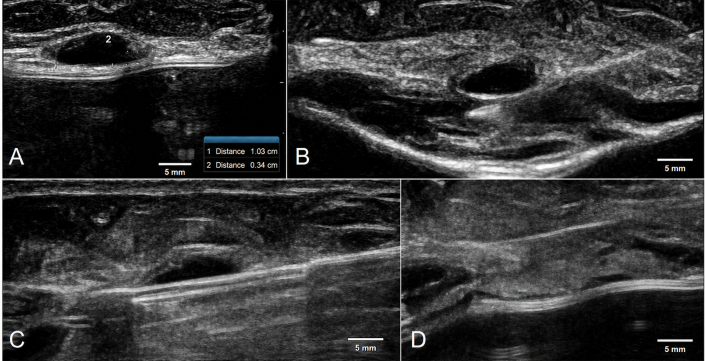
Figure 1: Ultrasound-guided rotational resection of nodules anterior to breast implants: Intraoperative procedure diagram (A) A 36-year-old female with a BI-RADS category 3 breast nodule anterior to the implant, with a maximum diameter < 1.5 cm, meeting indications for therapeutic resection. (B) Establishment of a safe distance (≥ 1.0 cm) between the glandular layer and the implant during the procedure. (C) Insertion of a 7-gauge rotational resection needle in a near-parallel approach beneath the nodule. (D) Complete resection of the nodule without damage to the breast implant. Please click here to view a larger version of this figure.
After resection, residual blood was aspirated under negative pressure, and the rotational resection needle was withdrawn. The surgical area was packed with folded sterile gauze and compressed with an elastic bandage for 24 h. All resected tissues were sent for pathological examination.
Statistical methods
Due to the limited number of cases of patients with breast implants undergoing US-VABB, the sample size of this study included all consecutive cases meeting the inclusion and exclusion criteria during the study period. Therefore, descriptive statistical methods were used to analyze the results. Statistical analysis was performed using statistical analysis software (See Table of Materials for details). Continuous variables were expressed as mean ± standard deviation, and categorical variables were expressed as frequencies and percentages. The 95% confidence intervals (Clopper-Pearson exact method) were calculated for the complete nodule resection rate, complication rate, and implant rupture rate. Due to the small sample size, no hypothesis testing or multivariate analysis was performed.
Access restricted. Please log in or start a trial to view this content.
Results
Study population and selection criteria
US-VABB was successfully completed for all 18 breast nodules in 18 patients. The maximum diameter of the nodules ranged from 0.5 to 1.5 cm, with an average of (1.1 ± 0.4) cm. Among the imaging classifications, there were 12 cases (66.7%) of BI-RADS category 3 nodules and 6 cases (33.3%) of BI-RADS category 4a nodules. According to the location of the nodules in the glandular tissue, Superficial nodules were defined as those located ...
Access restricted. Please log in or start a trial to view this content.
Discussion
Breast implant placement has become a common method to improve women's physical appearance3, and the diagnostic and therapeutic needs of patients with breast implants combined with breast nodules have become increasingly prominent. However, due to the presence of implants, such patients face many limitations in the selection of biopsy and minimally invasive resection methods for breast nodules. In this study, US-VABB achieved complete resection in all cases with no implant-related complication...
Access restricted. Please log in or start a trial to view this content.
Disclosures
The authors have no conflict of interest regarding any drugs, materials, or devices described in this study.
Acknowledgements
The authors would like to thank all patients who participated in this study for their cooperation and support. We also thank the colleagues in our department for their assistance with data collection and clinical management.
Data Availability:
The datasets generated and/or analyzed during the current study are publicly available in the Zenodo repository, accessible at: https://doi.org/10.5281/zenodo.19791301. All relevant data supporting the findings of this study are included within the article and its supplementary materials. Additional data may be obtained from the corresponding author upon reasonable request.
Access restricted. Please log in or start a trial to view this content.
Materials
| Name | Company | Catalog Number | Comments |
|---|---|---|---|
| 0.9% Sodium Chloride Injection (Normal Saline) | Baxter, USA | 2B0044 | 0.9% Sodium Chloride Injection, for anesthetic solution dilution and intraoperative irrigation |
| Disposable Sterile 23G Hypodermic Needle | BD, USA | 305122 | 23G × 1" (0.6×25mm), for local anesthetic infiltration |
| EnCor Vacuum-Assisted Breast Biopsy System | Bard, USA | DR ENCor | Rotating biopsy probe models: ECP01-7G or ECP01-10G, groove length 19 mm |
| Epinephrine Hydrochloride Injection | Hospira, USA | NDC 0409-1113-01 | 1:1000 Epinephrine Hydrochloride Injection, diluted to 1:100000 for local anesthesia admixture |
| Lidocaine Hydrochloride Injection | AstraZeneca, UK | NDC 0310-0313-10 | 1% Lidocaine Hydrochloride Injection, for local infiltration anesthesia |
| Mindray R9 Color Doppler Ultrasound Diagnostic System (Linear Array Probe) | Mindray Medical | R9 | Probe frequency: 9~12 MHz |
| SPSS 26.0 | IBM Corporation, USA | 26 | Statistical software for data analysis; RRID: SCR_002865 |