This protocol evaluates the effect of smoking on platelet-rich fibrin (PRF) by comparing growth factor release and blood parameters between smokers and non-smokers using a standardized and reproducible method.
Research Article
Effect of Smoking on Growth Factors in Platelet-Rich Fibrin: Ex-Vivo Pilot Study
In This Article
Summary
Abstract
Platelet rich fibrin (PRF) and growth factors are routinely used to accelerate hard and soft tissue regeneration in periodontal surgical procedures, dental implant applications and oral surgery for the last 30 years in the field of dentistry. The aim of this study was to evaluate the effect of smoking on autologous growth factors within PRF. The study population consisted of 20 male participants (10 smokers/10 non-smokers) and 20 female participants (10 smokers/10 non-smokers). Each participant provided two tubes of venous blood (approximately 7 ml each). The first tube was used for a complete blood count (CBC). The second tube was centrifuged at 708 x g for 12 min, and the resulting PRF was analyzed using enzyme-linked immunosorbent assay (ELISA) to determine the levels of growth factors (Platelet-derived growth factor (PDGF), Transforming growth factor-beta (TGF-β), Vascular endothelial growth factor (VEGF), Fibroblast growth factor (FGF) and Insulin-like growth factor (IGF)). The results showed that smoking resulted in a decrease in mean platelet volume (MPV), platelet distribution width (PDW) and platelet larger cell ratio (P-LCR) values (p<0.05) in the whole population. In females, smoking led to a decrease in red blood cell (RBC), MPV, PDW and P-LCR levels, while in male subjects, it resulted in an increase in hematocrit (HCT) values (p<0.05). It was also found that smoking led to an increase in FGF and TGFβ-1 within PRF (p<0.05). Although current results showed a slight increase in certain growth factors in smokers, the clinical relevance of this finding should be investigated in further studies.
Introduction
The utilization of autologous platelet concentrates in various fields of dentistry began in the 1980s and 1990s. Since then, platelet concentrates and growth factors have been routinely employed in periodontal surgical procedures, dental implant applications, and oral surgery to accelerate the regeneration of hard and soft tissues1. Platelets containing growth factors play crucial roles in cell migration, proliferation, differentiation, and angiogenesis, all of which are integral to tissue regeneration. Autologous platelet concentrates are produced by centrifuging venous blood at different speeds, with or without the use of thrombin and anticoagulants2. The growth factors present in platelet-rich fibrin (PRF) include platelet-derived growth factor (PDGF), transforming growth factor-beta (TGF-β), vascular endothelial growth factor (VEGF), and insulin-like growth factor (IGF), among others3,4. These growth factors are known to influence various cellular processes essential for tissue healing and regeneration5. Factors affecting the levels and efficacy of these growth factors in PRF include the patient's age and sex, PRF preparation method, platelet count in the collected blood, platelet distribution width (PDW), and mean platelet volume (MPV)as well as patient’s general health status, the presence of systemic conditions, and external influences such as smoking6.
Smoking poses a significant health hazard, particularly impacting coagulation physiology and directly affecting platelet function7. Cigarette smoke contains numerous toxic compounds, including nicotine, carbon monoxide, and reactive oxygen species, which are known to affect platelet behavior and vascular biology8,9 resulting in altered degranulation and release of growth factors8,10. Therefore, smoking may potentially influence the biological composition and functionality of platelet-rich fibrin (PRF) through multiple interconnected mechanisms such as disrupting cellular homeostasis and modulating cytokine production9,11 changing fibrin clot formation and architecture, including alterations in fibrin fiber thickness, density, and cross-linking12,13, modulating inflammatory responses and endothelial dysfunction which can further impact leukocyte activity and the regenerative microenvironment9,14. All of these factors may potentially affect the quality and quantities of bioactive molecules incorporated into the PRF matrix and influence the mechanical stability of PRF and the kinetics of growth factor release9,11.
The impact of smoking on the biological properties of platelet concentrates, including platelet-rich fibrin (PRF) has been investigated in recent studies. Comparative studies evaluating smokers and non-smokers have reported differences in growth factor release, fibrin architecture, and cellular content of PRF10,11. In particular, smoking has been associated with alterations in key growth factors such as transforming growth factor beta-1 (TGF-β1), platelet-derived growth factor (PDGF), and vascular endothelial growth factor (VEGF), as well as with structural changes in the fibrin network15,16. While Lara et al. (2023) reported that leukocyte- and platelet-rich fibrin (L-PRF) obtained from smokers and non-smokers exhibited similar fibrin architecture, cellular distribution, and uniaxial tensile properties, suggesting that smoking may not significantly affect the biomechanical behavior of PRF in vitro17. Srirangarajan et al. reported that smoking may influence the membrane properties of platelet-rich fibrin (PRF) and advanced platelet-rich fibrin (A-PRF), including their structural and morphological characteristics18.
Although previous studies have investigated the effects of smoking on platelet concentrates and PRF, the available evidence remains limited and inconsistent, and the underlying biological and molecular mechanisms have not been fully clarified17. These discrepancies may be attributed to variations in study design, differences in age and sex distribution across study populations, inconsistencies in PRF preparation protocols, and patient-related factors such as smoking intensity and duration11,19. In addition, most existing studies have focused primarily on general platelet behavior, fibrin architecture, membrane morphology, or broad biological characteristics of PRF, whereas quantitative data on specific PRF-derived growth factors remain scarce, inconsistent, and frequently restricted to single-time-point analyses. As a result, it is still unclear whether smoking alters the quantitative early release profile of PRF-derived growth factors under standardized conditions.
Furthermore, the time-dependent release pattern of these growth factors in smokers versus non-smokers, as well as the potential dose-related association with smoking intensity, has not been clearly established. Therefore, standardized and reproducible studies are needed to evaluate both the quantity and temporal release kinetics of PRF-derived growth factors in relation to smoking status20,21. Against this background, the rationale, aim, and hypothesis of the present study were formulated as follows. The positive effects of autologous growth factors in PRF on wound healing have been demonstrated in numerous studies, especially during surgical procedures in dentistry, although contradictory results also exist, probably related to individual differences in PRF characteristics. The aim of this study was to investigate the effect of smoking on the biological activity of platelet-rich fibrin by quantitatively evaluating growth factor levels at different time points and assessing their association with smoking intensity. The null hypothesis of the current study was that smoking does not alter the release kinetics and levels of growth factors in platelet-rich fibrin. It also states that smoking intensity is not associated with measurable differences in these growth factor levels between smokers and non-smokers.
Protocol
This study was conducted at the Department of Periodontology, Faculty of Dentistry, Cukurova University, with the participation of 20 male and 20 female volunteers. All participants provided written informed consent after being informed about the purpose, methodology, and procedures of the study. Ethical approval was obtained from the Ethics Committee of the Faculty of Medicine, Cukurova University (Approval No: 125/54, Date: 16.09.2022). All procedures, including essential biosafety guidance for handling human blood and waste disposal instructions for biohazardous materials and chemical reagents, were performed in accordance with NIH-CDC guidelines22. The reagents and the equipment used are listed in the Table of Materials.
1. Patient Selection
Participants included in the study were healthy individuals between 18 and 30 years of age, without any known systemic diseases, and who demonstrated adequate cooperation during the procedures. Individuals were excluded if they had any systemic disease, were pregnant or breastfeeding, had any bleeding disorders, or were using systemic medications that could affect hemostatic parameters. An equal number of male and female participants were included in the study to ensure balanced sex distribution between the groups. Smoking status was determined based on self-reported information obtained from the participants. Individuals who reported current daily cigarette smoking were classified as smokers, whereas individuals who reported no current smoking history were classified as non-smokers. Former smokers and occasional smokers were excluded to maintain clear group allocation. In addition, the number of cigarettes smoked per day was recorded for each smoker, and correlations between smoking intensity and the study outcome parameters were analyzed. No biochemical validation (e.g., cotinine measurement) was performed.
The sample size was determined based on feasibility considerations and the number of eligible subjects available during the study period. The present study was conceived as a preliminary exploratory clinical investigation.
2. Blood collection and group allocation
A total of four study groups were established: non-smoking female participants (n = 10), non-smoking male participants (n = 10), smoking female participants (n = 10), and smoking male participants (n = 10 Blood samples were collected from 20 healthy men (10 smokers and 10 non-smokers) and 20 healthy women (10 smokers and 10 non-smokers). Two blood collection tubes (approximately 7 mL each) were obtained from each participant. The first tube (sterile glass tube with ethylenediaminetetraacetic acid as an anticoagulant) was used for complete blood count analysis, including white blood cell (WBC), red blood cell (RBC), hemoglobin (HGB), hematocrit (HCT), platelet (PLT), mean platelet volume (MPV), platelet distribution width (PDW), platelet large cell ratio (P-LCR), and platelet crit (PCT) values. The second tube (sterile glass tube with no anticoagulant) was centrifuged for PRF preparation, and the resulting PRF was analyzed for growth factor levels using ELISA. The weight of the obtained PRF was measured and recorded.
3. PRF preparation
All PRF samples were prepared under strictly standardized conditions to reduce inter-sample variability. Venous blood was drawn from each participant using the same procedure and centrifuged immediately under identical settings. Centrifugation was performed at 708 × g for 12 min at room temperature using a centrifuge equipped with a 40° fixed-angle rotor and 10 mL glass tubes. Although the rotor radius of the device could be adjusted between 88 mm and 110 mm, a radius of 88 mm was consistently used in the present study to standardize the protocol. No anticoagulants were added at any stage of the procedure. Macroscopic evaluation of PRF formation was carried out immediately after centrifugation. Proper PRF formation was defined as the presence of a clearly identifiable fibrin clot located between the red blood cell layer and the plasma layer. In addition, the clot was required to exhibit a consistent gel-like structure with sufficient mechanical integrity to allow separation with sterile forceps. Only samples demonstrating a well-formed and clearly separated PRF clot were included in the study. Samples showing incomplete clot formation or poor layer separation were excluded from analysis, and when necessary, additional blood samples were collected from the participants during the same session.
4. Growth factor ELISA analysis
PRF samples were processed for growth factor analysis under strictly standardized experimental conditions to ensure comparability among participants. Following preparation, each PRF clot obtained from the 40 volunteers was separated from the red blood cell layer using sterile scissors and weighed on a precision balance. Cell culture medium was then added to each clot in proportion to its weight. After medium addition, the samples were incubated for 72 h on an orbital shaker operating at a slow speed. At the predefined evaluation time points of 24 h and 72 h, the media surrounding the PRF clots, representing PRF-derived exudates/supernatants, were collected and transferred into separate microcentrifuge tubes for subsequent ELISA analysis. The collected samples were stored at -80 °C until use. To minimize protein degradation and technical variation, all specimens were handled according to the same protocol, analyzed within the same experimental period, and thawed only once prior to measurement. Samples were kept on ice during relevant processing steps whenever applicable.
Quantitative analysis of IGF-1, PDGF, FGF, VEGF, and TGF-β1 levels was performed using commercially available ELISA kits in accordance with the manufacturer’s instructions. All measurements were carried out in duplicate, and the mean values were used for statistical analysis. Standard curves were prepared for each assay, and only readings within the linear detection range were included in the final evaluation. The same reagents, equipment, and incubation conditions were used throughout the study, and careful pipetting procedures were applied to reduce technical variability.
5. ELISA steps
For each assay, 100 µL of standards and sample aliquots were dispensed into the designated wells and incubated at 37 °C for 90 min. The wells were then washed three times with wash buffer. Subsequently, 100 µL of biotin-labeled antibody solution was added to each well, followed by incubation at 37 °C for 60 min and a further three washing steps. Next, 100 µL of horseradish peroxidase (HRP) solution was applied and incubated at 37 °C for 30 min. After three additional washes, 100 µL of substrate solution was added and allowed to react for 15 min. Finally, 50 µL of stop solution was added to each well, and absorbance was measured at 450 nm using a microplate reader.
6. Statistical analysis
The sample size per group was 20; therefore, the assumption of normality could not be ensured with sufficient confidence. Distributional properties of the variables were evaluated using graphical methods, including histograms and Q-Q plots. Considering the limited sample size and the uncertainty regarding normality assumptions, primary analyses were conducted using non-parametric tests, which are more appropriate for small samples and data that may not follow a normal distribution. The study groups were initially stratified by sex to ensure balanced distribution; however, statistical analyses were performed based on smoking status. Sex-based comparisons were not included in the primary analysis, as the study was not designed or powered to detect differences between male and female groups.
The Mann-Whitney U test was used for comparisons between two independent groups, while the Wilcoxon signed-rank test was applied for paired comparisons. Correlations between continuous variables were assessed using Spearman’s rank correlation coefficient23,24. To further evaluate the longitudinal effect of time and the interaction between time and smoking status, repeated-measures ANOVA was also performed as a secondary analysis. Despite the relatively small group size, repeated-measures ANOVA is generally considered robust to moderate deviations from normality when group sizes are similar. Therefore, repeated-measures ANOVA results were reported to support the consistency of the findings and to assess time and interaction effects. Statistical analyses were performed using a statistical software package. A significance level of 0.05 was used for all tests.
DATA AVAILABILITY:
All raw data generated and analyzed during this study have been deposited in the open-access repository Zenodo and are publicly available. The dataset can be accessed via the following DOI:10.528/zenodo.19204955.
Results
A total of 20 males (10 smokers and 10 non-smokers, aged between 18 and 25) and 20 females (10 smokers and 10 non-smokers, aged between 18 and 25) healthy participants were included in the study.
Evaluation of the Relationship Between Smoking and Blood Parameters
The comparison of blood parameters based on smoking status is presented in Table 1 and the correlation between the number of cigarettes smoked and blood parameters is shown in Table 2. Between-group comparisons were performed using the Mann–Whitney U test, while correlations were analyzed using Spearman’s rank correlation test. It was found that MPV, PDW and P-LCR were significantly lower in smokers, and the decreases in these parameters were significantly correlated with the number of cigarettes smoked.
Evaluation of the Relationship Between Smoking and Growth Factors
The comparison of growth factors based on smoking status and time is presented in Table 3 and the correlation between the number of cigarettes smoked and growth factors is shown in Table 4. Between-group comparisons at each time point were performed using the Mann–Whitney U test, and correlations were evaluated using Spearman’s rank correlation test. The inter-group comparisons showed that smokers had significantly higher FGF (Figure 1) and TGFβ-1 (Figure 2) levels (p = 0.005 and p = 0.019, respectively) on Day 1, and these levels were positively correlated with the number of cigarettes per day. In addition, intra-group comparisons over time were performed using the Wilcoxon signed-rank test, which indicated a significant decrease in FGF and TGFβ-1 levels and a significant increase in IGF-1 levels (Figure 3) from Day 1 to Day 3 in smokers compared to non-smokers. No significant inter- or intra-group differences were found for VEGF (Figure 4) and PDGF (Figure 5) between smokers and non-smokers.
Repeated-measures analysis of variance (ANOVA) was employed to evaluate the impact of time and the time-by-smoking interaction on each growth factor. A significant main effect of time was detected for FGF (p = 0.009), IGF-1 (p < 0.001), and TGF β-1 (p = 0.005), while no significant time effect was observed for VEGF (p = 0.182) and PDGF (p = 0.262). There was no significant time*smoking interaction for FGF (p = 0.112), VEGF (p = 0.535), IGF-1 (p = 0.622), or PDGF (p = 0.671). The interaction term for TGF-β1 approached statistical significance but did not reach the predefined threshold (p = 0.056).
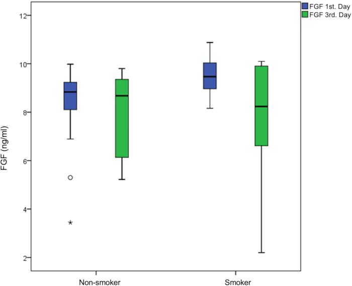
Figure 1: FGF Levels in PRF at 24 h (T1) and 72 h (T2). Box plots compare FGF concentrations in PRF samples obtained from non-smoker and smoker groups at day 1 and day 3. A significant increase was observed in the smoker group at day 1; however, FGF levels in smokers decreased significantly at day 3 compared with day 1. Data are presented as median and interquartile range, with outliers indicated. *p < 0.05 (n = 40). Please click here to view a larger version of this figure.
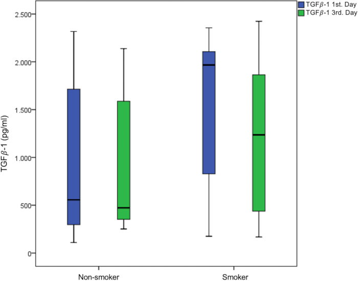
Figure 2: TGF-β1 Levels in PRF at 24 h (T1) and 72 h (T2). Comparison of TGF-β1 concentrations in PRF obtained from non-smoker and smoker groups at day 1 and day 3. A significant increase was observed in the smoker group at day 1; however, TGF-β1 levels in smokers decreased significantly at day 3 compared with day 1. Data are presented as median and interquartile range, with outliers indicated. *p < 0.05 (n = 40). Please click here to view a larger version of this figure.
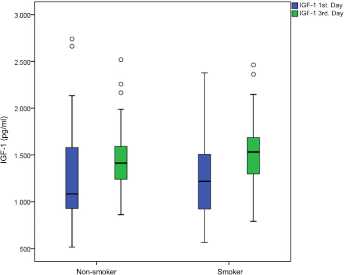
Figure 3: IGF-1 Levels in PRF at 24 h (T1) and 72 h (T2). IGF-1 concentrations in platelet-rich fibrin (PRF) were measured in samples obtained from non-smoker and smoker groups at day 1 and day 3. A significant increase was observed at day 3 in the smoker group. Data are presented as median and interquartile range, with outliers indicated. *p < 0.05 (n = 40). Please click here to view a larger version of this figure.
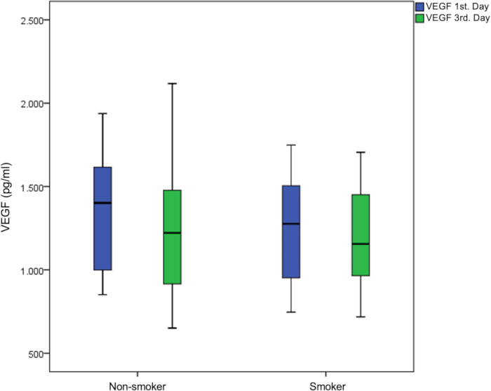
Figure 4: VEGF Levels in PRF at 24 h (T1) and 72 h (T2). VEGF levels in PRF showed no significant change over time or between groups. Bar plots display median and interquartile range for each time point *p < 0.05 (n = 40). Please click here to view a larger version of this figure.
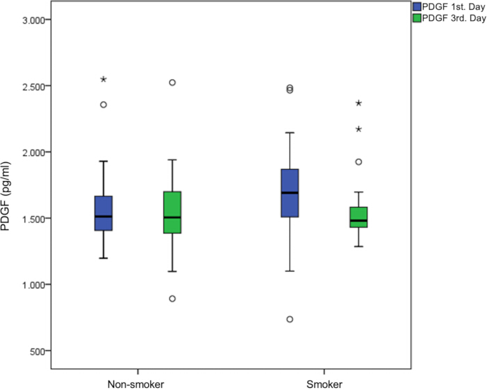
Figure 5: PDGF Levels in PRF at 24 h (T1) and 72 h (T2). Box plots compare PDGF concentrations in PRF samples obtained from non-smoker and smoker groups at day 1 and day 3. No significant differences were observed between groups or time points. Data are presented as median and interquartile range, with outliers indicated *p < 0.05 (n = 40). Please click here to view a larger version of this figure.
| Smoking status | |||
| Blood Parameters | Smokers | Non-smokers | P-value |
| WBC | 7.2±1.3 | 7.9±2.1 | 0.358 |
| 7.4(5-9.7) | 7.9(5-12.6) | ||
| RBC | 4.8±0.7 | 5.1±0.6 | 0.317 |
| 4.7(3.1-5.8) | 4.9(4.1-7.3) | ||
| HGB | 14.1±1.8 | 13.7±1.6 | 0.44 |
| 14.2(10.8-16.9) | 13.7(10.2-16) | ||
| HCT | 42.8±4.8 | 41.8±3.8 | 0.203 |
| 42.1(31.8-48.7) | 41.9(33.3-46.5) | ||
| PLT | 268.4±62.9 | 273.9±70.7 | 0.946 |
| 276(153-367) | 269(162-414) | ||
| MPV | 10.4±0.6 | 11.3±1 | 0.003* |
| 10.4(9.2-11.7) | 11.3(9.4-13.3) | ||
| PDW | 12±1.5 | 14.4±2.6 | 0.002* |
| 11.7(9.7-15.3) | 13.5(10.2-19.7) | ||
| P-LCR | 28±5.5 | 36±8.8 | 0.003* |
| 27.5(17.5-38.7) | 35.6(19.2-51.3) | ||
| PCT | 0.3±0.1 | 0.3±0.1 | 0.271 |
| 0.3(0.2-0.4) | 0.3(0.2-0.5) | ||
Table 1: Comparison of Blood Parameters by Smoking Status. Data are presented as mean ± standard deviation or median (minimum–maximum), depending on data distribution. Comparisons between smokers and non-smokers were performed using the Mann–Whitney U test. A p-value< 0.05 was considered statistically significant. Abbreviations: WBC, white blood cell; RBC, red blood cell; Hb, hemoglobin; Hct, hematocrit; PLT, platelet; MPV, mean platelet volume; PDW, platelet distribution width; P-LCR, platelet-large cell ratio.
| WBC | RBC | HGB | HCT | PLT | MPV | PDW | P-LCR | PCT | |
| Correlation coefficient (r) with number of cigarettes (piece) | -0.089 | -0.066 | 0.225 | 0.301 | -0.094 | -0.365 | -0.4 | -0.384 | -0.265 |
| P | 0.584 | 0.684 | 0.162 | 0.059 | 0.562 | 0.021* | 0.011* | 0.015* | 0.099 |
Table 2: Correlation Between Number of Cigarettes Smoked and Blood Parameters. Correlation analyses were performed using Spearman’s rank correlation coefficient. Correlation coefficients (r) and corresponding p-values are presented. A p-value< 0.05 was considered statistically significant. Abbreviations: WBC, white blood cell; RBC, red blood cell; Hb, hemoglobin; Hct, hematocrit; PLT, platelet; MPV, mean platelet volume; PDW, platelet distribution width; P-LCR, platelet-large cell ratio.
| Smoking Status | Day 1 | Day 3 | p | p value (time) | p value (time*smoking) | |
| FGF | Smokers | 9.4±0.7 | 7.8±2.3 | 0.015* | 0.009 | 0.112 |
| 9.5(8.2-10.9) | 8.2(2.2-10.1) | |||||
| Non-Smokers | 8.3±1.6 | 7.9±1.7 | 0.36 | |||
| 8.8(3.4-10) | 8.7(5.2-9.8) | |||||
| P | 0.005* | 0.534 | ||||
| VEGF | Smokers | 1246.8±324.6 | 1200.5±308.5 | 0.55 | 0.182 | 0.535 |
| 1276.4(746-1748.5) | 1155.6(718.5-1705.2) | |||||
| Non-Smokers | 1348.7±351.3 | 1223.4±371.9 | 0.351 | |||
| 1401(851-1937.7) | 1221.4(651-2117.7) | |||||
| P | 0.425 | 0.989 | ||||
| IGF-1 | Smokers | 1296.1±483.7 | 1562.4±403.1 | 0.001* | <0.001 | 0.622 |
| 1217.8(564.6-2377.1) | 1530.9(789.6-2462.1) | |||||
| Non-Smokers | 1299.3±627 | 1507.3±427.9 | 0.057 | |||
| 1082.8(514.6-2740.9) | 1412.8(860.9-2518.4) | |||||
| P | 0.705 | 0.482 | ||||
| PDGF | Smokers | 1694.8±403 | 1569.8±279 | 0.218 | 0.262 | 0.671 |
| 1690.9(736.4-2483.6) | 1480.9(1285.5-2368.2) | |||||
| Non-Smokers | 1603.6±337 | 1547±336.8 | 0.514 | |||
| 1511.8(1197.3-2548.2) | 1505(891.8-2523.6) | |||||
| P | 0.13 | 0.914 | ||||
| TGFβ-1 | Smokers | 1571.8±788.7 | 1217.8±786.6 | 0.014* | 0.005 | 0.056 |
| 1966.8(175.2-2356) | 1236.4(167.5-2424.5) | |||||
| Non-Smokers | 919.2±753.4 | 847±687.2 | 0.455 | |||
| 556.8(109.9-2317.5) | 473.3(251.4-2138.3) | |||||
| P | 0.019* | 0.204 |
Table 3: Comparison of Growth Factors by Smoking Status and Time. Data are presented as median (minimum–maximum). Between-group comparisons (smokers vs. non-smokers) were performed using the Mann–Whitney U test. Within-group comparisons (Day 1 vs Day 3) were analysed using the Wilcoxon signed-rank test. A p-value< 0.05 was considered statistically significant. FGF levels showed a significant decrease over time in smokers (p = 0.015), where as no significant temporal change was observed in non-smokers. Abbreviations: IGF, insulin-like growth factor; PDGF, platelet-derived growth factor; FGF, fibroblast growth factor; VEGF, vascular endothelial growth factor; TGF-β1, transforming growth factor beta-1.
| FGF | FGF | VEGF | VEGF Day 3 | IGF-1 Day 1 | IGF-1 Day 3 | PDGF Day1 | PDGF Day 3 | TGFβ-1 Day 1 | TGFβ-1 Day 3 | |
| Day 1 | Day 3 | Day 1 | ||||||||
| Correlation coefficient (r) with number of cigarettes (piece) | 0.454 | 0.12 | -0.09 | -0.032 | 0.056 | 0.07 | 0.244 | -0.007 | 0.343 | 0.187 |
| P | 0.003* | 0.46 | 0.58 | 0.847 | 0.732 | 0.669 | 0.129 | 0.968 | 0.030* | 0.247 |
Table 4: Correlation Between Number of Cigarettes Smoked and Growth Factors. Correlation analyses were performed using Spearman’s rank correlation coefficient. Correlation coefficients (r) and p-values are presented. A p-value< 0.05 was considered statistically significant. A significant positive correlation was observed between smoking intensity and FGF levels at Day 1, as well as TGF-β1 levels at Day 1. Abbreviations: IGF, insulin-like growth factor; PDGF, platelet-derived growth factor; FGF, fibroblast growth factor; VEGF, vascular endothelial growth factor; TGF-β1, transforming growth factor beta-1.
Discussion
In this study, we aimed to evaluate the impact of smoking on autologous growth factors within Platelet-Rich Fibrin (PRF), a widely used material in dental surgeries to enhance wound healing. The findings indicated that smoking slightly affects blood parameters. In addition, the evaluation of the effects of smoking on the growth factors levels at different time points revealed that smoking influences the release profile of growth factors with these effects being correlated with the number of cigarettes smoked. Each of these findings directly supports the rejection of the null hypothesis that smoking does not affect the biological properties of PRF. Specifically, the significant alterations in platelet indices (MPV, PDW, P-LCR) demonstrate that smoking affects baseline hematological characteristics. The increased early release of FGF and TGF-β1 indicates that smoking modifies the initial biological activity of PRF. Furthermore, the observed temporal changes in growth factor levels (decrease in FGF and TGF-β1 and increase in IGF-1 over time) confirm that smoking alters the release kinetics of PRF-derived growth factors. Together, these results provide consistent evidence that smoking has a measurable and multifaceted impact on PRF biology.
The impact of smoking on hematological parameters is controversial. Various studies have compared the hematological parameters of smokers and non-smokers, finding changes in total WBC, RBC, HGB, HCT, and PLT values25. A non-significant increase in PLT values was also noted among smokers in the current study. Misra et al. (2018) reported increased platelet counts in smokers, consistent with our findings26. Conversely, Sandhya et al. (2015) found reduced platelet counts in chronic smokers, associating prolonged smoking with decreased platelet counts27. Lupia et al. suggested that increased thrombopoietin levels in smokers stimulate platelet production, raising platelet counts28. Kario et al. (1992) reported increased MPV values in smokers, which decreased upon smoking cessation, contrary to our findings of a non-significant decrease in MPV while Butkiewicz et al. found no effect of smoking on MPV, highlighting conflicting results in the literature25.
Few studies have examined the effects of smoking on physical properties and growth factor content of PRF. Srirangarajan et al. (2020) noted morphological differences in PRF membranes, with smokers showing more disorganized fibrin structures while Lara et al. have shown that smoking does not alter the tensile properties of PRF membranes17,18. It has also been demonstrated that cotinine, the main nicotine metabolite, is present in PRF29. However, recent studies showed comparable levels of growth factors and biomolecules released from PRF from both smokers and nonsmokers29,30 with similar effects on angiogenesis and osteoblast differentiation31. The clinical relevance of increased levels of FGF and TGFβ-1 found in the current study on wound healing and regeneration should further be investigated.
The increased early release of FGF and TGF-β1 in smokers may be attributed to smoking-induced platelet activation and enhanced degranulation during PRF formation. Nicotine-related mechanisms have been shown to stimulate platelet activation pathways and growth factor release8,9. However, the subsequent decrease in these growth factors over time suggests a depletion of growth factor reserves, resulting in a shift from sustained release toward an early burst release pattern. In contrast, the increase in IGF-1 over time may reflect delayed release dynamics within the fibrin matrix10,16. The time points of 24 h and 72 h were selected to represent the early and intermediate phases of growth factor release from platelet-rich fibrin (PRF). Previous studies have shown that PRF exhibits a sustained release profile, with an initial release occurring within the first 24 h followed by a continued release over several days10,16. Therefore, the 24 h time point reflects the early release phase, while 72 h represents a later stage of growth factor release. These time points were chosen to evaluate temporal changes and better characterize the release kinetics of PRF.
Smoking-related oxidative stress likely contributes to these findings. Reactive oxygen species may enhance platelet activation while simultaneously altering fibrin structure, resulting in a less organized matrix that affects growth factor retention and release 8,14. These changes may explain the altered release kinetics observed in smokers. Previous studies have primarily focused on the structural and mechanical characteristics of PRF. For instance, Lara et al. reported no significant differences in the tensile properties of PRF between smoker sand non-smokers, suggesting that smoking may not affect its biomechanical behavior17. In contrast, the present study revealed differences in growth factor release, indicating that biological activity may also be affected even when mechanical properties remain unchanged. This suggests that structural integrity alone may not fully reflect the functional capacity of PRF. Similarly, Srirangarajan et al. demonstrated alterations in the morphological characteristics of PRF membranes in smokers, indicating that smoking may influence fibrin structure18. In addition, studies evaluating platelet function have shown that smoking can alter platelet activation and growth factor release, supporting the biological basis of the present findings8,9.
The discrepancies between studies may be attributed to several factors. First, differences in PRF preparation protocols, including centrifugation speed, duration, and relative centrifugal force, can significantly influence the cellular composition and growth factor content of PRF20. Second, variations in study design, such as the selection of time points, may affect the detection of growth factor release patterns. While some studies evaluate PRF at a single time point, the present study analysed temporal changes (24 h and 72 h), providing a more comprehensive understanding of release kinetics10,16. Additionally, population characteristics such as smoking intensity, duration of smoking, and individual biological variability may contribute to inconsistent findings. Environmental and systemic factors, including oxidative stress and inflammatory status, may also influence platelet function and fibrin network formation8,14.
The present study provides several important contributions to the current literature. Unlike many previous studies that focused primarily on structural or mechanical aspects of PRF, this study specifically evaluated the quantitative and temporal release of growth factors. Furthermore, the inclusion of multiple growth factors and the assessment of their changes over time offer a more detailed understanding of the biological behavior of PRF. Another strength of this study is the evaluation of the relationship between smoking intensity and growth factor levels, which remains insufficiently explored in the literature. This approach provides additional insight into dose-dependent effects of smoking on PRF biology19. From a clinical perspective, these findings are relevant for the use of PRF in regenerative procedures in smokers. Even if PRF appears structurally intact, alterations in growth factor release may compromise its regenerative potential. Therefore, clinicians should consider the potential impact of smoking when planning PRF-based therapies and interpreting treatment outcomes.
This study has several limitations. First, the sample size was relatively small, and the findings should be confirmed in larger populations. Second, although participants were systemically healthy, donor-related physiological factors such as age, sex, hormonal status, stress during blood collection, and time of blood draw may have influenced platelet activity and growth factor content, as reported for platelet-derived products6,8.Third, growth factor levels were assessed only at 24 h and 72 h, whereas PRF is known to exhibit a sustained release of bioactive molecules over a longer period, potentially up to 10–14 days10,16. Therefore, longer observation periods are required to fully characterize PRF release kinetics. Fourth, the mechanical and ultrastructural properties of PRF were not evaluated in the present study. However, fibrin architecture, cellular distribution, and membrane density are known to influence growth factor retention and release behavior. Fifth, smoking exposure was assessed based on self-reported cigarette consumption, which may not fully reflect individual variability in nicotine intake, cigarette composition, or cumulative exposure. Differences in smoking duration and intensity may influence platelet activation, oxidative stress, and fibrin architecture, thereby affecting PRF characteristics8,14. Finally, PRF preparation remains highly technique-sensitive. Variations in centrifugation speed, duration, rotor angle, tube material, and processing time may lead to differences in growth factor release and limit comparability between studies. Previous studies have demonstrated that centrifugation protocols—including relative centrifugal force, rotor type, and tube orientation (e.g., horizontal vs. fixed-angle centrifugation)—can significantly affect PRF composition and biological performance20,32 .In particular, horizontal centrifugation has been shown to improve the uniform distribution of cells and fibrin structure, potentially enhancing the biological quality of PRF. Standardization of PRF preparation protocols is therefore essential for improving reproducibility and clinical applicability20.
This study observed that smoking leads to an increase in certain growth factors within Platelet-Rich Fibrin (PRF) without causing significant differences in platelet counts. The increase in growth factors among smokers might be associated with the general enhancement of thrombosis and platelet function seen in these individuals. However, the clinical implications of this increase in growth factors on oral surgical procedures remain unclear. To determine whether these changes have a meaningful impact on clinical outcomes in oral surgery, further research is necessary. Future studies should include detailed histological, immunological, and clinical evaluations across diverse patient populations. These studies will be crucial in understanding the true clinical significance of the observed changes and in optimizing treatment protocols for smokers undergoing oral surgical procedures.
Disclosures
The authors declare no conflicts of interest.
Acknowledgements
The authors wish to thank the Department of Biochemistry at Cukurova University Faculty of Medicine for their support in conducting the ELISA analysis.
Materials
| Name | Company | Catalog Number | Comments |
|---|---|---|---|
| Centrifuge (40° fixed-angle rotor) | N/A (Duo Centrifuge (Hettich)) | 708 x g, 12 min | |
| ELISA Kit - FGF | Fine Test (Wuhan Fine Biotech) | EH0541 | |
| ELISA Kit - IGF-1 | Fine Test (Wuhan Fine Biotech) | ER0030 | |
| ELISA Kit - PDGF | Fine Test (Wuhan Fine Biotech) | ER1240 | |
| ELISA Kit - TGFβ-1 | Fine Test (Wuhan Fine Biotech) | EH0287 | |
| ELISA Kit - VEGF | Fine Test (Wuhan Fine Biotech) | EH0327 | |
| Eppendorf Tubes | N/A | ||
| Glass Blood Collection Tubes (10 mL) | N/A | No anticoagulant | |
| HRP Solution | Included in ELISA kit | ||
| Micropipettes & Tips | N/A | ||
| Microplate Reader | N/A | 450 nm | |
| Orbital Shaker | N/A | Slow program, 72 h | |
| Precision Scale | N/A | ||
| RPMI-1640 Cell Culture Medium | HyClon | ||
| Statistical analyses | IBM SPSS Statistics Version 20.0 (Armonk, NY: IBM Corp.) | ||
| Sterile Scissors | N/A | ||
| Stop Solution | Included in ELISA kit | ||
| Substrate Solution | Included in ELISA kit | ||
| Wash Buffer | Included in ELISA kit |
References
- Chieruzzi, M., et al. Nanomaterials for tissue engineering in dentistry. Nanomaterials. 6, 134 (2016).
- Cakir, S., et al. Histological evaluation of the effects of growth factors in a fibrin network on bone regeneration. J Craniofac Surg. 30, 1078-1084 (2019).
- Del Fabbro, M., Bortolin, M., Taschieri, S., Weinstein, R. Is platelet concentrate advantageous for the surgical treatment of periodontal diseases?. J Periodontol. 82, 1100-1111 (2011).
- Dohan Ehrenfest, D. M., de Peppo, G. M., Doglioli, P., Sammartino, G. Slow release of growth factors in platelet-rich fibrin. Growth Factors. 27, 63-69 (2009).
- Feigin, K., Shope, B. Use of platelet-rich plasma and platelet-rich fibrin in dentistry and oral surgery. J Vet Dent. 36, 109-123 (2019).
- Verma, R., Kandwal, A., Negi, G., Chandra, H. Factors affecting platelet-rich plasma and platelet-derived growth factor-BB. J Bio-X Res. 4, 67-70 (2021).
- Padmavathi, P., Damodara Reddy, V., Maturu, P., Varadacharyulu, N. Smoking-induced alterations in platelet membrane fluidity and Na/K-ATPase activity. J AtherosclerThromb. 17, 123-129 (2010).
- Qasim, S. B., Al-Salihi, A. R., Al-Nuaimi, A. S. Effects of cigarette smoking on platelet function and oxidative stress. Platelets. 31, 728-735 (2020).
- Ambrose, J. A., Barua, R. S. The pathophysiology of cigarette smoking and cardiovascular disease. J Am Coll Cardiol. 77, 1918-1931 (2021).
- El-Sharkawy, H., et al. Platelet-rich fibrin: growth factors and pro-angiogenic properties. J Periodontol. 91, 790-800 (2020).
- Ghanaati, S., et al. Fifteen years of platelet-rich fibrin. J Clin Med. 9, 976 (2020).
- Barua, R. S., et al. Cigarette smoke exposure alters fibrin structure. Thromb Res. 126, 426-430 (2010).
- Stępień, E., et al. Smoking cessation improves fibrin clot properties. J Thromb Thrombolysis. 32, 177-182 (2011).
- Apatzidou, D. A. Role of cigarette smoking in periodontal disease and implant therapy. Periodontol 2000. 90, 45-61 (2022).
- Miron, R. J., et al. Platelet-rich fibrin and soft tissue wound healing. Tissue Eng Part B Rev. 27, 456-472 (2021).
- Kobayashi, E., et al. Comparative release of growth factors from platelet-rich fibrin. Clin Oral Investig. 24, 747-755 (2020).
- Lara, C., et al. Leukocyte- and platelet-rich fibrin obtained from smokers and nonsmokers shows similar tensile response. Biomedicines. 11, 3286 (2023).
- Srirangarajan, S., Sindhu, V., Rao, R., Prabhu, S., Rudresh, V. Effect of cigarette smoking on morphologic characteristics of platelet-rich fibrin membranes. Int J Oral Maxillofac Implants. 35, 275-280 (2020).
- Castro, A. B., et al. Regenerative potential of platelet-rich fibrin in smokers. Clin Oral Investig. 25, 1231-1241 (2021).
- Choukroun, J., Ghanaati, S. Reduction of relative centrifugation force in platelet-rich fibrin preparation. Eur J Trauma Emerg Surg. 46, 243-253 (2020).
- Temmerman, A., et al. The use of platelet-rich fibrin in oral surgery. Clin Oral Investig. 25, 1421-1434 (2021).
- Richardson, J., Barkley, W. E., Richmond, D. J., McKinney, R. W. . Biosafety in microbiological and biomedical laboratories. , (2009).
- Conover, W. J. . Practical nonparametric statistics. , (1999).
- Gibbons, J. D., Chakraborti, S. . Nonparametric statistical inference. , (2011).
- Kario, K., Matsuo, T., Nakao, K. Cigarette smoking increases mean platelet volume. Clin Lab Haematol. 14, 281-287 (1992).
- Misra, J., Venkatesh, K. Comparison of platelet count in smokers versus non-smokers. J Evid Based Med Healthc. 5 (19), 1522-1528 (2018).
- Sandhya, M., Satyanarayana, U., Mohanty, S., Basalingappa, D. R. Impact of chronic cigarette smoking on platelet aggregation and coagulation profile in apparently healthy male smokers. Int J Clin Exp Physiol. 2 (2), 128-133 (2015).
- Lupia, E., et al. Thrombopoietin contributes to enhanced platelet activation. J Am Coll Cardiol. 48, 2195-2203 (2006).
- Ríos, S., et al. Smoking habits and biological responses induced by platelet-rich fibrin. J Oral Pathol Med. 52, 169-180 (2023).
- Srirangarajan, S., Sindhu, V., Prabhu, S., Rao, R., Rudresh, V. Cigarette smoking and biological properties of platelet-rich fibrin. Int J Periodontics Restorative Dent. 41, e213-e221 (2021).
- Ríos, S., et al. Platelet-rich fibrin secretome stimulates angiogenesis and osteoblast differentiation. Biomedicines. 12, 874 (2024).
- Farshidfar, N., et al. Advantages of horizontal centrifugation of platelet-rich fibrin in regenerative medicine and dentistry. Periodontology 2000. , (2025).