A rare case of ultra-delayed intracardiac bone cement embolism detected 2.5 years after vertebroplasty is presented. Diagnostic imaging, surgical removal under cardiopulmonary bypass, and concurrent coronary artery bypass grafting are demonstrated.
Case Report
Ultra-Delayed Intracardiac Cement Embolism After Percutaneous Vertebroplasty: A Case Report and Review of Management Strategies
July 7th, 2026
Opens in a new tab
In This Article
Summary
Loading...
$$\rightleftharpoonup{xx}$$
$$\longleftharp{xx}$$,
$$\longrightharp{xx}$$,
Abstract
Loading...
$$\rightleftharpoonup{xx}$$
$$\longleftharp{xx}$$,
$$\longrightharp{xx}$$,
Cardiac cement embolism is a rare but potentially life-threatening complication of percutaneous vertebroplasty, usually occurring during or shortly after the procedure. Delayed presentations remain poorly recognized. Here, we report a rare case of intracardiac bone cement embolism 2.5 years after vertebroplasty. A 76-year-old man with a history of lumbar vertebroplasty presented with progressive exercise intolerance and intermittent chest pain. Transthoracic echocardiography and cardiac computed tomography angiography revealed three rod-shaped, high-density foreign bodies in the right ventricle, one of which was embedded in the ventricular myocardium. Coronary angiography demonstrated severe stenosis of the left anterior descending artery. Given the risks of cardiac perforation and coexisting coronary artery disease, simultaneous surgical removal of the cement emboli and coronary artery bypass grafting were performed under cardiopulmonary bypass. All foreign bodies were successfully removed, and the right ventricular wall was reinforced. The patient recovered uneventfully, with resolution of symptoms and preserved cardiac function during 6 months of follow-up. This report highlights the possibility of ultra-delayed and asymptomatic intracardiac cement embolism after vertebroplasty. Long-term cardiovascular surveillance may be warranted in patients with a history of vertebroplasty presenting with unexplained cardiopulmonary symptoms. Although earlier post-vertebroplasty imaging was unavailable, the embolic route likely traversed the pulmonary circulation before lodging in the right ventricle. Given the asymptomatic latency, selective chest imaging may aid early detection in patients with high-risk leakage patterns, though routine screening remains impractical.
Introduction
Loading...
$$\rightleftharpoonup{xx}$$
$$\longleftharp{xx}$$,
$$\longrightharp{xx}$$,
Percutaneous vertebroplasty (PVP) is a widely used minimally invasive procedure for the treatment of osteoporotic vertebral compression fractures. By injecting polymethylmethacrylate bone cement into the collapsed vertebral body, PVP effectively restores vertebral stability and provides rapid pain relief1. Despite its clinical benefits, cement leakage remains a recognized complication, with reported incidences varying widely depending on detection methods and cement properties2.
Most cement leakage events are clinically silent and confined to paravertebral soft tissue, intervertebral discs, or the spinal canal3. However, in rare circumstances, cement can enter the vertebral venous plexus and migrate through the inferior vena cava to the pulmonary arteries or cardiac chambers, resulting in pulmonary or cardiac cement embolism4. Among these complications, cardiac cement embolism is particularly rare, accounting for approximately 0.1%–0.5% of PVP-related adverse events, but it carries a high risk of severe outcomes such as cardiac perforation, tamponade, arrhythmia, and shock5. Diagnosis hinges on integrating procedural history with imaging hallmarks: cement emboli appear as linear/rod-shaped hyperdensities (≥1,000 Hounsfield units) on computed tomography (CT), distinct from thrombus, myxoma, or calcified vegetation. Differential diagnosis includes right-sided thromboembolism, metastatic seeding, or retained catheter fragments, but vertebroplasty history and fragment morphology prioritize cement embolism6.
Previous reports indicate that cardiac cement embolism typically presents acutely or subacutely, occurring intraoperatively or within hours to several months after PVP6. Clinical manifestations may include sudden chest pain, dyspnoea, or hemodynamic instability, often prompting emergency intervention4,5. In contrast, delayed or asymptomatic intracardiac cement embolism remains poorly recognized, and cases discovered years after the initial procedure are extremely rare. Potential mechanisms for such ultra-delayed presentation remain underexplored but may involve gradual fragmentation and slow migration of residual paravertebral cement, alongside myocardial adaptive changes, including fibrosis and encapsulation, that transiently mitigate arrhythmogenic or perforation risks despite chronic foreign-body presence7,8.
In addition, there is currently no consensus regarding optimal diagnostic strategies or management algorithms for intracardiac cement embolism. Treatment options reported in the literature range from conservative observation and anticoagulation therapy to percutaneous retrieval or open surgical removal, depending on the size, location, and clinical impact of the cement fragment9,10,11,12.
Unlike most reported cases of acute or subacute embolism, this report documents an ultra-delayed (>2 years), initially asymptomatic intracardiac cement embolism managed surgically in combination with coronary artery bypass grafting (CABG). Our objectives are to (1) detail the multimodal diagnostic pathway for late-presenting intracardiac cement emboli; (2) demonstrate a one-stage surgical strategy for concomitant cement removal and CABG, avoiding staged interventions; and (3) highlight the need for long-term vigilance even years after vertebroplasty, an aspect less emphasized in prior literature focused on perioperative complications.
Case presentation:
This study describes a 76-year-old man with a history of lumbar vertebroplasty who presented with progressive exercise intolerance and intermittent chest pain 2.5 years post-procedure. Initial evaluation revealed no acute cardiopulmonary decompensation, but subsequent imaging identified unexpected intracardiac foreign bodies alongside significant coronary artery disease (CAD).
Diagnosis, treatment, and plan:
Multimodal imaging, comprising transthoracic echocardiography, cardiac CT angiography, and coronary angiography, identified three rod-shaped cement emboli within the right ventricle, with one embedded in the myocardium, alongside severe stenosis of the left anterior descending (LAD) artery. Following multidisciplinary consultation, a single-stage surgical strategy was devised: removal of intracardiac cement under cardiopulmonary bypass (CPB) combined with CABG to address both pathologies simultaneously.
Protocol
Loading...
$$\rightleftharpoonup{xx}$$
$$\longleftharp{xx}$$,
$$\longrightharp{xx}$$,
This study was conducted in accordance with the Declaration of Helsinki and approved by the Ethics Committee of The Second People's Hospital of Liaocheng (approval number: XY2024-LC-038). Written informed consent was obtained from the patient for publication of this case report and accompanying images. The steps of the protocol are presented in Supplementary Figure 1.
1. Preoperative evaluation
- Transthoracic echocardiography was performed to identify intracardiac foreign bodies and assess cardiac function.
- Cardiac CT angiography was conducted to determine the size, shape, and location of cement emboli. As illustrated in Figure 1, intracardiac cement embolism was confirmed, and thrombus or tumor was ruled out by these CT findings.
- Coronary angiography was performed to evaluate concomitant CAD. The dual pathology was established in Figure 2, and severe LAD artery stenosis alongside radiopaque cement fragments was shown.
- A multidisciplinary consultation was conducted, involving cardiology, cardiac surgery, radiology, and orthopedics. The embolic origin was traced to paravertebral cement leakage adjacent to the lumbar spine in Figure 3.
- Embolus mobility and myocardial involvement were assessed on imaging; whether symptoms correlated more with cement irritation or underlying coronary disease was evaluated; and the risks of combined versus staged surgery were compared. Single-stage cement removal plus CABG was performed if the embolus showed high-risk embedding and the coronary lesions were suitable for concurrent repair.
2. Surgical preparation
- Standard preoperative sedation was administered, and general anesthesia was induced with endotracheal intubation; adequate ventilation and oxygenation were confirmed via .capnography and pulse oximetry.
- Invasive hemodynamic monitoring, including an arterial blood pressure catheter, a central venous catheter, and continuous electrocardiography, was established, and activated clotting time (ACT) was monitored throughout the procedure.
- CPB equipment, including the oxygenator, arterial filter, and cardioplegia delivery system, was prepared and tested, and the ready availability of microsurgical instruments, bipolar cautery, and ventricular wall repair materials was ensured.
- A median sternotomy was performed by dividing the sternum vertically; the sternal edges were retracted widely to achieve clear exposure of the pericardium and anterior mediastinal structures.
- The pericardium was incised longitudinally and suspended to the sternal edges to achieve full exposure of the ascending aorta, both venae cavae, the right atrium, and the right ventricle.
- The left internal mammary artery (LIMA) was harvested as a bypass graft with careful dissection to preserve blood flow and avoid injury to adjacent structures; the harvested LIMA was wrapped in moist, papaverine-soaked gauze to prevent spasm.
3. Cardiopulmonary bypass and cardiac arrest
- Systemic heparin sodium was administered at a dose of 300–400 IU/kg to achieve an ACT > 480 s; the ACT was confirmed with point-of-care testing before cannulation.
- The ascending aorta was cannulated with an arterial cannula; bicaval cannulation was performed via. the right atrium, targeting the superior and inferior venae cavae, to establish full CPB.
- CPB was initiated with gradual blood flow adjustment to maintain stable systemic perfusion; the patient was cooled to 32–34 °C (systemic hypothermia) for myocardial protection.
- An aortic cross-clamp was placed across the ascending aorta proximal to the arterial cannula; cold crystalloid cardioplegia (4 °C) was administered into the aortic root at a dose of 15–20 mL/kg to induce rapid electromechanical cardiac arrest.
- The cardioplegia infusion was repeated every 20–30 min, or as needed, to maintain myocardial hypothermia and electromechanical silence; myocardial temperature was monitored continuously.
4. Removal of intracardiac cement emboli
- The location of the right ventricular (RV) cement fragments was confirmed using direct visual inspection and intraoperative transoesophageal echocardiography (TEE); the embedded fragment was mapped against the RV free wall.
- A limited right ventriculotomy was made at a site remote from the embedded cement fragment to avoid additional myocardial injury; gentle traction and retraction were used to expose the three rod-shaped, high-density cement emboli.
- For the fragment embedded in the ventricular myocardium, the surrounding myocardial fibers were dissected sharply with microscissors to free the cement without forceful manipulation; scraping or avulsing the adjacent myocardium was avoided.
- All three cement emboli were extracted completely using microsurgical forceps; complete removal was verified by direct inspection and TEE to rule out residual fragments. The successful retrieval of all intracardiac cement emboli was validated in Figure 4, with morphology matching preoperative imaging.
- The RV wall was repaired by placing interrupted mattress sutures with felt reinforcement to prevent bleeding and strengthen the area of previous cement embedding; the ventriculotomy was closed securely in layers.
- The tricuspid valve apparatus and RV cavity were inspected carefully to confirm no damage to valvular structures or residual foreign material.
5. Coronary artery bypass grafting
- The severely stenosed segment of the LAD artery distal to the lesion was identified; the target vessel was prepared by opening the arteriotomy with a microknife and dilating gently.
- An end-to-side anastomosis was performed between the distal end of the harvested LIMA graft and the LAD artery using continuous 7–0 polypropylene sutures; precise suture placement was ensured to avoid graft stenosis or bleeding.
- The anastomosis was tested for patency and hemostasis by releasing gentle blood flow through the graft; no leakage at the anastomotic site was confirmed.
- Myocardial protection was maintained during grafting with intermittent cold cardioplegia as needed; the surgical field was kept clear and bloodless for accurate anastomosis.
6. Weaning from bypass and closure
- The patient was rewarmed gradually to 36–37 °C (systemic normothermia) while on CPB; spontaneous cardiac electrical activity was restored by defibrillation if necessary.
- Inotropic and vasoactive medications were adjusted to maintain adequate blood pressure and cardiac output; the patient was weaned from CPB slowly under TEE guidance.
- Protamine sulfate was administered to reverse heparinization, typically at a 1:1 ratio based on the initial heparin dose, and the ACT was normalized to confirm adequate hemostasis.
- One pericardial drainage tube and one mediastinal drainage tube were placed to evacuate residual fluid and prevent tamponade; all tubes were secured firmly.
- The sternum was closed with multiple stainless steel wires; the subcutaneous tissue and skin were approximated in layers with absorbable and non-absorbable sutures, respectively.
7. Postoperative management and follow-up
- The patient was admitted to the cardiac intensive care unit (ICU) for continuous hemodynamic, electrocardiographic, and respiratory monitoring. Hourly vital signs, chest tube output, and urine output were recorded. Cardiac biomarkers, blood count, coagulation, and electrolytes were checked daily.
- Long-term aspirin and short-term clopidogrel were prescribed for graft patency. Prophylactic antibiotics, beta-blockers, and statins were used as routine postoperative therapy.
- Transthoracic echocardiography was performed within 24 h postoperatively to confirm complete cement removal and normal cardiac function. Chest radiographs were obtained until drain removal.
- The patient was discharged when clinically stable. Follow-up was scheduled at 6 months with clinical assessment and echocardiography. Prompt reporting of any new cardiopulmonary symptoms was advised.
Results
Loading...
$$\rightleftharpoonup{xx}$$
$$\longleftharp{xx}$$,
$$\longrightharp{xx}$$,
Diagnostic imaging confirmed three rod-shaped intracardiac cement emboli, with one embedded in the RV myocardium, and severe LAD artery stenosis, with paravertebral cement suggesting a venous migration route. All emboli were successfully removed via. CPB-assisted right ventriculotomy, followed immediately by LIMA-to-LAD grafting. The total operative duration was 215 min, with a CPB time of 112 min and an aortic cross-clamp time of 78 min. The patient was extubated on postoperative day 1 and transferred from the ICU on day 3. Chest pain resolved completely, and exercise tolerance improved progressively; however, this symptomatic relief is attributed to the combined effect of cement removal and coronary revascularisation, as ischemia and mechanical irritation likely contributed synergistically to preoperative symptoms. No arrhythmias, pericardial effusion, or hemodynamic instability occurred during hospitalization. At the 6-month follow-up, transthoracic echocardiography showed preserved biventricular function, with a left ventricular ejection fraction of 55%–60% and an RV fractional area change of 45%, no residual foreign bodies, and normal tricuspid valve function. Cardiac CT angiography at 6 months confirmed graft patency without stenosis or recurrence. Right ventricular wall motion remained stable, with no regional hypokinesia at the prior embedding site. Serial troponin levels normalized by discharge and remained unremarkable thereafter.
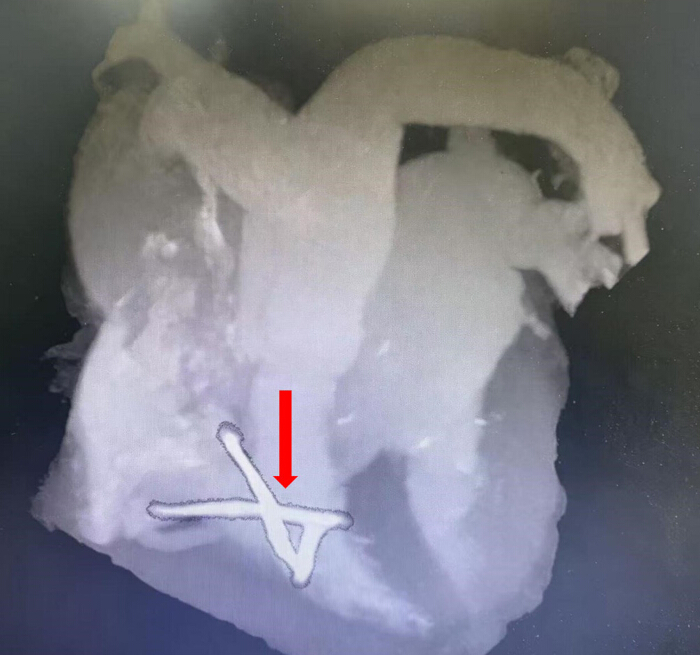
Figure 1: Cardiac computed tomography demonstrating three high-density, rod-shaped cement emboli within the right ventricle. Please click here to view a larger version of this figure.
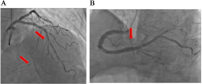
Figure 2: Coronary angiography showing severe stenosis of the left anterior descending artery and radiopaque cement fragments. (A) Coronary angiography shows 50% localized stenosis at the proximal end of the left anterior descending artery, and 80%–90% diffuse stenosis at the middle segment. The arrow above: the stenosis site at the proximal end of the left anterior descending artery; The arrow below: The image of bone cement under X-ray fluoroscopy. (B) Coronary angiography shows a tumor-like dilation in the middle segment of the right coronary artery. The arrow indicates the site of the tumor-like dilation in the right coronary artery. Please click here to view a larger version of this figure.
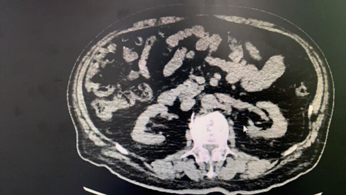
Figure 3: Abdominal computed tomography revealing paravertebral cement leakage adjacent to the lumbar spine. Please click here to view a larger version of this figure.
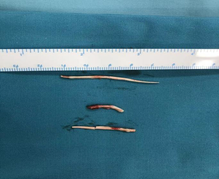
Figure 4: Gross appearance of the removed intracardiac cement emboli. Please click here to view a larger version of this figure.
Supplementary Figure 1: The steps of the protocol.Please click here to download this file.
Discussion
Loading...
$$\rightleftharpoonup{xx}$$
$$\longleftharp{xx}$$,
$$\longrightharp{xx}$$,
Cardiac cement embolism is a rare but serious complication of PVP. Most reported cases occur acutely or within a short period after surgery and are associated with obvious cardiopulmonary symptoms4,5,7. In contrast, the present case demonstrates that intracardiac cement embolism can remain asymptomatic for an extended period and be detected more than 2 years after the initial procedure, expanding the known clinical spectrum of this complication. Computed tomography pulmonary angiography, using a pulmonary embolism (PE) protocol, confirmed isolated intracardiac emboli with no detectable pulmonary cement, directing surgical planning towards the right heart.
The diagnostic challenge in this case was the overlap of symptoms caused by cement embolism and concomitant CAD. Exercise intolerance and chest pain are common manifestations of both conditions, increasing the risk of misdiagnosis or delayed diagnosis if the history of vertebroplasty is overlooked10,11. Multimodal imaging, including echocardiography, cardiac CT angiography, and coronary angiography, played a decisive role in identifying the intracardiac foreign bodies and guiding clinical decision-making.
The morphology and location of the cement emboli in this patient were distinctive. The rod-shaped fragments and myocardial embedding suggest continuous venous migration of bone cement through a cortical defect into the paravertebral venous system, rather than embolization of a single mass10,12. According to the Yeo classification, this pattern corresponds to type C leakage, which is the only subtype capable of causing cardiopulmonary embolism10. Long-term mechanical irritation of the myocardium may explain the absence of acute symptoms despite the potential for catastrophic complications.
Compared with prior reports of delayed intracardiac cement embolism, the present case exhibits distinct chronological and management features. Unlike the systematic review by Lubbad et al.13, where 67 of 71 patients presented symptomatically, mostly within the first postoperative day, this patient remained asymptomatic for 2.5 years, aligning with the rare subgroup of incidentally detected cases (4/71) but extending the reported latency period. Similar to the 5-year delayed case by Cianciulli et al.14, presenting with arrhythmia, this patient developed insidious cardiopulmonary symptoms. However, the embolic burden here comprised multiple rod-shaped fragments with myocardial embedding, differing from the single linear embolus traversing the tricuspid valve in that report. Management entailed open removal under CPB, aligning with the 100% success rate in 46 surgical cases. The decision was driven by embedding-related perforation risk, paralleling the rationale in the previous case. Crucially, the current case uniquely demonstrates simultaneous CABG during cement extraction, a combined approach scarcely described in existing literature for ultra-delayed presentations. Postoperative recovery was uneventful with symptom resolution, reinforcing that timely intervention yields favorable outcomes even after prolonged latency.
There is no standardized treatment strategy for intracardiac cement embolism. Conservative observation may be appropriate for small, asymptomatic fragments, whereas surgical removal is recommended for large, mobile or myocardium-embedded foreign bodies due to the risk of perforation, rupture, or tamponade7,9,12. In the present case, the choice of open removal over percutaneous retrieval was compelled by myocardial embedding and fragment rigidity, which posed unacceptable perforation risk with endovascular manipulation. Single-stage surgery (cement removal plus CABG) was preferred to avoid repeated sternotomy and CPB in an elderly patient, leveraging the same ischaemic period to address both threats. Intraoperatively, the embedded fragment required sharp dissection from fibrous tissue under direct vision to prevent myocardial tear, underscoring the need for CPB backup even if hybrid approaches are considered.
Symptom attribution remains challenging: preoperative chest pain and exercise intolerance likely reflected synergistic contributions from myocardial irritation and coronary ischemia, making it impossible to isolate the effect of cement alone. Consequently, the apparent benefit of combined surgery, while pragmatic for this patient, cannot be extrapolated to isolated cement embolism without CAD. Generalisability is further limited by the unique surgical context; this strategy prioritizes efficiency in dual-pathology scenarios but does not establish superiority over staged interventions in broader populations.
Beyond acute perioperative risks, cement entering vertebral veins may traverse the pulmonary circulation before reaching the right heart, establishing PE as a potential precursor to intracardiac migration, especially with larger or rigid fragments evading capillary trapping15,16,17. In this complex case, simultaneous removal and CABG proved feasible and safe, yet broader endorsement of one-stage strategies requires comparative data. Consequently, we advocate risk-stratified surveillance rather than universal annual imaging: a baseline postoperative chest radiograph and symptom screen suffice for most; echocardiography or chest CT is reserved for patients with high-risk leakage patterns (cortical defects or venous filling) or new cardiopulmonary symptoms. Although migration risk peaks early, delayed embolization years later remains possible; thus, patient education on symptom recognition proves more feasible than routine CT, given radiation and cost constraints.
Disclosures
Loading...
$$\rightleftharpoonup{xx}$$
$$\longleftharp{xx}$$,
$$\longrightharp{xx}$$,
The authors declare no competing financial interests.
Acknowledgements
Loading...
$$\rightleftharpoonup{xx}$$
$$\longleftharp{xx}$$,
$$\longrightharp{xx}$$,
The authors thank the multidisciplinary team members involved in the diagnosis, surgical treatment, and postoperative care of this patient.
Materials
| Name | Company | Catalog Number | Comments |
|---|---|---|---|
| Aspirin | Bayer | www.bayer.com | Antiplatelet agent used to maintain coronary graft patency |
| Beta-blocker | Institution-specific supplier | www.astrazeneca.com | Used for postoperative cardiovascular protection |
| Cardiopulmonary bypass system | Getinge | www.getinge.com/int/products/extracorporeal-life-support/cardiopulmonary-bypass/ | Full perfusion system with oxygenator and arterial filter |
| Clopidogrel | Sanofi | www.sanofi.com | Used for short-term dual antiplatelet therapy |
| Cold crystalloid cardioplegia solution | Institution-specific supplier | N/A | Used for myocardial protection and induction of cardiac arrest |
| Heparin sodium | Pfizer | NDC 0069-0057-01* | Systemic anticoagulant used during cardiopulmonary bypass |
| Left internal mammary artery harvest set | Medtronic | www.medtronic.com | Instruments used for harvesting the left internal mammary artery |
| Microknife | Fine Science Tools | 10056-12 | Used for coronary arteriotomy and fine surgical dissection |
| Microscissors | Fine Science Tools | 14060-09 | Used for sharp dissection around embedded cement fragments |
| Microsurgical forceps | Fine Science Tools | 11003-12 | Used for extraction of intracardiac cement emboli |
| Papaverine hydrochloride | Pfizer | www.pfizer.com | Applied to prevent left internal mammary artery graft spasm |
| Polymethylmethacrylate (PMMA) bone cement | Heraeus Medical | 66004906 (PALACOS R+G) | Bone cement used during vertebroplasty |
| Polypropylene suture, 7-0 | Ethicon | 8701H (PROLENE 7-0) | Used for left internal mammary artery-to-left anterior descending artery anastomosis |
| Protamine sulfate | Pfizer | NDC 0069-0925-01 | Used to reverse heparinization after cardiopulmonary bypass |
| Statin | Institution-specific supplier | www.pfizer.com | Used for long-term secondary prevention after coronary artery bypass grafting |
| Surgical felt pledgets | B. Braun | 1021355 | Used for reinforcement of right ventricular wall repair |
| Transesophageal echocardiography system | GE HealthCare | www.gehealthcare.com/products/ultrasound/cardiovascular-ultrasound | Used intraoperatively to localize cement emboli and confirm complete removal |
Reprints and Permissions
Request permission to reuse the text or figures of this JoVE article
Request Permission