This review evaluates pediatric lung isolation techniques, emphasizing age-specific strategies: double-lumen tubes for children >8 years, intraluminal blockers for ages 2–8 years (ETT ID ≥4.5 mm), and extraluminal blockers or mainstem intubation for infants <2 years. It highlights how 3D printing and protective ventilation optimize safety.
A subscription to JoVE is required to view this content. Sign in or start your free trial.
Review Article
One-lung Ventilation in Pediatric Patients: Lung Isolation Techniques and Clinical Management
In This Article
Summary
Abstract
Implementing one-lung ventilation (OLV) in pediatric patients presents unique clinical challenges due to age-dependent physiological and anatomical constraints. As minimally invasive thoracic surgery expands in pediatrics, the demand for precise lung isolation has increased. This narrative review evaluates current techniques—single-lumen tubes (SLT), double-lumen tubes (DLT), and bronchial blockers (BB)—with a focus on their safety profiles and clinical indications. Findings indicate that DLTs remain the benchmark for older children but are anatomically prohibitive for those under 8–10 years or shorter than 130 cm. BBs are the primary alternative for children aged 2–8 years (ETT ID ≥4.5 mm), although traditional devices such as the Arndt-Fogarty blockers have design limitations. For infants under 2 years, the primary strategies remain SLT mainstem intubation or extraluminal blocker placement, both of which carry a risk of right upper lobe (RUL) obstruction. Emerging innovations, particularly 3D-printed anatomically customized blockers, are highlighted as potential solutions to resolve device “fit” challenges in infants. Perioperative management remains non-standardized; however, evidence-based recommendations emphasize lung-protective ventilation to reduce pulmonary complications. Hypoxemia occurs in approximately 26% of pediatric OLV cases and requires a prioritized management framework that includes fiberoptic verification, secretion clearance, and optimized PEEP/CPAP application. By synthesizing current evidence, this review provides a decision-making algorithm to guide optimal device selection and improve safety outcomes in this vulnerable pediatric population.
Introduction
For anesthesiologists, managing one-lung ventilation (OLV) in children poses significantly greater challenges than in adults. As minimally invasive thoracoscopic and robotic surgical techniques continue to evolve and expand, the demand for OLV in pediatric thoracic surgery is increasing. In parallel, advances in the understanding of pediatric physiology and lower respiratory tract anatomy, along with the development of bronchial blocking devices and related technologies, have contributed to progress in pediatric lung isolation.
Under physiological conditions, the human body maintains a homeostatic balance between ventilation and perfusion (V/Q). In adult thoracic anesthesia, the lateral decubitus position utilizes gravitational and hydrostatic pressure gradients to optimize oxygenation within the dependent lung. In contrast, pediatric patients exhibit a heightened vulnerability to mismatch and subsequent hypoxemia during OLV1,2. Several factors contribute to this disparity. First, the high compliance of the pediatric chest wall provides insufficient support for the dependent lung in the lateral position, reducing functional residual capacity (FRC) and predisposing the ventilated lung to airway closure and atelectasis following anesthesia induction3. Second, children derive less mechanical advantage from the diaphragm than adults. Additionally, the diminished hydrostatic pressure gradient in smaller bodies results in relatively decreased perfusion to the ventilated lung. Finally, the combination of reduced FRC and a metabolic rate approximately three times higher than that of adults (6–8 mL/kg/min vs. 2–3 mL/kg/min) markedly increases the risk of desaturation.
Anatomical disparities between pediatric and adult patients are equally critical in the context of lung isolation. The mean airway diameter in children follows a linear growth pattern from infancy through adolescence4, expanding from approximately 5 mm in neonates to 8 mm by 10 years of age5, and ultimately reaching adult dimensions of 15–20 mm6. Clinically, the left main bronchus is narrower than the right, necessitating the use of smaller endotracheal tubes for left-sided intubation. This constraint is particularly evident in infants aged 0–3 months, in whom the left main bronchus is often too small to accommodate even a 3.0 mm uncuffed tube2. Furthermore, the “margin of safety” for the precise placement of isolation devices is severely limited in younger patients. In children under 8 years old, the distance from the carina to the orifice of the right upper lobe (RUL) is typically less than 1 cm. If a device inadvertently bypasses this orifice, it may result in ventilation of only the right middle and lower lobes, potentially causing severe hypoxemia or obstructing the operative field7. In contrast, the distance from the carina to the left upper lobe orifice is generally three times greater, providing a larger margin for error during left mainstem isolation8.
Therefore, the objective of this narrative review is to synthesize current evidence on pediatric lung isolation techniques, evaluate their clinical profiles, and provide a structured framework for perioperative management.
Access restricted. Please log in or start a trial to view this content.
Review and Perspective
Pediatric lung isolation techniques
Although double-lumen tubes (DLTs) serve as the definitive benchmark for one-lung ventilation (OLV) in adult thoracic surgery, pediatric lung isolation lacks a universally accepted gold standard9. Current practice relies on a tailored selection of single-lumen endotracheal tubes (SLTs), DLTs, or bronchial blockers (BBs), depending on the patient’s age and anatomy10 (Table 1).
| Device Category | Device Name / Model | Outer Diameter / Size | Cuff Characteristics | Central Lumen Availability | Clinical Indications & Limitations | ||
| Bronchial Blockers (BB) | Arndt Blocker | 5F (Smallest) | Low-Pressure / High-Volume (Safe) | Yes (Internal wire loop) | Preferred for children 2-6 years (if intraluminal). | ||
| • Size Req: Intraluminal use requires ETT ID ≥ 4.5 mm. | |||||||
| • Risk: Complex setup; potential wire entrapment. | |||||||
| Fogarty Catheter | 4F, 5F | High-Pressure / Low-Volume (Risk) | No | Used for infants < 2 years (off-label). | |||
| • Limitation: Cannot suction or provide CPAP; no guide channel. | |||||||
| • Risk: High risk of bronchial mucosal ischemia/injury. | |||||||
| Univent Tube | 3.5, 4.5, 5.5 (ID) | High-Pressure / Low-Volume (Risk) | Yes (Separate channel) | Limited to older children (> 6-8 years). | |||
• Size Req: 3.5 ID Univent has large OD ( 8.0 mm), equivalent to 6.0 ETT. 8.0 mm), equivalent to 6.0 ETT. | |||||||
| • Risk: High airflow resistance; thick shaft may injure glottis. | |||||||
| 5F Fuji Blocker | 5F | Low-Pressure / High-Volume (Safe) | No | Alternative for young children. | |||
| • Feature: Pre-formed distal curve aids placement stability. | |||||||
| • Limitation: No central lumen for suction/CPAP. | |||||||
| EZ-Blocker | 7F | Low-Pressure / High-Volume (Safe) | No | Restricted to children > 6 years. | |||
| • Size Req: Requires ETT ID ≥ 5.5 mm (minimum). | |||||||
| • Feature: Y-shape provides stability on carina. | |||||||
| Single-Lumen Tubes (SLT) | Standard ETT | 2.5 - 6.0 mm (ID) | Cuffed / Uncuffed | Yes (Main lumen) | Emergency / Infants < 2 years. | ||
| • Indication: When airways are too small for other devices. | |||||||
| • Limitation: Poor lung isolation seal; cannot suction operative lung. | |||||||
| • Risk: Obstruction of RUL bronchus if placed too deep. | |||||||
| Double-Lumen Tubes (DLT) | Smallest DLT | 26F (8.7 mm OD) | Low-Pressure / High-Volume (Safe) | Yes (Dual lumens) | Restricted to older children (> 8-10 years). | ||
| • Size Req: Corresponds to 6.5 mm ID ETT. | |||||||
| • Advantage: Gold standard for isolation & independent ventilation. | |||||||
| • Risk: Large diameter risks glottic trauma in small airways. | |||||||
Table 1: Comparison of physical parameters and clinical applications of pediatric lung isolation devices. This table summarizes the physical specifications, including outer diameter, cuff characteristics, and central lumen availability, along with the clinical profiles of common lung isolation devices, including bronchial blockers (Arndt, Fogarty, Univent, Fuji, and EZ-Blocker), single-lumen tubes, and double-lumen tubes. Please click here to download this Table.
Single-lumen endotracheal intubation
Endobronchial intubation with an SLT is the simplest method for achieving pediatric OLV and is particularly suitable for children under 2 years of age11. It is often selected when appropriate DLTs or BBs are unavailable due to its sufficient length12. A case reported by Koo et al. (2020) illustrated successful OLV in a 4-year-old child using an SLT guided by an adult fiberoptic bronchoscope (FOB) in the absence of pediatric-sized equipment13. Typically, the tube advances naturally into the right main bronchus. During right-sided intubation, care must be taken to avoid excessive advancement, which may obstruct the right upper lobe bronchus14. For left-sided intubation, rotating the tube bevel 180° and turning the patient’s head to the right can facilitate correct placement15. For children under 8 years, the tube size should be reduced by 0.5 mm inner diameter (ID) for left bronchial intubation2.
Although effective and simple in emergency settings, SLTs have significant limitations1˒2˒12: they may provide an inadequate seal, fail to achieve sufficient lung collapse, and do not allow suctioning or application of continuous positive airway pressure (CPAP) to the non-ventilated lung. Additionally, the endotracheal tube is susceptible to obstruction by secretions and hemorrhagic debris. In clinical practice, a technique employing two conventional endotracheal tubes for selective bronchial intubation and independent lung ventilation has been attempted. While this approach addresses some limitations of SLTs, particularly regarding suctioning and CPAP delivery, it is rarely used due to significant drawbacks, including increased respiratory resistance, difficulty in airway clearance, and the risk of injury to the vocal cords and lower respiratory tract16.
Recent innovations include the “tube-in-tube” technique reported by Mohan et al. (2023), in which a thinner catheter is coaxially placed through a larger endotracheal tube into the left main bronchus. However, this approach depends on the successful passage of the inner tube through the outer tube to achieve effective isolation17.
Double-lumen tubes (DLT)
To overcome the functional limitations of SLTs, the double-lumen tube (DLT) is used as the gold standard in adult thoracic surgery due to its ability to provide independent lung ventilation and suctioning. The DLT is designed with two lumens: one angled, longer lumen for placement within the main bronchus and a shorter lumen positioned within the trachea12. DLTs are available in left- and right-sided configurations, with left-sided DLTs more commonly used to avoid obstruction of the right upper lobe bronchus.
In adults, insertion depth correlates with patient height; however, no corresponding standardized method has been established for children1. When positioning relies solely on auscultation rather than fiberoptic bronchoscopy, accuracy decreases significantly, leaving insertion depth largely dependent on clinical experience.
DLTs offer several advantages, including relatively straightforward intubation, high-quality lung isolation when correctly positioned, the ability to apply CPAP and suction to the operative lung, and rapid transition between one-lung and two-lung ventilation. However, their large size renders them anatomically unsuitable for most pediatric patients. The smallest available DLT is 26F, corresponding to a 6.5 mm endotracheal tube2˒18, and is generally unsuitable for children under 8–10 years of age16.
A specialized DLT designed for neonates and infants (Marraro), consisting of two independent uncuffed endotracheal tubes of different lengths, has been successfully used but has not achieved widespread adoption9,19. For older children and adolescents approaching adult size, height- and sex-based sizing recommendations (e.g., 35F–41F) exist in the adult literature2,20,21; however, these fall outside the practical scope of routine pediatric practice. Consequently, the large size of DLTs creates a critical gap in airway management for younger patients, necessitating alternative strategies2. A structured overview of device selection is illustrated in Figure 1.
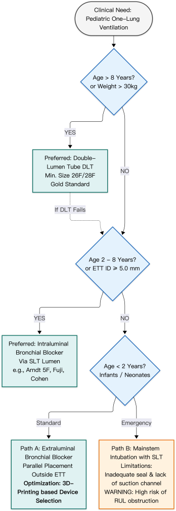
Figure 1: Decision Algorithm for Pediatric Lung Isolation Device Selection. This flowchart illustrates a step-by-step clinical decision strategy based on patient age, weight, and airway anatomy. For older children (>8 years), double-lumen tubes (DLTs) are the gold standard for lung isolation. In children aged 2–8 years (or with an endotracheal tube (ETT) inner diameter (ID) ≥5.0 mm), intraluminal bronchial blockers are preferred via a single-lumen tube. For infants (<2 years), options are limited. Path A indicates extraluminal blocker placement, with 3D–printed device selection highlighted as an optimization strategy to ensure a precise anatomical fit. Path B represents traditional mainstem intubation, with attention to the high risk of right upper lobe (RUL) obstruction. While age-based categorization provides a general framework, device selection is more accurately guided by airway size, specifically ETT inner diameter and the narrow anatomical “margins of safety” in pediatric patients. Abbreviations: ETT, endotracheal tube; ID, inner diameter; RUL, right upper lobe; DLT, double-lumen tube. Please click here to view a larger version of this figure.
Bronchial blockers
Bronchial blockers (BBs) serve as the preferred alternative for children under 6 years of age, offering effective lung isolation with reduced airway trauma12. Traditional devices include the Fogarty embolectomy catheter and the Arndt blocker. The Fogarty catheter lacks a central lumen for lung deflation or CPAP application22 and carries a risk of bronchial injury due to its high-pressure balloon23. In contrast, the Arndt blocker features a high-volume, low-pressure cuff and a wire loop for FOB-guided placement2, making it suitable for pediatric thoracic procedures24. However, neither device is ideal due to size and design limitations, highlighting the need for age-matched devices with ultra-thin membranes25.
Newer devices include the 5F Fuji blocker, the Univent tube, and the EZ-Blocker. The 5F Fuji blocker has a pre-formed distal curve and a stiffer shaft but lacks a central lumen26˒27. When used within a 4.5 mm endotracheal tube, increased airway resistance may occur; in such cases, transitioning to pressure-controlled ventilation–volume guaranteed (PCV-VG) mode is recommended to maintain tidal volume while limiting peak inspiratory pressures. If peak pressures persistently exceed 30–35 cmH₂O despite optimization, switching to an extraluminal approach is advised to prevent barotrauma.
The EZ-Blocker, characterized by a “Y”-shaped design, requires a minimum 5.5 mm ID endotracheal tube, limiting its use in younger children28. The Univent tube integrates a blocker within the endotracheal tube but has a large outer diameter (e.g., 3.5 mm ID corresponds to 8 mm OD), restricting its use to older pediatric patients2. Overall, current devices remain suboptimal for pediatric applications.
There is a clear need for precision-engineered BBs with anatomically tailored designs. A 2022 study by Xiaomin D et al. described the development of a 3D printing–based bronchial blocker specifically for infants and young children29. Although this device has obtained a patent and is undergoing regulatory registration, barriers to widespread clinical use include high time costs and stringent material safety requirements.
At present, the most practical application of 3D printing lies in preoperative simulation. Patient-specific CT-based airway models can be used to test the fit of available devices in vitro, reducing intraoperative trial-and-error and improving safety in complex cases.
For infants under 2 years, the small endotracheal tube diameter often precludes simultaneous passage of a bronchoscope and a blocker. In these cases, extraluminal placement—positioning the blocker outside the endotracheal tube—is often the only viable approach. This technique preserves airway diameter for ventilation. Technical refinements include shaping the blocker tip for rotational guidance30 and using guidewire-assisted placement31. Although combining supraglottic airway devices (SADs) with BBs may reduce airway injury and hospital stay, risks of displacement and aspiration remain significant32.
Perioperative management of pediatric OLV
Despite the absence of standardized guidelines, emerging evidence suggests that lung-protective ventilation strategies reduce postoperative pulmonary complications3. A 2022 clinical trial by Zhu et al. demonstrated that pressure-controlled ventilation–volume guaranteed (PCV-VG) mode reduces peak airway pressure and improves pulmonary compliance compared with traditional volume-controlled ventilation33.
Hypoxemia is a common complication, occurring in approximately 26% of pediatric OLV cases7. A prioritized, stepwise management approach is recommended. Initial interventions include notifying the surgical team, increasing the fraction of inspired oxygen (FiO₂), and clearing airway secretions. Accurate verification of device position is essential. When fiberoptic bronchoscopy is limited, point-of-care ultrasound (POCUS) provides a reliable alternative. The absence of lung sliding combined with the presence of a lung pulse on the operative side serves as a practical indicator of successful isolation. Changes in end-tidal CO₂ (EtCO₂), including sudden decreases or waveform alterations, may indicate device malposition. A summary of complications and associated risk factors across techniques is provided in Table 2.
| Technique | Common Complications | Clinical Risk Factors / Etiology |
| Single-Lumen Tube (SLT) Endobronchial Intubation | Mucosal injury; inability to suction secretions or blood from the operative lung; suboptimal for right-sided isolation. | Distal tip of the SLT prone to tissue trauma; inaccessible operative airway for suctioning; extremely short carinal-to-RUL distance in infants leading to RUL obstruction and lung collapse failure. |
| Double-Lumen Tube (DLT) | Laryngeal and tracheal mucosal injury; vocal cord edema. | Large outer diameter and inherent stiffness of DLTs, which predispose the narrow and delicate pediatric airway to trauma. |
| Intraluminal Bronchial Blocker (BB Inside-ETT) | High airway resistance; hypoxemia; malposition, displacement, or dislodgement of the cuff. | Significant increase in ventilatory resistance when used with small-diameter ETTs; cuff migration or dislodgement triggered by positional changes or surgical manipulation. |
| Extraluminal Bronchial Blocker (BB Outside-ETT) | Insertion failure; device malposition; cuff displacement or dislodgement. | Difficulty in passing small, flexible blockers through the glottis; displacement caused by positional changes or surgical traction. |
| Insufflation of CO2 into the Operative Hemithorax | Hypoxemia; hypercapnia; myocardial depression. | Ventilation/perfusion (V/Q) mismatch in the lateral decubitus position; reduction in functional residual capacity (FRC); lack of continuous cuff pressure monitoring. |
Table 2: Summary and comparison of common complications across pediatric lung isolation techniques. This table provides a comparative synthesis of the primary complications associated with each isolation modality and outlines the underlying anatomical and physiological risk factors to assist clinicians in preoperative risk assessment and technique selection. Please click here to download this Table.
If hypoxemia persists despite correct positioning, secondary interventions include applying positive end-expiratory pressure (PEEP) to the ventilated lung or continuous positive airway pressure (CPAP) to the non-ventilated lung2˒20. As a final rescue measure, intermittent two-lung ventilation or conversion to open thoracotomy may be required34. Continuous vigilance and a low threshold for reverting to two-lung ventilation are essential for patient safety35.
Access restricted. Please log in or start a trial to view this content.
Conclusions
The management of one-lung ventilation (OLV) in pediatric patients remains one of the most demanding tasks in modern anesthesiology and is fundamentally distinct from adult practice due to unique physiological and anatomical constraints. This review highlights that, despite increasing demand for pediatric thoracic surgery, a unified “gold standard” for lung isolation remains elusive. Device selection is dictated by age and airway dimensions: while double-lumen tubes (DLTs) provide optimal isolation in older c...
Access restricted. Please log in or start a trial to view this content.
Disclosures
The authors declare no conflict of interest.
Acknowledgements
This work was supported by Capital’s Funds for Health Improvement and Research (No. 2022-2Z-2098 to Xin Zhao), Beijing Hospitals Authority Clinical Medicine Development of Special Funding Support (No. XMLX202145 to Xin Zhao), the Beijing Municipal Science & Technology Commission Proof of Concept Platforms (No. 2024030119), and the Beijing Municipal Science & Technology Commission Technology Transfer Office (No. 20250481320).
Access restricted. Please log in or start a trial to view this content.